
Filchneria mongolica ( Klapálek, 1901 ), Klapalek, 1901
|
publication ID |
https://doi.org/10.5281/zenodo.199585 |
|
DOI |
https://doi.org/10.5281/zenodo.6209671 |
|
persistent identifier |
https://treatment.plazi.org/id/8F7787D6-EC22-E342-8AB4-FDF3FF5191C2 |
|
treatment provided by |
Plazi |
|
scientific name |
Filchneria mongolica ( Klapálek, 1901 ) |
| status |
|
Filchneria mongolica ( Klapálek, 1901) View in CoL
( Figs. 1–19 View FIGURE 1 View FIGURES 2 – 4 View FIGURES 5 – 9 View FIGURES 10 – 15 View FIGURES 16 – 19 )
Dictyogenus mongolica Klapálek, 1901: 13 , Fig. 10 View FIGURES 10 – 15 .
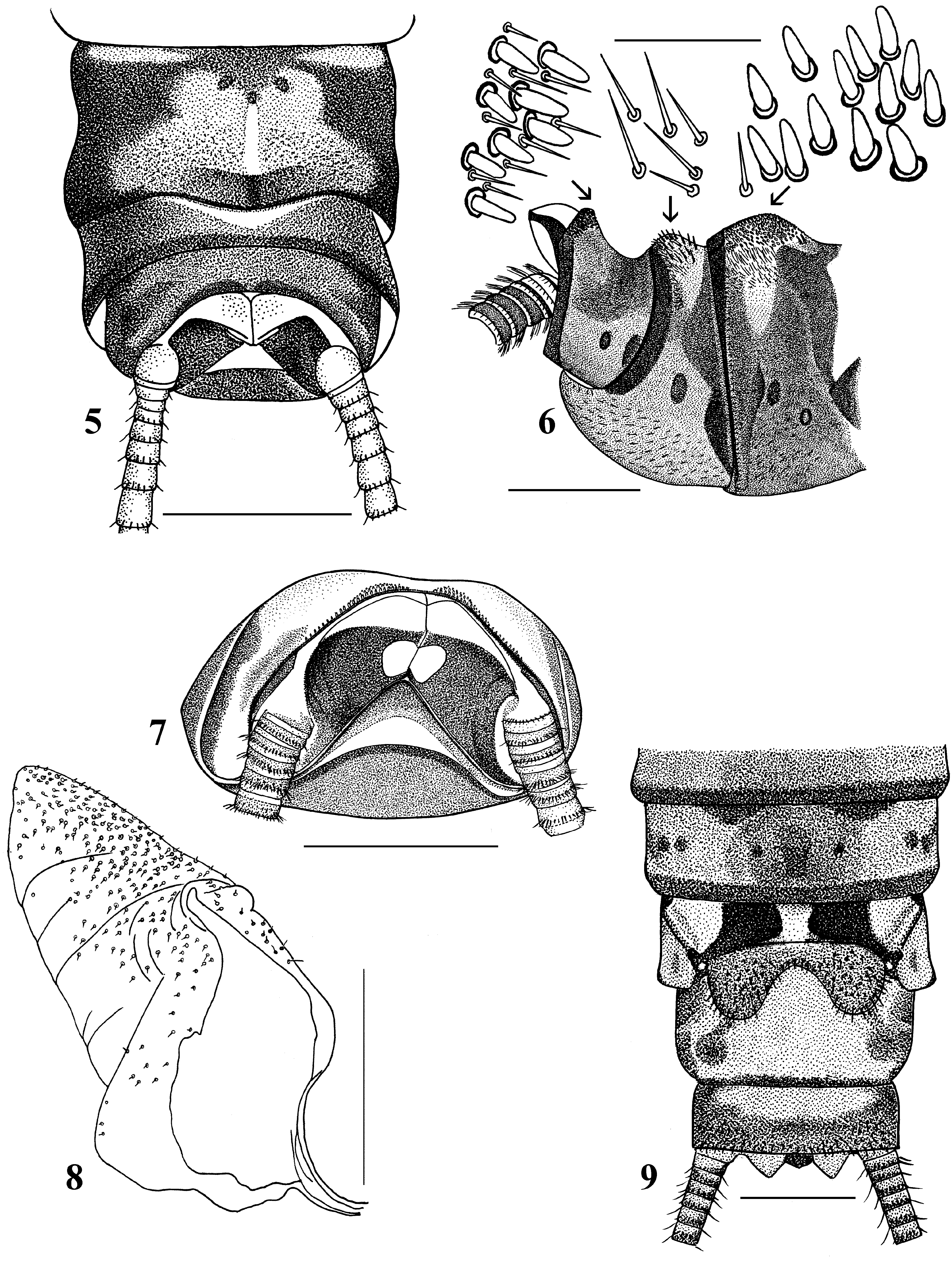
Diagnosis. The narrow medially interrupted strip of short, blunt thick spines on the posterior margin of tergum 10 distinguishes the male of F. mongolica ( Fig. 7 View FIGURES 5 – 9 ). The female is distinguished by a deep notch between the lobes of the subgenital plate ( Fig. 9 View FIGURES 5 – 9 ). The egg of F. mongolica is triangular in cross section ( Fig. 10, 12 View FIGURES 10 – 15 ). In addition to longitudinal ridges, a transverse ridge is found near the posterior pole ( Fig. 11, 12 View FIGURES 10 – 15 ). The collar is formed by projections of the three longitudinal ridges, which are flat and triangular in lateral view ( Fig. 14 View FIGURES 10 – 15 ). The surface of the chorion is granular ( Fig. 13, 15 View FIGURES 10 – 15 ).
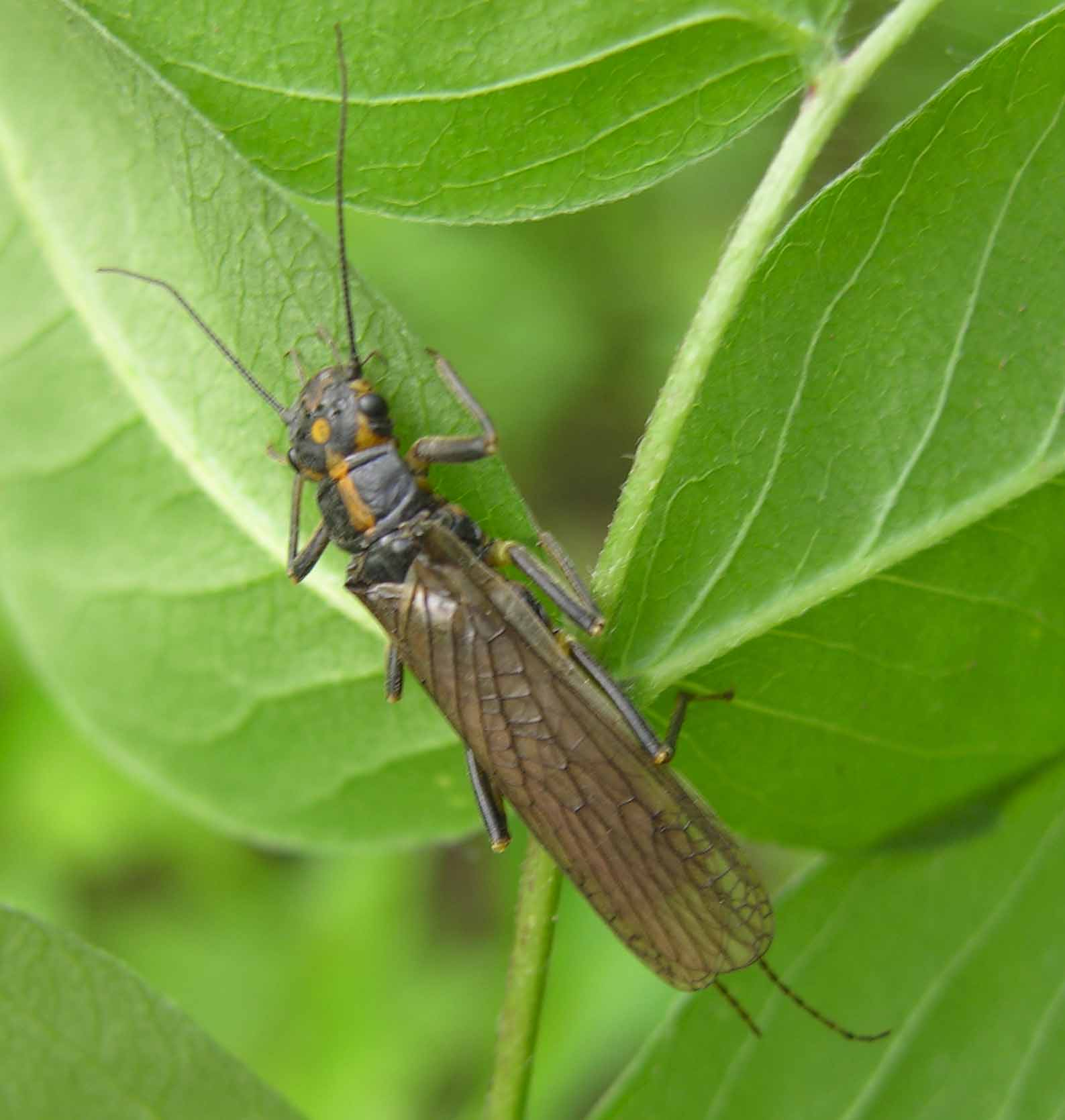
Adult habitus. Males are brachypterous, while females are macropterous. Wings have a brownish tint and brown veins ( Fig. 1 View FIGURE 1 ). The general body colour is dark, males are mostly black. The head is dark, wider than the pronotum. In front of the black, distinct M-line a yellow spot projects onto the clypeus; in females this spot includes a dark centre ( Figs. 2, 3 View FIGURES 2 – 4 ). The interocellar area carries a small tear-shaped yellow spot which is widest anteriorly. The anterolateral margins of the head are dark from the compound eyes to across the clypeus. A yellow U-shaped band extends across the occiput and forms a triangular medial projection along the epicranial stem ( Figs. 2, 3 View FIGURES 2 – 4 ). Behind each compound eye is a dark posterolateral spot. Submental gills are lacking. Antennae are brown and palpi pale.
The pronotum is brown with a broad, yellow median band that is widest in its basal third ( Fig. 2 View FIGURES 2 – 4 ). The pronotal rugosites are dark brown, meso- and metascuta are dark brown. The mesosternal Y-arms reach the posterior corners of the furcal pits.
The abdomen is covered by short colourless clothing hairs. Abdominal segments 1–3 are divided laterally by hairless pleural membranes, the remaining segments are undivided. Legs are brown, with a basal dark brown spot on the tibia. Cerci pale, each cercal segment darker in its distal half.
Male. Mean body length 13.5 ± 1.0 (sd) mm (n=15). Wings short, not reaching the posterior margin of tergum 2. Mean forewing length is 3.5 ± 0.3 mm, wingspan 9.6 ± 0.8 mm.
Abdominal tergum 8 with a butterfly-shaped brownish spot which is caudally expanded and divided by a longitudinal, pale, median line; two submedial swellings densely covered by sensillae basiconica and colourless hairs close to the posterior margin, especially obvious in lateral view ( Figs. 5, 6 View FIGURES 5 – 9 ). Abdominal tergum 9 with two similar but smaller posterior swellings which are covered by acute sensilla basiconica and short fine conspicuous colourless hairs posterolaterally ( Fig. 6 View FIGURES 5 – 9 ); anterior tergal margin medially with a membranous, triangular light spot which is almost hidden under tergum 8 ( Fig. 5 View FIGURES 5 – 9 ). Sternum 9 light brown medially, posterior margin tongue-shaped, extended backward and upcurved, corresponding to 2/3rd length of sternum 10 ( Fig. 6 View FIGURES 5 – 9 ). Tergum 10 in lateral view, arcuate and upcurved ( Fig. 6 View FIGURES 5 – 9 ), its posterior margin obtusely angled with a narrow medially interrupted strip of short, blunt, thick spines ( Figs. 5–7 View FIGURES 5 – 9 ). Paraproct lobes in repose are dorsally pale, dorsomedially touching each other and pressed against the inner surface of tergum 10. Laterally, lobes are slightly angular and project under abdominal tergum 10 ( Fig. 5 View FIGURES 5 – 9 ). The paraproct sclerite is wide and well sclerotized basally, distally it narrows rapidly, the thin apex resembling a slightly rounded claw ( Fig. 8 View FIGURES 5 – 9 ). In caudal view each paraproctal sclerite surrounds a pale oval spot ( Fig. 7 View FIGURES 5 – 9 ). The eversible paraproct lobe (EPL) emerges from a fold near the thin apex of the sclerite. The everted lobe is bell-shaped, unpigmented, soft, and is covered by many fine sensory scales and small thin spinules, especially dorsally ( Fig. 8 View FIGURES 5 – 9 ).
Female. Mean body length 17.0 ± 4.6 mm (n=15). Forewing length is 16.7 ± 0.9 mm, wingspan 36.7 ± 2.0 mm. Front wing tinted, fumose, hind wing paler. The venation includes an irregular net near the apex sometimes consisting of three rows of cells. Four cross veins between C and Sc; five apical veins between Sc and R1 ( Fig. 4 View FIGURES 2 – 4 ). Rs with five apical branches. Eight veins between М and Cu2; three anal veins. Hind wing anal area large, A2 and A5 forked.
Abdominal terga brown, with three pairs of small, black spots in a row; tergum 10 is pale brown with a small dark brown medial spot. Abdominal sterna yellowish-brown, anterior margin of abdominal sterna 2–7 with medially interrupted dark brown stripe, and one median and three lateral pairs of dark spots, the median pair smallest. Sternum 8 with a large subgenital plate extending half the length of sternum 9 ( Fig. 9 View FIGURES 5 – 9 ), posterior margin of the plate with a parabolic notch separating two large, rounded lateral lobes covered by red spinules. An arched transverse fold separates the lobes from two broad angled dark bands ( Fig. 9 View FIGURES 5 – 9 ) on the anterior part of the sternum. Abdominal sternum 9 is medially pale, with a pair of large, dark, rounded spots posterolaterally. Abdominal sternum 10 yellow anteriorly, dark posteriorly.
Egg. Trilateral ( Figs. 10–12 View FIGURES 10 – 15 ), 462 X 295 µm, depth 302 μm. Longitudinal ridges delimit the three sides of the egg. Each side has additionally a transverse ridge close to the posterior pole ( Figs. 11, 12 View FIGURES 10 – 15 ). Collar long, formed by medially projecting extensions of the three longitudinal ridges, each flat and triangular laterally, crest-like, inner edges slightly curved ( Fig. 14 View FIGURES 10 – 15 ). A row of 3–6 micropyles is found near the transverse ridge ( Fig. 12 View FIGURES 10 – 15 ) on each of the three sides. Anchor mushroom-shaped with short pedicel ( Fig. 11 View FIGURES 10 – 15 ) with single globular bodies on the whole anchor plate. The margin of the anchor covers the collar completely ( Figs. 11, 12 View FIGURES 10 – 15 ). The structure of the chorion surface is rough with small light tubercles ( Figs. 13, 15 View FIGURES 10 – 15 ).
Larvae. Mature nymphs are 19.0±1.0 mm long (n=15). The general colour is grey-brown with a pale pattern ( Fig. 18 View FIGURES 16 – 19 ). Body covered by hardly visible short colourless clothing hairs. Antennae and cerci grey, brownish apically. The ventral body side is grey. The head is slightly wider than the pronotum with a large, triangular, pale spot in front of the anterior ocellus extending onto the clypeus ( Fig. 18 View FIGURES 16 – 19 ). M-line brown, indistinct; interocellar area brown with small triangular central pale spot, closed posteriorly. A small triangular pale spot laterally from each posterior ocellus is connected with a narrow pale band extending anterolaterally to the area of the tentorial callosities. Occiput with triangular pale spot around the epicranial stem with two pairs of pale spots bordered by sinuate brownish rows behind each eye. No postocular spinule row. Lacinia ( Fig. 19 View FIGURES 16 – 19 ) bidentate, apically narrow, basal half dramatically expanded. No submarginal setae except two small transparent ones below the base of the subapical tooth. There are three setae at the juncture of the apical teeth. Galea reaches the base of the subapical tooth. Mandible ( Fig. 16 View FIGURES 16 – 19 ) simple, not deeply cleft, with medial setae; mandibular teeth without serrations; a patch of acanthae basally from the last small tooth. The submental gills are short ( Fig. 17 View FIGURES 16 – 19 ). Pronotum rectangular with rounded angles, brownish with wide pale lateral margins; pale, median, dorsal stripe narrow and wider posteriorly, laterally merging with pale reticulate markings inside the brown lateral fields, all margins without fringe of bristles ( Fig. 18 View FIGURES 16 – 19 ). The mesosternal Yarms meet the posterior corners of the furcal pits. Meso- and metanota anteriorly with six longitudinal narrow short pale stripes in front of an arched brown transverse band. Pale spots at the base of the wingpads extend backward and narrow posteriorly ( Fig. 18 View FIGURES 16 – 19 ). Legs grey, femur with a basally incomplete elongate light brown mark that darkens distally. Outer margins of femur, tibia and tarsus with a fringe of colourless silky hairs. The surface of femur and tibia with few scattered brown spinules.
Abdominal segments 1–3 divided by hairless pleural membranes, remainder undivided, having a continuous posterior fringe of small brown setae. Terga at posterior margin with a few colourless long hairs medially. Abdominal terga brownish, each with a transverse row of six small, black spots grouped in three pairs; two rows of large, oval, pale paramedial spots separated by a narrow median brown stripe, this stripe being incomplete on terga 9 and 10 so as to form heart-shaped spots ( Fig. 18 View FIGURES 16 – 19 ). All abdominal terga have a few short, stout, brown intercalary spinules that become more numerous on abdominal sterna. Tergum 10 of mature male nymph not modified. Paraprocts long, apex rounded. Cerci grey with dorsal fringe of fine, silky, colourless hairs ( Fig. 18 View FIGURES 16 – 19 ), increasing in length towards apical cercal segments. Each cercal segment with an apical whorl of short brown setae.
Material examined: Slide with eggs from the female holotype: N. Mongolei Lederer 92/ mongolica Klapálek /Mus. Vienna; pinned, macerated abdomen in microvial. 1 male, 1 female, Mongolei, Central aimak: Songino, 24 km SW von Ulan Bator, 1300 m, 7.VI.1966. Am Ufer des Flusses Tola unter Steinen geeinzelt; Z. Kaszab, No. 502 (Hungarian Natural History Museum, Budapest); Russia: Buryatiya: 1 nymph, Selenga River, set. Kolesovo, 52.0838 N, 106.3885 E, drift, 01.V.2005, coll. N. Bazova; 3 females, the same place, 30.IV.2007, coll. A. Bazov; 7 males, 9 females, 9 eggs masses, the same place, 07–13.V.2009, coll. A. Bazov, N. Bazova; 1 male, Chikoi River, Selenga R. Basin, set. Ust’-Kiran, 50.1951 N, 106.5122 E, 27.V.2008, coll. M. Proshchelykin; 1 nymph, Chikoi R., set. Char’yasta, 50.4317 N, 106.3857 E, drift, 27.XII.2008, coll. A. Bazov; 7 males, 3 females, Chikoi R., Selenga R. Basin, 25 km from the mouth, 05.V.2009, coll. A. Bazov, N. Bazova; Amurskaya oblast’: 1 female, Meun R., Nora R. Basin, Selemdzha R. Basin, Zeya R. Basin, Amur R. Basin, 52.5686 N, 130.0737 E, 18.VI.2003, coll. Eu. Dimitruyk; Primorskyi krai: 1 female, Bolshaya Ussurka River, Ussuri R. Basin, Amur R. Basin, set. Zvenigorodka, 45.5859 N, 133.5918 E, 08.VI.2004, coll. V. Teslenko; 1 female, Ussuri River, Amur R. Basin, 4 km from set. Stepanovka, apiary, 44.5619 N, 133.3282 E, 15.VI.2005, coll. T. Tiunova, fot. M. Tiunov.
Distribution. The species inhabits large rivers: the transfrontier Selenga River in Mongolia and Russia (southern Siberia), and the Amur River Basin (Meun River, Bolshaya Ussurka River, Ussuri River) in the south of the Russian Far East.
Remarks. We studied the presumed F. mongolica material described by Zhiltzova (1971):
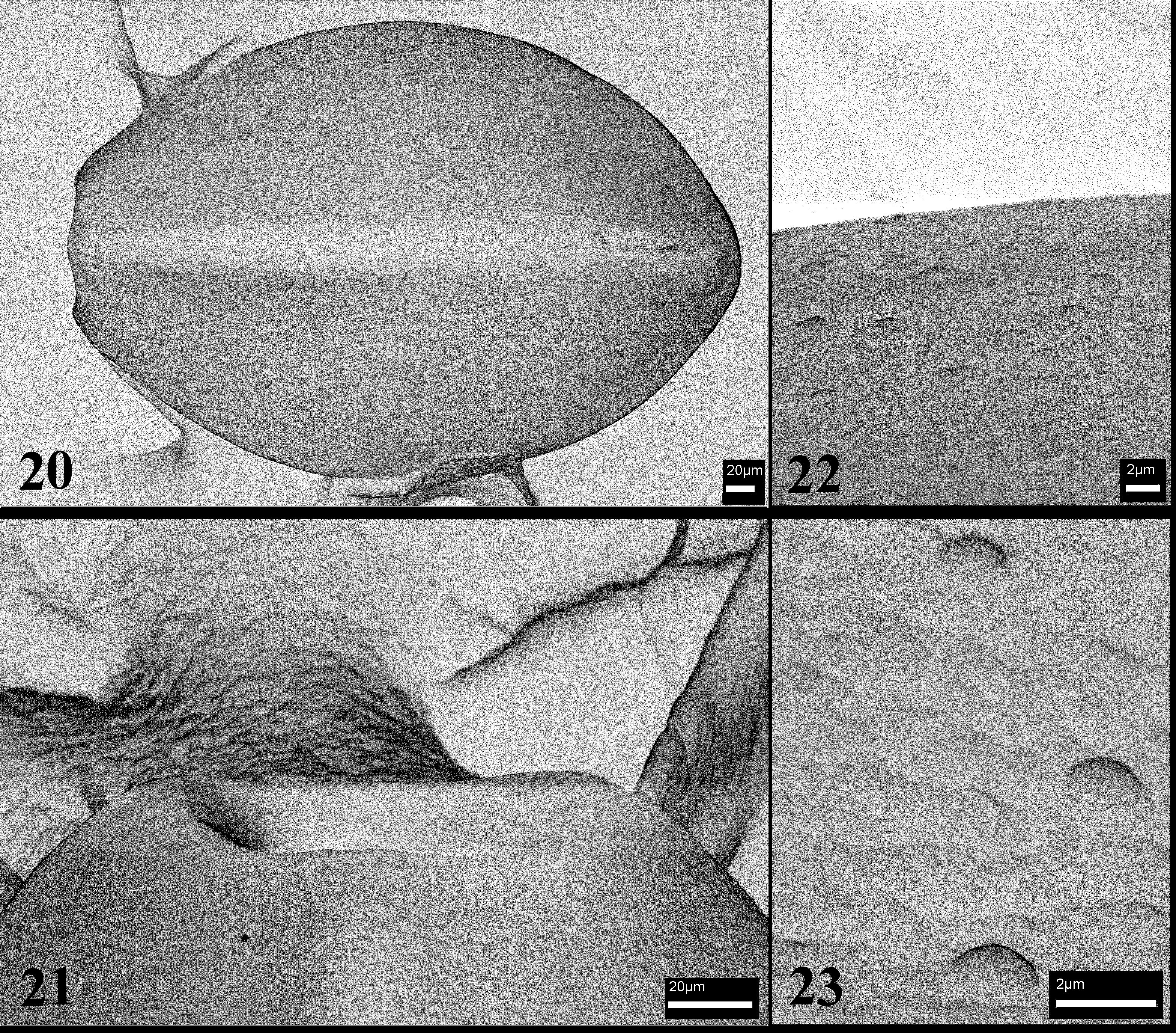
Kyrgyzstan: 4 females, Kurumdu River, Susamyr River Basin, 29.VI.1966, coll. L. Zhiltzova; 3 males, 1 female, 2 nymphs, Karakolka River, Susamyr River Basin, 19.X.1967, coll. L. Zhiltzova. They are distinct from F. mongolica . The female subgenital plate is similar but the notches between the lobes are less deep than in F. mongolica . Only a female from the Karakolka River contains mature eggs which differ distinctly from eggs of F. mongolica . In particular, the transverse ridge close to the posterior egg pole is absent and the micropyles are situated close to the equator ( Fig. 20 View FIGURES 20 – 23 ). Instead of a collar there are only very short extensions of the three longitudinal ridges ( Fig. 21 View FIGURES 20 – 23 ). The structure of the chorion surface is rough, with low raised tubercles ( Figs. 22, 23 View FIGURES 20 – 23 ). The artificial eversion of the EPLs in males from the Karakolka River was unsuccessful but the stripe of short, blunt thick spines on the posterior margin of tergite 10 is different, not narrow and medially interrupted as in true F. mongolica .
Males from the Klapálek collection regarded as F. mongolica by Raušer (1968) were not available but the very detailed and careful illustrations of the last tergites show no short blunt thick spines on the posterior margin of tergum 10 ( Raušer 1968, his figs. 62–66).
The first author studied P. stigmata ( Ra, Kim, Kang, Ham, 1994) : South Korea: 3 males, 1 female, north of Chonchon, 13.IV.1995, coll. V. Kuznetzov. The eggs of F. mongolica are similar to eggs of the Korean species ( Figs. 24–27 View FIGURES 24 – 29 ), but in P. stigmata the collar is wider and its three ridges have almost straight medial edges directed towards the longer anchor pedicle. The edge of the anchor does therefore not cover the collar completely; in F. mongolica it does. The paraprocts of male P. stigmata are longer than those of F. mongolica , and crossed. The paraproct sclerite of P. stigmata narrows gradually towards the apex and forms a thin rod rounded at the tip; the EPLs are short, resembling small bulbs situated at an almost right angle to the paraproct sclerite ( Fig. 28 View FIGURES 24 – 29 ). The female subgenital plate of P. stigmata has a weak, shallow notch and two concave stripes extend from the posterolateral side of the sternum ( Ra et al. 1994). There are distinct differences in the shape of spots in front of the M-line and in the interocellar area on the head ( Fig. 29 View FIGURES 24 – 29 ). Considering the main specific characters such as egg morphology, male EPLs, and the fact that abdominal segments 1–3 are divided by a membranous pleura, the first author assumes that P. stigmata should be removed to the genus Filchneria , but study of the holotype is required.


No known copyright restrictions apply. See Agosti, D., Egloff, W., 2009. Taxonomic information exchange and copyright: the Plazi approach. BMC Research Notes 2009, 2:53 for further explanation.
|
Kingdom |
|
|
Phylum |
|
|
Class |
|
|
Order |
|
|
Family |
|
|
Genus |
Filchneria mongolica ( Klapálek, 1901 )
| Teslenko, Valentina A., Zwick, Peter & Bazova, Natalya V. 2010 |
Dictyogenus mongolica Klapálek, 1901 : 13
| Klapalek 1901: 13 |