
Gaeolaelaps queenslandicus ( Womersley, 1956 )
|
publication ID |
https://doi.org/ 10.24349/acarologia/20184266 |
|
persistent identifier |
https://treatment.plazi.org/id/912387E7-F425-FFA3-40E5-FB6EAC22FAAC |
|
treatment provided by |
Marcus |
|
scientific name |
Gaeolaelaps queenslandicus ( Womersley, 1956 ) |
| status |
|
Gaeolaelaps queenslandicus ( Womersley, 1956) and G. angustus ( Karg,
1965)
Gaeolaelaps queenslandicus ( Womersley, 1956) originally described as a species of Androlaelaps , based on a single specimen collected from leaf debris in Taringa, South Queensland, Australia. Womersley (1956) described it as a rather small species with dorsal shield not covering entire idiosomal dorsum, bears about 46 setae longer on posterior margin. Based on Womersley’s (1956) figure 48–B (p. 578), the left lateral margin of the dorsal shield presents a small inward curvature at level of Z3 seta, while the margin is almost straight on the opposite side. Karg (1965) described Hypoaspis (Hypoaspis) angustus based on specimens collected from meadow soil, Berlin, Kleinmachnow, which have dorsal shield with straight lateral margins in the opisthonotal region. Many morphological characters not described in original descriptions of Karg (1965) and Womersley 1956). Ten years after the original description of Hypoaspis queenslandicus , Costa (1966) redescribed it and rectified some morphological characters of this species based on type material and Israel specimens, especially concerning the dorsal shield margins. Redrawn figures ( Figure 1 p View Figure 1 . 142) of Costa (1966) based on mentioned specimens of G. queenslandicus show a dorsal shield with lateral margins that are clearly curved inwards at level of Z3– S3, and angled posteriorly at level of Z4– S4 ( Figures 2A, B View Figure 2 ).
Costa (1966) pointed out the morphological differences between G. queenslandicus and G. angustus , and presented these two species as distinct based on the following differences:
1) Opisthonotal region of dorsal shield with lateral margins concave G. ( queenslandicus ) vs straight G (. angustus ).
2) The distribution of dorsal shield setae.
3) Relative lengths of dorsal shield setae (longer in the first species).
4) The shape of deutosternal groove G (. queenslandicus with discontinuous lateral margins of deutosternal groove while the second one with contiguous lateral margins) and their relative sizes (longer in the first one).
5) The smaller number of teeth on the cheliceral fixed digit G in. angustus .
All mentioned characters and some other morphological traits examined as follow.
1 — Shape of dorsal shield
The lateral margins of the dorsal shield G in. angustus (based on type materials and Iranian specimens with straight opisthonotal margins) tend to be converged in podonotal region at level of r3–r4 as an almost straight line and without curvature along it with nearly rounded posterior end of the shield ( Figures 1 View Figure 1 A-B, 6 and 8).
Podonotal lateral margins of dorsal shield in G. queenslandicus sensu Costa (1966)
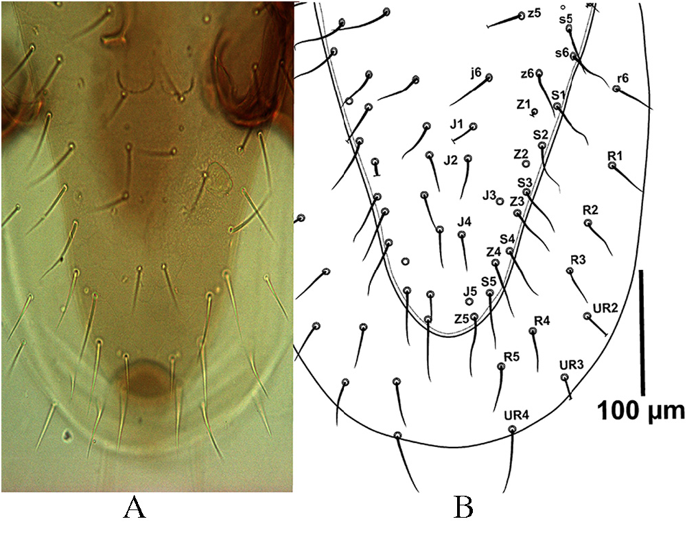
and examined Australian specimens are similar to those in G. angustus , but the margins of opisthonotal part with concave at the level of Z3– S4, then tapered near the level of Z4– S5 as a straight line in both sides of shield with posterior end rounded ( Figures 2A, B View Figure 2 ).
Based on the redescription of Costa (1966) – Figure 1 p View Figure 1 . 142- this is one of the main morphological difference between these species. Our observations on Australian specimens and extensive microslides of specimens previously identified as G. angustus ( Figures 1A,B View Figure 1 ) and
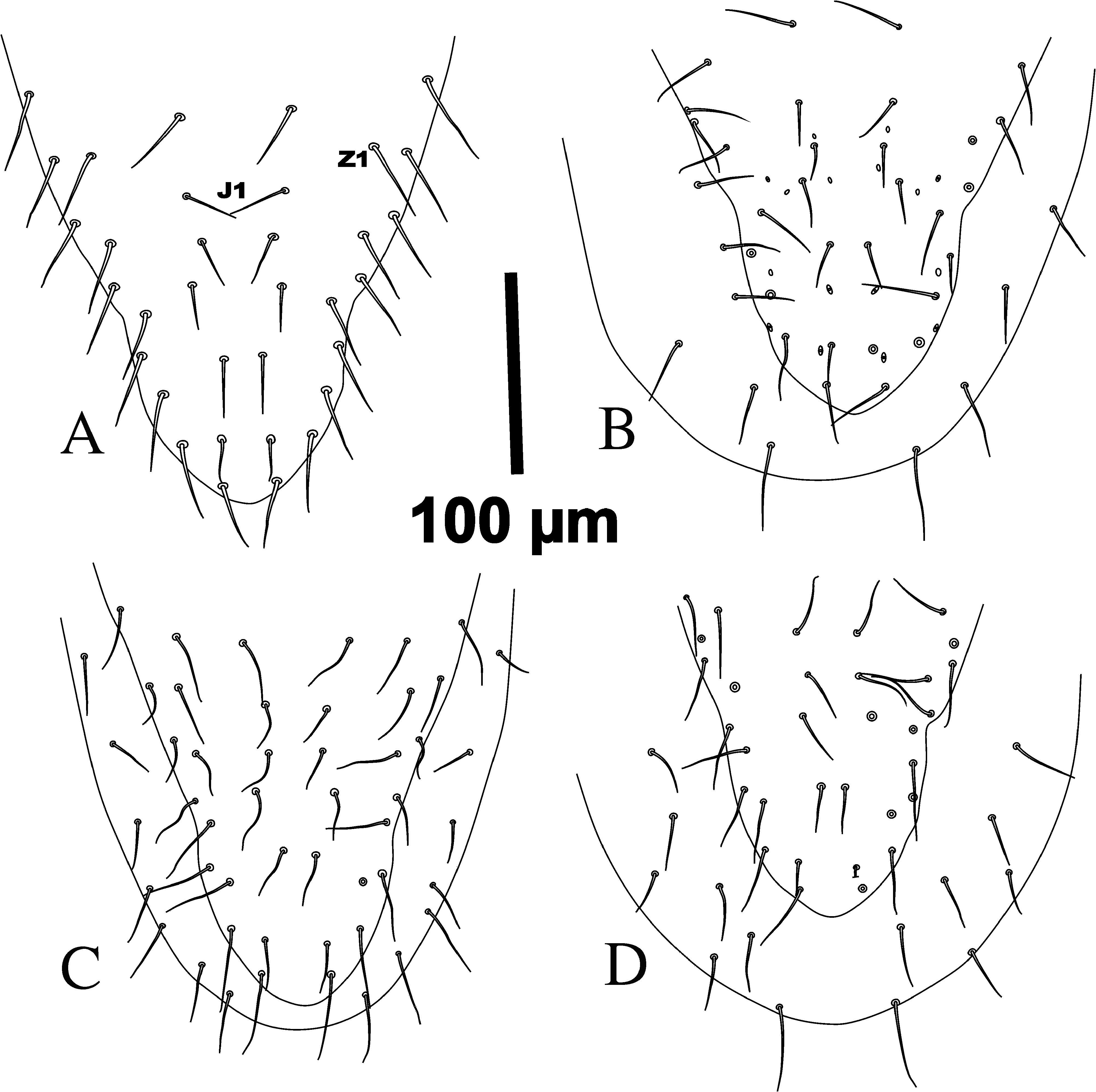
G. queenslandicus ( Figures 2 View Figure 2 , 3 View Figure 3 ), showed considerable variation in this character that could be considered in different groups as follows: (1) specimens with dorsal shield with straight lateral margins (as in Figures 1A, B View Figure 1 ) which considered G as. angustus . (2) specimens having dorsal shield margins with clear depressions at level of S3– Z3 [as in Figures 2 View Figure 2 , 3 View Figure 3 A-C and similar to some (two microslides) Australian specimens] which considered as G. queenslandicus . (3)
specimens with only slightly depressed margins at level of S3-Z3 ( Figures 4 View Figure 4 A-D). (4) specimens with asymmetrical dorsal shield, A: having nearly deep depression on one side and slightly concave on other side ( Figure 8 and two slides of Australian specimens); B: having lateral margin slightly concave on one side and nearly straight margin in other side ( Figures 5A, B View Figure 5 ).
The above mentioned variations in lateral margins of dorsal shield in opisthonotal part shows that this character with inconstancy condition and worthless taxonomically.
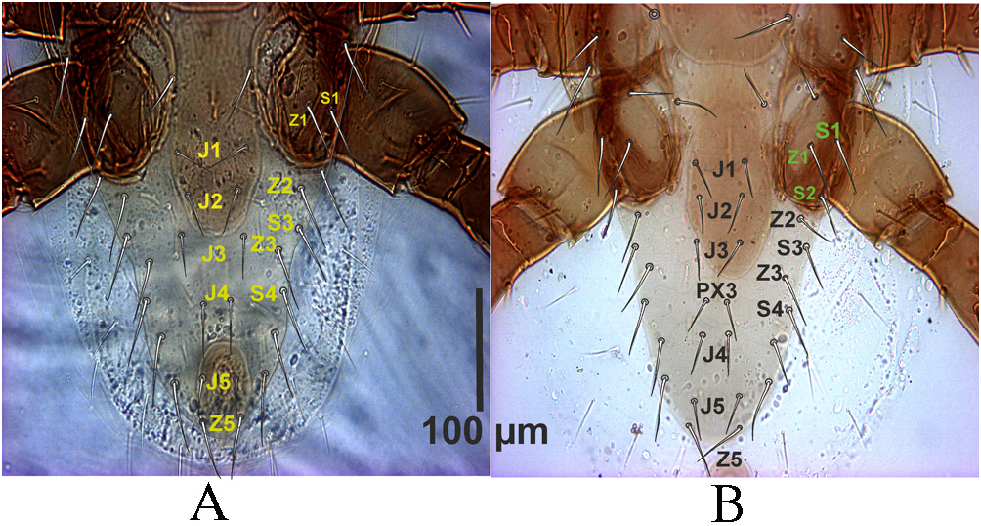
2 — Number and distribution of dorsal shield setae
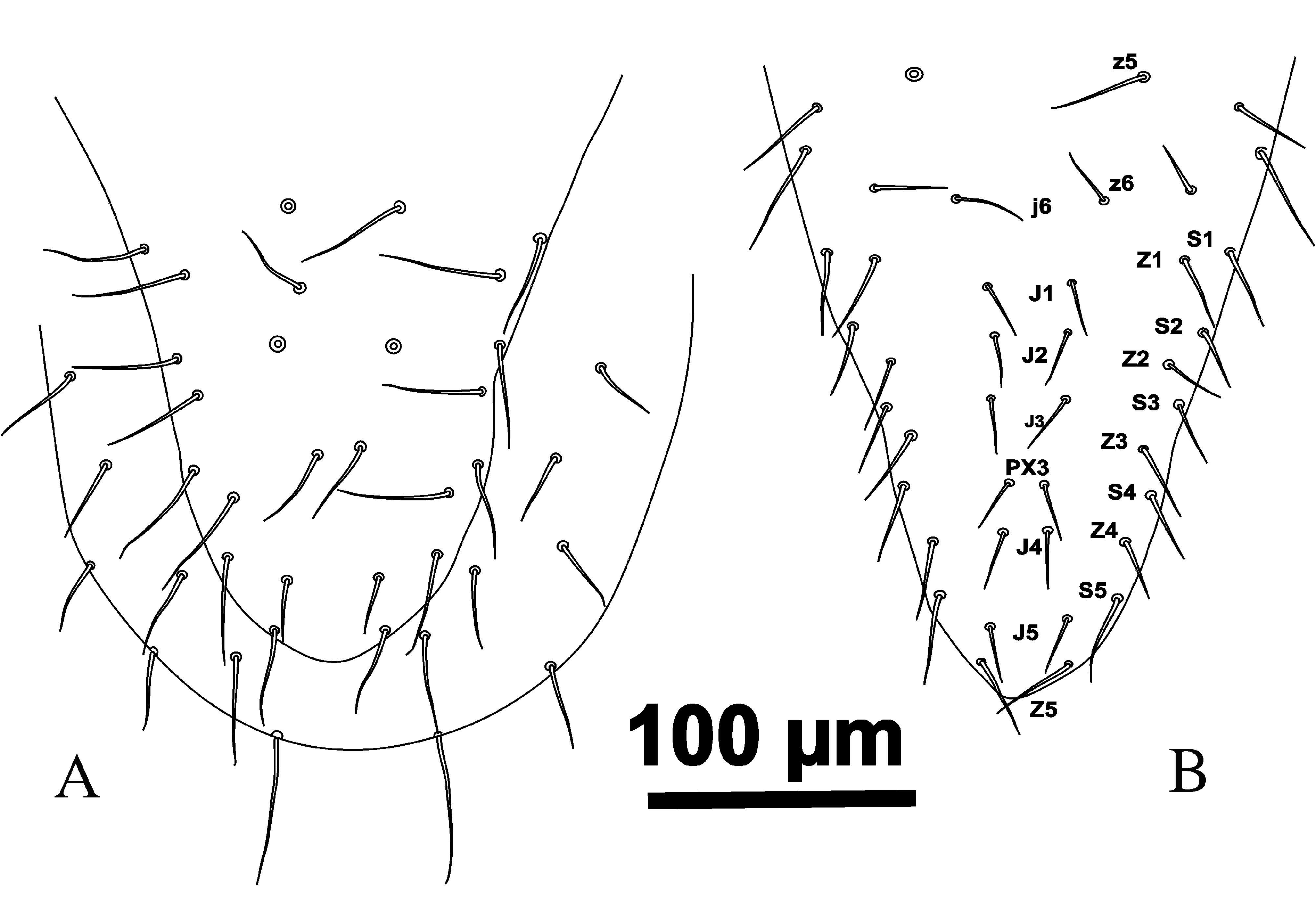
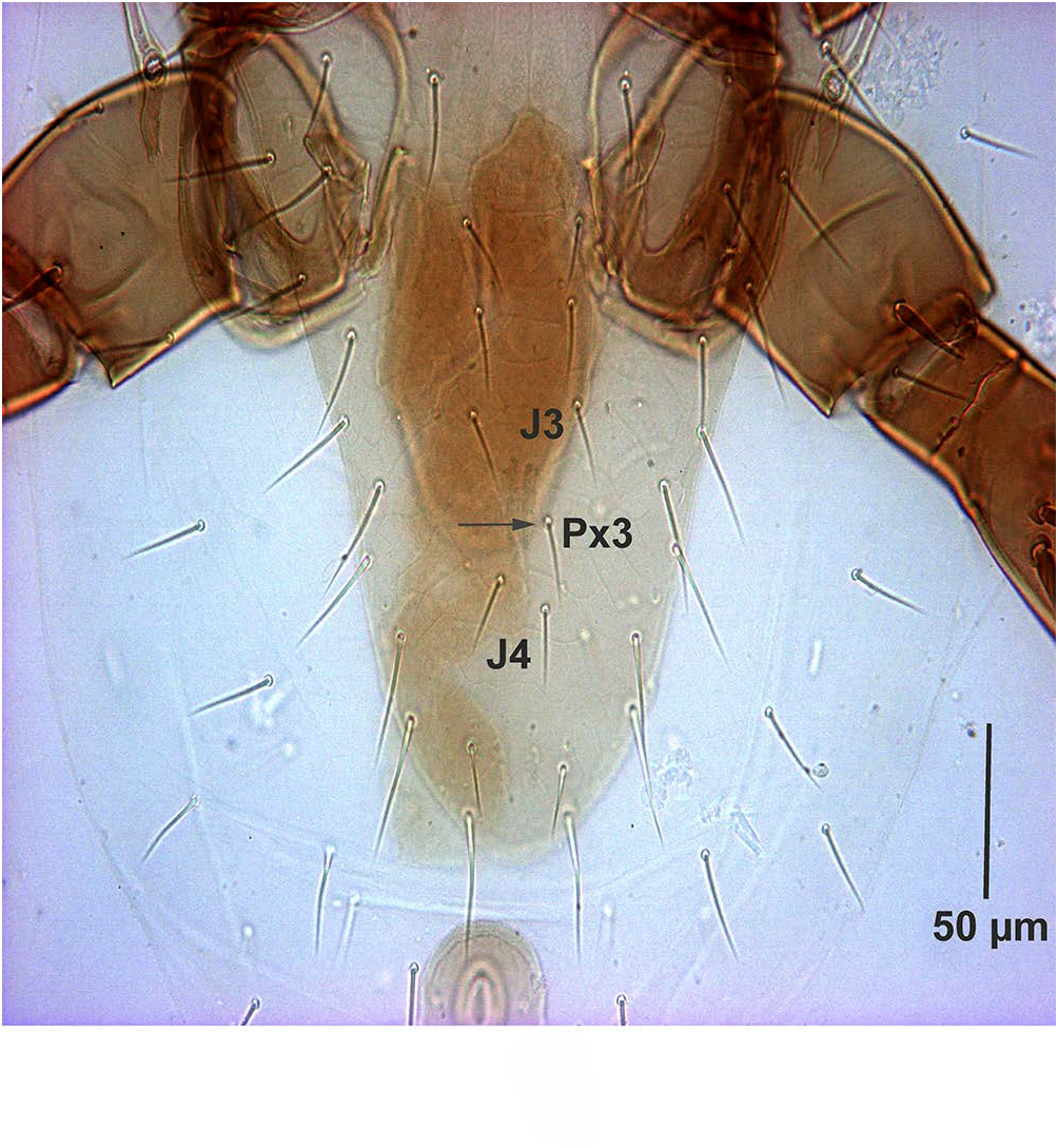
The number of dorsal shield setae in G. queenslandicus and G. angustus (as in literature and the most examined specimens in this survey) normally is 37 pairs of simple acicular setae with similar distribution. However, dorsal shield setae also vary in number in both species ( Figures 4A View Figure 4 , 5B View Figure 5 , 6 View Figure 6 and 7B View Figure 7 ) based on some Iranian specimens. Some specimens with one pair of PX3
setae between J3– J4 with totally 38 pairs of dorsal shield setae ( Figures 4A View Figure 4 , 5B View Figure 5 and 6 View Figure 6 ), some with one PX 3 in right or left side at the same place ( Figure 6 View Figure 6 ).
The distribution of dorsal shield setae of G. angustus holotype is the same as Figure (8).
The most specimens of intermediate forms, with some characters intermediate between G. angustus holotype and G. queenslandicus description by Costa (1966) which were checked in Iran and all specimens which considered as G. angustus based on straight lateral margins of dorsal shield in opisthonotal part in Germany (Karg collection, Berlin) have such situation of setae distribution with 37 pairs of setae on dorsal shield ( Figure 8). Note that Costa (1966) considered z2 missing on the dorsal shield of G. angustus ( Figure 13 p View Figure 13 . 145). The seta z3 typically appears at the deutonymphal stage in Gamasina, and z2 appears at the larval stage.
The theory is that setae appearing later during development have higher probabilities of being missing (i.e. and not appearing at all) in the adult stage. In other words, larval setae present in the larval stage tend to be the most stable i.e (. are not often lost later on), and setae appearing at the protonymphal stage are more stable than setae added in the deutonymphal and adult stages ( Evans and Till 1965; Lindquist and Evans 1965; Faraji and Halliday 2009). Therefore if there is a need for suppress, z3 is most often considered missing because it appears during development later than z2 and seta z2 has theoretically more chances of being present. Overall setae mentioned as z3, s3, r3, r4, r5 and r6 on dorsal shield of G. angustus in mentioned figure of Costa (1966) should be replaced with z2, z3, s3, r3, r4 and r5 respectively. Seta r6 is located on soft integument in ventral side, slightly posteriorly to s6. Based on above explanation we do not agree with the statement of Costa (1966) concerning the difference between dorsal setae distribution between G. angustus and G. queenslandicus .
3 — Relative lengths of dorsal shield setae
As mentioned before, different variations have been observed in populations of G. angustus and G. queenslandicus and intermediate forms which were collected from different parts of Iran and Australia. The lengths of dorsal shield setae in these specimens in addition G. of angustus sensu Costa (1966) and holotype and G. tripodiger shown in Tables (1, 4). The ranges of these measurements for nearly all dorsal shield setae are similar except for z1, s3 and r2 which perhaps with more specimens those also would overlaps.
4 — Shape of deutosternal groove and lengths of hypostomal setae
The deutosternal groove of G. angustus and G. queenslandicus are quite similar (Figure
9), both having the three posterior rows of denticles narrower than the three anterior rows. However, the difference between anterior and posterior rows is stronger for the illustration of G. queenslandicus [as illustrated by Costa (1966)]. Our observations indicate that such difference is not dichotomic among specimens examined, and it is not correlated with other characters previously assigned to G. angustus or G. queenslandicus , such as the shape of the lateral margins of the dorsal shield. Hypostomal setae h1 (-3) vary within groups and locations,
typical for both specimens that were assigned to either Gaeolaelaps queenslandicus or G. angustus .
and broadly overlap between them as in Table (3). According to the specimens, which have been studied herein, these characters states overlap.
5 — Fixed cheliceral digit
The chelicerae of all samples examined, including types of G. angustus , and all specimens from Iran, Australia and Italy, shared the following characteristics ( Fig 10 View Figure 10 ): chelate-dentate, with fixed digit bearing 11–13 teeth including a small proximal tooth followed by an enlarged one, then by 6–8 small teeth, ending up with the largest tooth at level of pilus dentilis and two teeth subapically including the offset and most distal tooth (gabelzhan). It bears terminal hook similar to thumb nail ( Figures 10A, B View Figure 10 ), including well-developed gabelzhan. The number and shape of small teeth between the two large teeth on fixed digit varies from six ( Figures 10B, C View Figure 10 ) to eight ( Figure 10D View Figure 10 ) within each groups angustus of -like and queenslandicus -like specimens [based on opisthonotal lateral margins and deutosternal grooves (sensu Costa, 1966)] from Iran and Australia. In some specimens, the number of fixed digit teeth varies between left and right chelicerae ( Figures 10B, C View Figure 10 ). Movable digit of chelicera with two enlarges teeth.
Some other morphological characters
Karg (1979) considered the Gaeolaelaps genus as a subgenus of Hypoaspis s. lat. and divided it into four species-groups based on various attributes. Hypoaspis (Geolaelaps) angustus -Group with posterior end of dorsal shield resembles wedge shape, opisthonotal lacks Zx setae and legs II in female possess spur or spine-like setae, including: H. (G.) queenslandicus , H. (G.)
angustus , H. (G.) elongata and H. (G.) angustiscutatus. The last one has conspicuous knob at basal part of dorsal setae and transferred to Cosmolaelaps genus by Nemati and Gwiazdowicz
(2016). Karg (1979) in a key to the species of this group separated G. queenslandicus and G.
angustus according to the length of their first legs relative to their idiosomal length. So that the first legs of the first species are longer than the length of the idiosoma while in the second one those are shorter. For this purpose we studied some other morphological characters including the sizes of different parts of body.
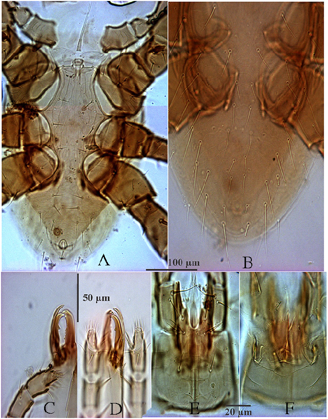
Presternal area with narrow granulate stripe adjacent to anterior margin of sternal shield and two presternal plates close together basally and with granulated surfaces. Sternal shield surface reticulated throughout, except small medio-posterior portion, anterior margin with variations and in some specimens medially concave, straight or tend to concave antero-laterally.
Some morphological character measurements of Gaeolaelaps angustus (type materials), G.
queenslandicus (specimens from Australia), G. angustus -like from Iran, G. queenslandicus -like from Iran, G. tripodiger (type) and male specimens from Iran have been shown in Table (4).
The length and width of sternal shield G in. angustus -like and G. queenslandicus -like populations (specimens from Iran) are larger than the others. The sizes of other characters do not differ among populations.
Epigynal shield in different specimens of G. queenslandicus -like (with different variation in lateral margins of dorsal shield as in Figures 3-5 View Figure 3 View Figure 4 View Figure 5 ) with different shape and reticulation (Figure
11). Abnormality observed in one specimen with small plate at posterolateral part ( Figure 11D View Figure 11 ).
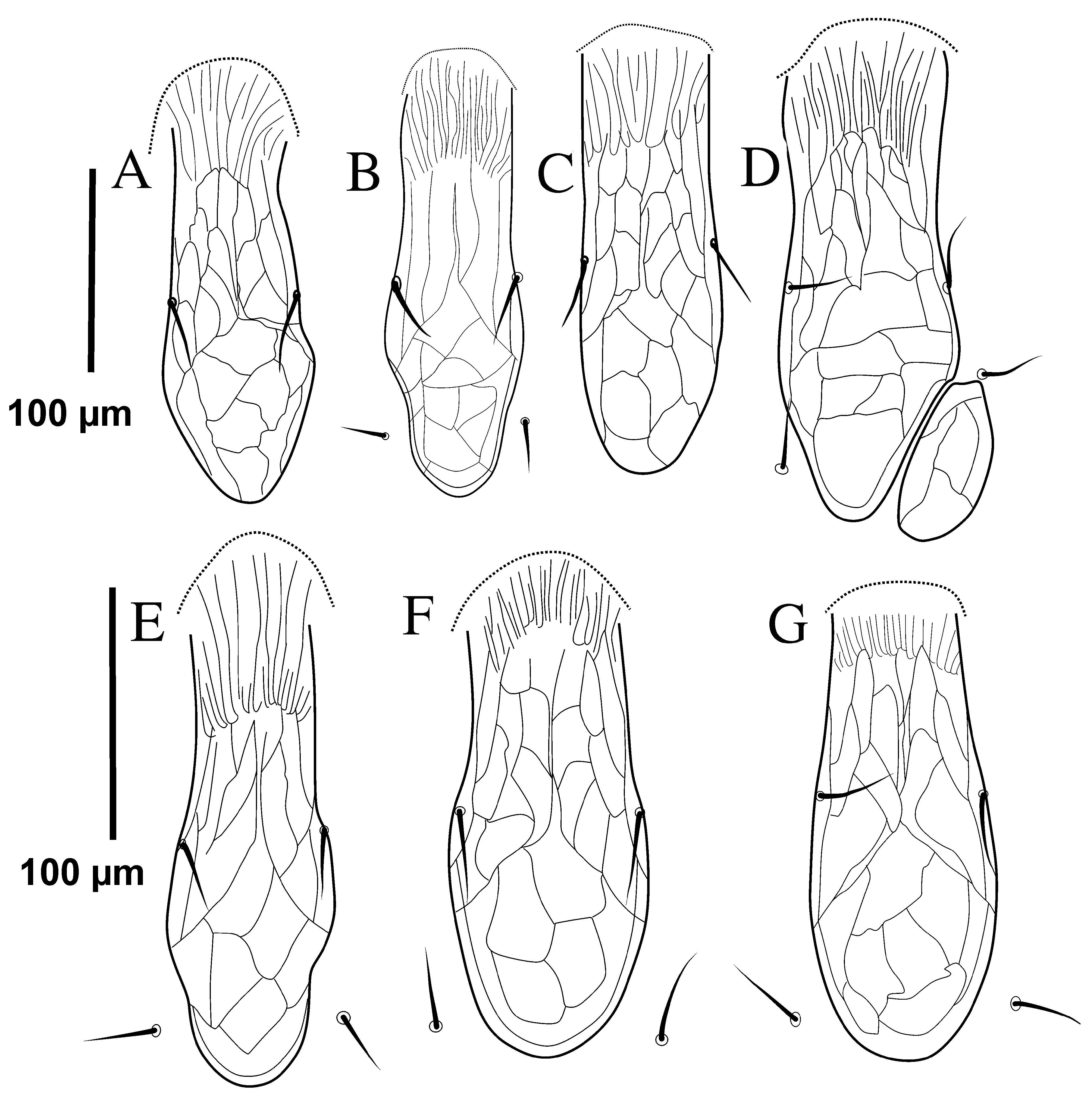
Epigynal shield measurements shown in Table (4). Palp chaetotaxy is normal for Gamasida
(sensu Evans 1963b), palp tarsal claw three-tined. Palp segments and setation were similar in all specimens, which studied here. Anterior margin of epistome is densely denticulate in all members. The length of denticles and the extensions of teeth vary among members of
G. queenslandicus -like and G. angustus -like in Iran. In some specimens, there are two long projected teeth with different shapes and different numbers of teeth at apex at lateral margins of epistome ( Figure 12 View Figure 12 ). Some specimens have a small protuberance in median part of epistome ( Figure 12C View Figure 12 ).
The corniculi well sclerotised, horn-like with different length [in specimens which were considered as G. angustus and angustus-like (53–60) and for G. queenslandicus and queenslandicus -like (55–69)]; internal malae with two pairs of separated median fringed projections extended beyond the tip of curniculi and one pair of lateral projections fringed and smaller than curniculi. Pilose labrum is longer than median internal malae projections.
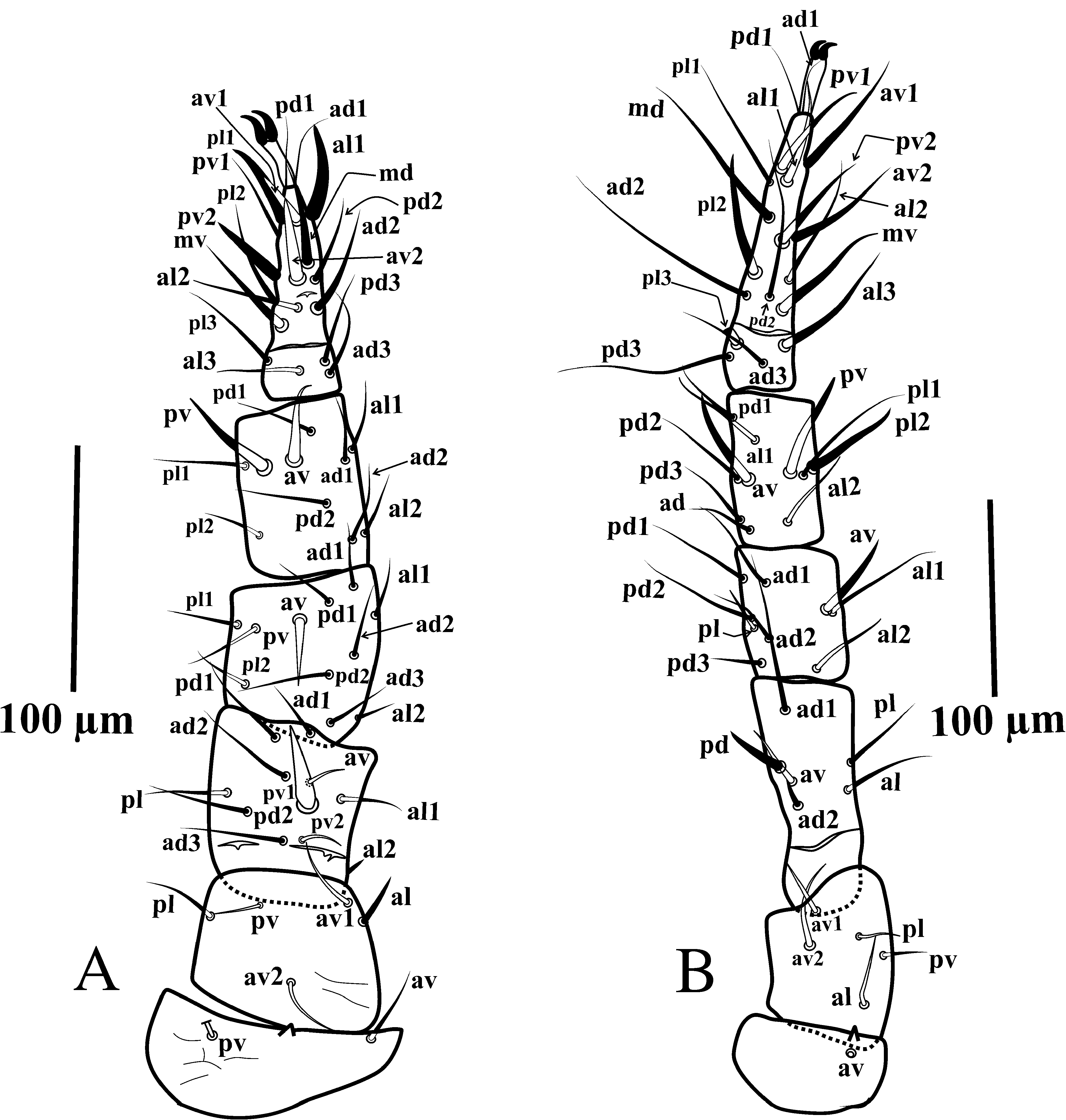
Legs Chaetotaxy. The leg chaetotaxy is essentially identical for all specimens that we examined including G. tripodiger type material. The situation of legs II and IV chaetotaxy could be seen in Figure (13) respectively.
The formulae and some explanation concerning all leg chaetotaxy of mentioned species and all relevant forms in Iran are as follows: Leg I: coxa 0 0/1 /1 0; trochanter 1 0/2 1/1 1; femur 2 2/1 3/3 2 (pd3 slightly thicker than the others); genu 2 3/2 3/1 2; tibia 2 3/2 3/1 2. Leg
II ( Figure 13A View Figure 13 ): coxa 0 0/1 0/1 0; trochanter 1 0/2 0/1 1; femur 2 3/1 2/2 1(pv1 spine-like and thick); genu 2 3/1 2/1 2 (av slightly thicker than other setae on the segment); tibia 2 2/1 2/1
2 (av and pv thicker than other setae on the segment); tarsus 3 3/2 3/2 3 + mv, md (pl1, al1,
pv2, av1–2, md and mv thicker than other setae on the segment). Leg III: coxa 0 0/1 0/1 0;
trochanter 1 0/2 0/1 1; femur 1 2/1 1/0 1 (setae pd and pl thicker than the others); genu 2 2/1
2/1 1 (setae av and pv thicker than the others); tibia 2 1/1 2/1 1 (setae av and pv thicker than the others); tarsus 3 3/2 3/2 3 + mv, md (setae mv, av1–2, pv1–2, md, pd2, al1 and pl1 thicker than the others). Leg IV ( Figure 13B View Figure 13 ): coxa 0 0/1 0/0 0; trochanter 1 0/2 0/1 1 (av2 slightly thicker than the other setae on the segment); femur 1 2/1 1/0 pd 1 (slightly thicker); genu 2
2/1 3/0 1 (av thicker than other setae on segment); tibia 2 1/1 3/1 2 av (, pv and pl2 thicker than other setae on the segment); tarsus 3 3/2 3/2 3 + mv, md (setae md, ad2 and pd3 slightly longer than the others on the segment; al1, pl2, av1– 2, pv1–2, mv, md, al3 and pl3 slightly thicker than other setae on the segment). All setae fine and needle-like unless otherwise noted.
Male ( Figure 14 View Figure 14 ). Short description of G. queenslandicus male was given by Ryke (1963)
and Yan and Ma (1999). In this study, males were collected with different samples from
Iran (see materials examined). The morphological characters of males were similar between populations, and that here is a description of the main characters of the male that differ from those of the female.
Idiosoma smaller than in female: dorsal shield length (405-450), dorsal idiosomal length
(418-457), dorsal shield width (226-253). Dorsal shield in all male specimens without curvature.
This situation could be observed in male specimens, which have been collected with female specimens of G. queenslandicus and queenslandicus -like population. In other words, the changes in the female dorsal shield, seen in different G. queenslandicus -like population, cannot be seen in male specimens. The length of dorsal, some ventral idiosomal setae and hypostomal setae of Gaeolaelaps queenslandicus (Womersley) male shown in Table (5). Sternitogenital,
anal and endopodal shields fused in a well-developed holoventral shield (378-396), with anterior margin well defined, prominent at level of genital opening ( Figure 14A View Figure 14 ). Holoventral shield well reticulated, bears 10 pairs of smooth acicular setae st (1–st5, Zv1–Zv2, Jv1–Jv3),
including para and post-anal setae. The width of shield at level st ofsetae 1 86-91, at level of
st2 89–91, between coxae II-III 154–156, at level of st3 98–100, widest part slightly posterior to coxae IV 135–170, the distances between st1–st1 (54–61), st2–st2 (64–66), st3–st3 (83–85),
the lengths of ventral setae: Zv1–Zv2, Jv1–Jv3 (24–30), post-anal seta (27–30). Movable digit of chelicera with one large tooth, arched spermatodactyl finger-like, longer than movable digit and with rounded tip; fixed digit multidentate ( Figures 14C, D View Figure 14 ). Other morphological characters including Hypostome ( Figure 14E View Figure 14 ), epistome ( Figure 14F View Figure 14 ), legs and palp chaetotaxy as in female.


No known copyright restrictions apply. See Agosti, D., Egloff, W., 2009. Taxonomic information exchange and copyright: the Plazi approach. BMC Research Notes 2009, 2:53 for further explanation.
|
Kingdom |
|
|
Phylum |
|
|
Class |
|
|
Order |
|
|
Family |
|
|
Genus |
Gaeolaelaps queenslandicus ( Womersley, 1956 )
| Nemati, Alireza, Gwiazdowicz, Dariusz J. & Khalili-Moghadam, Arsalan 2018 |
Hypoaspis (Hypoaspis) angustus
| Karg 1965 |
G. angustus
| Karg 1965 |
Androlaelaps
| A.Berlese 1903 |