
Ascidia sideralis, Bonnet, Nadia Y. K., Rocha, Rosana M. & Carman, Mary R., 2013
|
publication ID |
https://doi.org/ 10.11646/zootaxa.3691.3.4 |
|
publication LSID |
lsid:zoobank.org:pub:855A00F4-2E4B-4D0C-A458-B8111BFB7762 |
|
DOI |
https://doi.org/10.5281/zenodo.5665670 |
|
persistent identifier |
https://treatment.plazi.org/id/9152C60F-FF93-6670-9980-8127FDCA6FBD |
|
treatment provided by |
Plazi |
|
scientific name |
Ascidia sideralis |
| status |
sp. nov. |
Ascidia sideralis sp. nov. Bonnet & Rocha
( Figs. 7–8 View FIGURE 7 View FIGURE 8 )
Material examined. Holotype: MZUSP 0 0 0 34 – 1 ind.; Pluto, Isla Canales de Tierra; 5.0 m; 12/i/2009; col. J. Dijkstra.
Paratypes: DZUP ASC 148 – 1 ind.; Isla Taboguilla, Panama City; 09/xii/2008; col. R.M. Rocha. DZUP ASC 149 – 6 ind.; Isla Taboga, Panama City; floating dock; 05/xii/2008; col. R.M. Rocha.
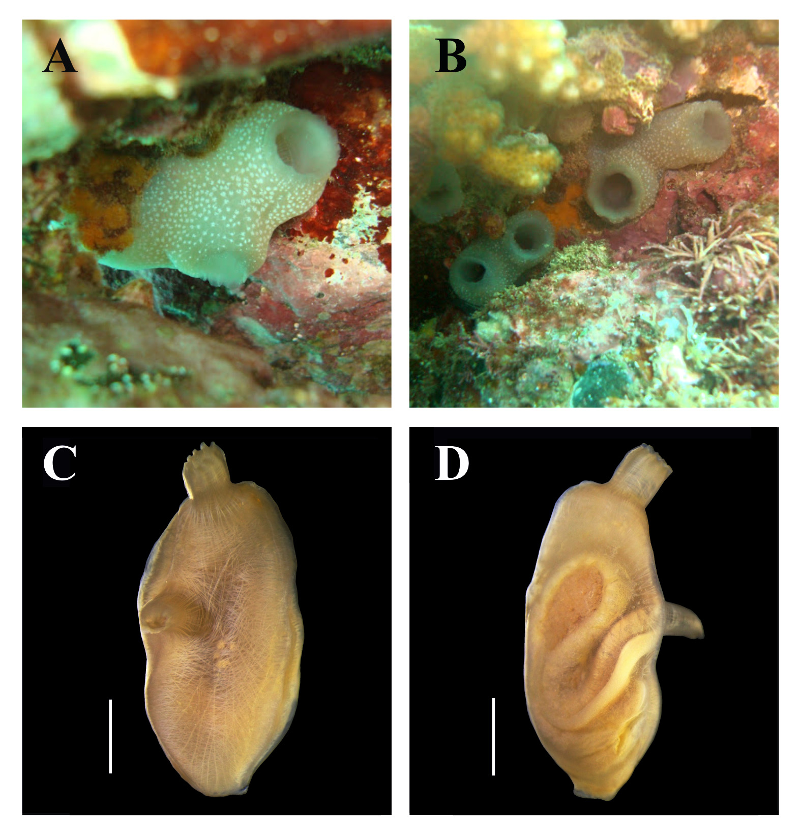
Etymology. The name is in reference to the white dots present on the entire surface of the tunic in living animals, giving the impression of a stellated night.
Diagnosis. Living animals are grayish with conspicuous white dots on the surface of the tunic; body musculature as a net is on the right side of the body, with complete transverse fibers close to the oral siphon; oral tentacles are connected by a thick membrane; large papillae are in the prebranchial area; 8–10 stigmata per mesh; primary papillae are trilobed in the pharynx; the intestine is isodiametric; and the ovary is lobed inside both intestinal loops.
Individuals are up to 5.7 cm total length and attached to the substrate by the left side of the body. The free surface usually has some epibionts (hydrozoans and bryozoans). Living specimens are grayish with numerous white dots on the entire surface of the tunic; the dots correspond to the terminations of blood vessels, which do not project from the surface of the animal. In formaldehyde, the gray coloration disappears, but the white dots are visible at least after ten months in preservative. The tunic is translucent and slightly wrinkled, with rigid or cartilaginous consistency (0.6–2.5 mm thick). Siphons are side-by-side and apical.
The body is oblong, 2.0– 3.5 cm long (without the oral siphon) and 1.1–1.9 cm wide. The oral siphon is 0.4–1.0 cm long and has eight lobes, while the atrial siphon is 0.4–1.6 cm long and has 7–8 lobes. The atrial siphon is displaced posteriorly 0.7–1.5 cm away from the ring of oral tentacles. Lobes on both siphons have smooth margins. The neural ganglion is closer to the oral siphon.
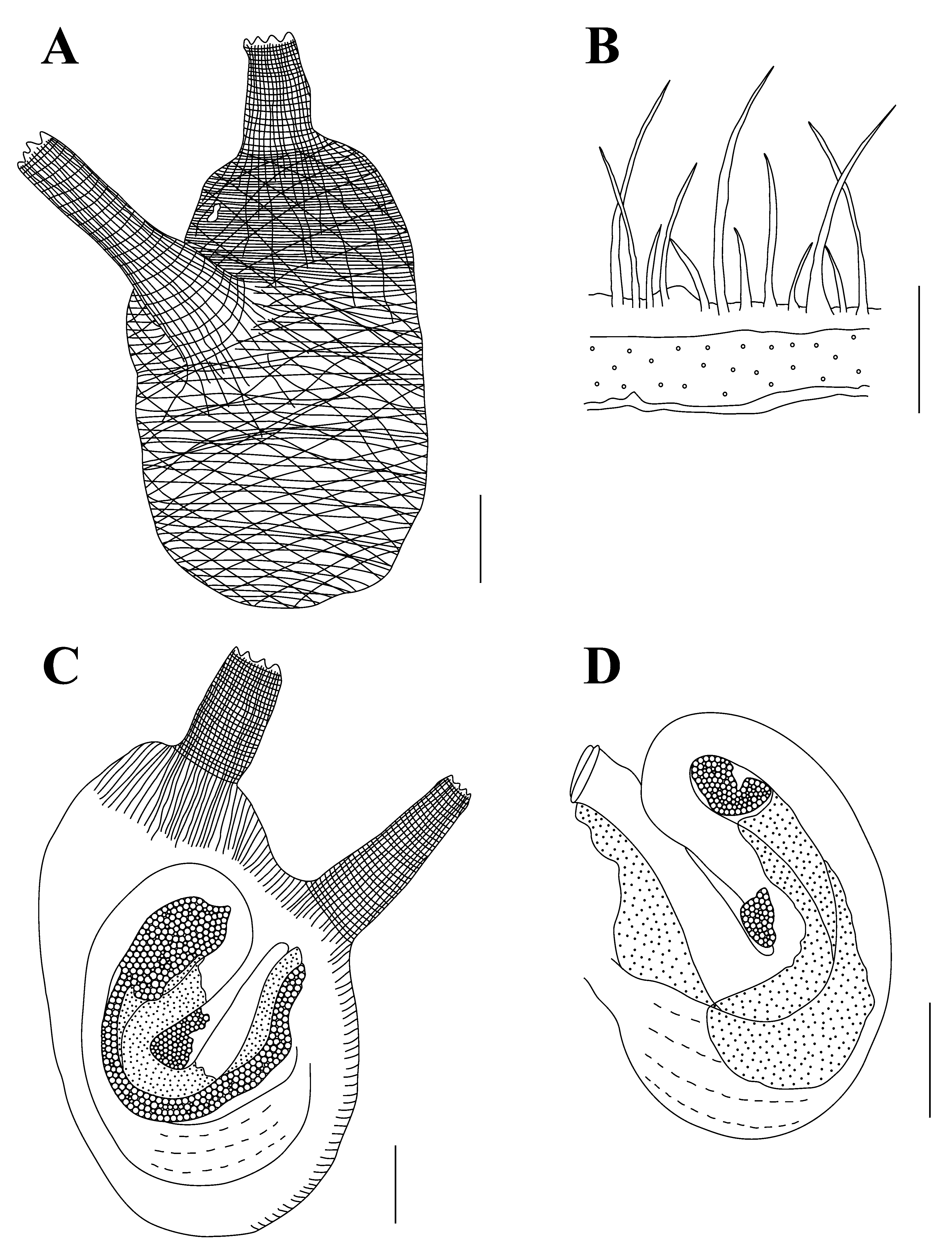
A dense net of fibers (0.08–0.16 mm thick) forms the body wall musculature on the right side, but close to the oral siphon horizontal fibers predominate. On the left side of the body there are short longitudinal fibers extending out of the oral siphon and some short fibers on the dorsal margin. The longitudinal muscle fibers in the siphons are not organized in bands.
There are 56–96 oral tentacles, of four sizes, and the largest is 1.7–5.3 mm long, with a thick membrane connecting them. The smooth prepharyngeal groove is double; the distance between the line of tentacles and the prepharyngeal groove is short (0.4–0.8 mm long), with large but not abundant papillae (0.04–0.06 mm in diameter). The dorsal tubercle is U-shaped, with enrolled ends; the peritubercular area is U-shaped. The dorsal lamina is double and smooth anteriorly, but it is toothed posteriorly due the elongation of the left transverse vessels plus small projections between them. The dorsal lamina passes by the esophageal aperture on its left to the end of the pharynx, close to the stomach. There are no papillae on the right side of the dorsal lamina near the esophageal aperture, but there is a wide lamina on the right side of the esophageal aperture. The pharynx has 35–47 longitudinal vessels on the right side, 31–41 on the left side and 86–142 transverse vessels; it is very pleated and there are 8–10 stigmata per mesh. The primary papillae are slightly trilobed and no intermediate papillae or parastigmatic vessels were found.
The alimentary canal occupies half or more of the left side of the body. The stomach is elongated, with 6–10 internal longitudinal folds. The isodiametric intestine forms two loops and the bilobed anus is located 6.2–12.5 mm from the oral tentacles. Renal vesicles that are 0.1–0.16 mm in diameter cover the intestine and stomach. There are conspicuous conical projections on the peritoneum covering the alimentary canal.
The ovary is lobed, included in the primary and secondary intestinal loops, and visible mostly from the external side of the body wall. The oocytes are 0.15–0.18 mm in diameter. The testis follicles are elongated, but not very ramified, and overlie the stomach and most of the intestine. Gonoducts open just posterior to the anus aperture.
Remarks. The only species of Ascidia whose living specimens have brilliant dots in the tunic are A. decepta Kott, 1985 , A. latesiphonica Hartmeyer, 1922 and A. ornata Monniot & Monniot, 2001 . Ascidia decepta also has eight lobes on the oral siphon and 6–7 lobes on the atrial siphon; body wall musculature as a net, 60–80 oral tentacles, 8–10 stigmata per mesh, and an isodiametric intestine. However, A. decepta can be differentiated from A. sideralis sp. nov. by the presence of sand embedded in the tunic, pigmented stripes in the siphon lining in some individuals, and the ovary ramified and restricted to the primary intestinal loop (Kott 1985). Similar to A. sideralis , A. latesiphonica has 40–100 oral tentacles, a very pleated pharynx, isodiametric intestine and lobed ovary (Hartmeyer 1922; Kott 1985). The differences with A. sideralis sp. nov. are the yellow dots in the tunic, body wall musculature on the right side that is formed by long longitudinal fibers in the middle of the body, short perpendicular fibers along the dorsal and ventral margins, longitudinal fibers in the siphons that are organized in bands, 6–8 stigmata per mesh, and ovary restricted to the primary intestinal loop (Hartmeyer 1922; Kott 1985). Similar to A. sideralis sp. nov., A. ornata has a net of muscular fibers on the right side of the body, papillae covering the prepharyngeal area, 6–10 stigmata per mesh and a lobed ovary (Monniot & Monniot 2001). However, the presence of 20 lobes on the oral siphon, white pigment lines in the margin of both siphons, 30 oral tentacles, descending limb of the intestine dilated, and ovary restricted to the primary intestinal loop (Monniot & Monniot 2001) distinguish A. ornata from A. sideralis sp. nov.
Phallusia julinea Sluiter, 1915 , a common species in the Pacific Ocean, is characterized by the conspicuous dots on the surface of the tunic in living animals and, although classified in the genus Phallusia , the absence of accessory openings in some individuals has been reported (Monniot 1987). However, the presence of 10–13 toothed lobes on the siphons, complete transverse muscle fibers close to the oral siphon on the left side of the body, a toothed lamina on the right side of the esophageal aperture, 70–90 longitudinal vessels in each side of the pharynx, intestine with the descending part dilated, and opening in the multilobed anus, are characteristics that distinguish P. julinea from A. sideralis sp. nov.
If the living aspect is disregarded, two other species of Ascidia also have a similar network pattern of body musculature and an isodiametric intestine. The dorsal tubercle in Ascidia occidentalis Kott, 1985 , is also close to the neural gland and it has 8–10 stigmata per mesh (Kott 1985). The differences are the toothed lobes on the siphons, the reduced number of oral tentacles (approximately 30) and the few internal folds in the stomach (Kott 1985). Ascidia melanostoma Sluiter, 1885 also has 36–90 oral tentacles, papillae in the prepharyngeal area, 26–48 longitudinal vessels on each side of the pharynx and a bilobed anus (Kott 1981; Nishikawa 1986). Nevertheless, the smaller number of stigmata per mesh (6–8) and the ramified ovary (Kott 1981; Nishikawa 1986) distinguish A. melanostoma from A. sideralis sp. nov.
No known copyright restrictions apply. See Agosti, D., Egloff, W., 2009. Taxonomic information exchange and copyright: the Plazi approach. BMC Research Notes 2009, 2:53 for further explanation.