
Ascidia ceratodes (Huntsman, 1912)
|
publication ID |
https://doi.org/10.11646/zootaxa.3691.3.4 |
|
publication LSID |
lsid:zoobank.org:pub:855A00F4-2E4B-4D0C-A458-B8111BFB7762 |
|
DOI |
https://doi.org/10.5281/zenodo.5665664 |
|
persistent identifier |
https://treatment.plazi.org/id/9152C60F-FF99-6678-9980-8179FE6C6FB9 |
|
treatment provided by |
Plazi |
|
scientific name |
Ascidia ceratodes (Huntsman, 1912) |
| status |
|
Ascidia ceratodes (Huntsman, 1912)
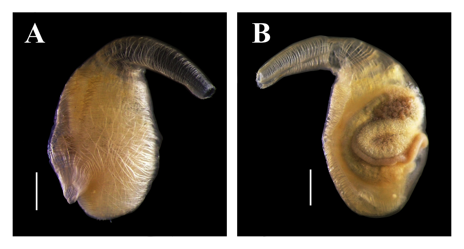
( Fig. 2 View FIGURE 2 )
Material examined. DZUP ASC 52 – 5 ind.; STRI dock, Panama City; 1.5 m, inside bricks; 07/xii/2008; col.: R.M. Rocha; DZUP ASC 187 – 6 ind.; 04/i/2009; col.: R.M. Rocha. DZUP ASC 51 – 3 ind.; Marina Flamenco , Panama City; 1.5 m, inside bricks; 06/xii/2008; col.: R.M. Rocha; DZUP ASC 53 – 4 ind.; 1.5m, inside bricks; 04/ i/2009; col.: R.M. Rocha. DZUP ASC 54 – 1 ind.; Pluto, Canales de Tierra Island; 13.0 m, under rocks; 10/i/2009; col.: R.M. Rocha. DZUP ASC 184 – 2 ind.; Playa Venados, Veracruz; intertidal, under rocks; 30/ix/2011; N.Y.K. Bonnet.
The animals are usually small (up to 3.3 cm total length) and are fixed to the substrate by the left side of the body. Usually there is no incrustation, but occasionally there are tubes of polychaetes and hydroids on the free surface. The living animal is yellowish with orange siphons, but after fixation these colors disappear. The tunic is translucent, with cartilaginous consistency ( 0.2–2.2 mm thick).
The body is oval, 0.8–2.0 cm long (including the oral siphon) and 0.4–2.0 cm wide, without the tunic. Usually both siphons are small (about 0.1–0.5 cm), but the oral siphon may be longer (up to 1.2 cm). The oral siphon has 8– 9 lobes and the atrial has 6 lobes; both with orange dots between the lobes, and without any projections on the margin. The atrial siphon is directed towards the posterior region of the body. The neural ganglion is closer to the oral siphon.
A net of 0.03–0.15 mm thick fibers forms the musculature on the right side. On the left side, the longitudinal muscles extending from the oral siphon are short and a band of short parallel transverse fibers is found on the dorsal region. The longitudinal fibers in the siphons are not organized in bands.
There are 83–217 oral tentacles, of three sizes; the largest is 1.3–2.1 mm long. The prepharyngeal groove is double and the anterior membrane of the prepharyngeal groove usually has projections. The distance between the line of tentacles and the prepharyngeal groove is short (up to 0.3–1.1 mm), with or without papillae. The peritubercular area is short and the dorsal tubercle aperture is U-shaped, with or without enrolled ends. The dorsal lamina is double anteriorly; when it merges, the margin is toothed (teeth not related with the ends of the transverse vessels). The dorsal lamina passes by the left of the esophagus aperture to the end of the pharynx, close to the stomach; absence of papillae on the right side of the dorsal lamina at the level of the esophagus. There is a narrow lamina on the right of the esophageal aperture. The pharynx has 24–38 longitudinal vessels on the right side, 23–35 longitudinal vessels on the left, and 47–81 transverse vessels; it is pleated, with 5–7 stigmata per mesh. The primary papillae in the pharynx could be simple, bi or trilobed. There are secondary papillae in some parts of the pharynx. Parastigmatic vessels are absent.
The alimentary canal occupies half or more of the left side of the body. The stomach is oval and large, with 9– 11 internal longitudinal folds. The isodiametric intestine forms a primary and a secondary loop. The anus is located approximately 3.0– 8.9 mm from the oral tentacles; it has a smooth or bilobed rim. Renal vesicles are very conspicuous ( 0.1–0.25 mm in diameter) and cover the stomach and the ascendant loop of the intestine.
The ovary is compact and localized inside the primary intestinal loop, where it is visible both from the outside and atrial cavity side. Oocytes are approximately 0.1 mm in diameter. The testis follicles are elongated and overlying part of the stomach and intestine. Gonoducts open just posterior to the anus aperture.
Remarks. Van Name (1945) described A. ceratodes as similar to juveniles of A. interrupta Heller, 1978 . We do not agree because the absence of projections in the tunic surface, the larger number of oral tentacles, the smaller number of longitudinal vessels in the pharynx, the isodiametric intestine and the conspicuous renal vesicles are characteristics which distinguish A. ceratodes from A. interrupta . Van Name (1945) cited the geographical distribution of this species from British Columbia south to California and comments about doubtful specimens from Ecuador and north Chile. Later, Tokioka (1972) reported the species in Costa Rica and Carman et al. (2011) extended its range to Panama. It is uncertain whether these tropical populations are in their native region and the species is expected to be found along all Central America Pacific coast or whether the southern populations were introduced by human transport.
No known copyright restrictions apply. See Agosti, D., Egloff, W., 2009. Taxonomic information exchange and copyright: the Plazi approach. BMC Research Notes 2009, 2:53 for further explanation.
|
Kingdom |
|
|
Phylum |
|
|
SubPhylum |
Tunicata |
|
Class |
|
|
Order |
|
|
Family |
|
|
Genus |