
Ascidia cf. liberata Sluiter, 1887
|
publication ID |
https://doi.org/10.11646/zootaxa.3691.3.4 |
|
publication LSID |
lsid:zoobank.org:pub:855A00F4-2E4B-4D0C-A458-B8111BFB7762 |
|
DOI |
https://doi.org/10.5281/zenodo.5665668 |
|
persistent identifier |
https://treatment.plazi.org/id/9152C60F-FF9D-6675-9980-8486FA0E6F95 |
|
treatment provided by |
Plazi |
|
scientific name |
Ascidia cf. liberata Sluiter, 1887 |
| status |
|
Ascidia cf. liberata Sluiter, 1887
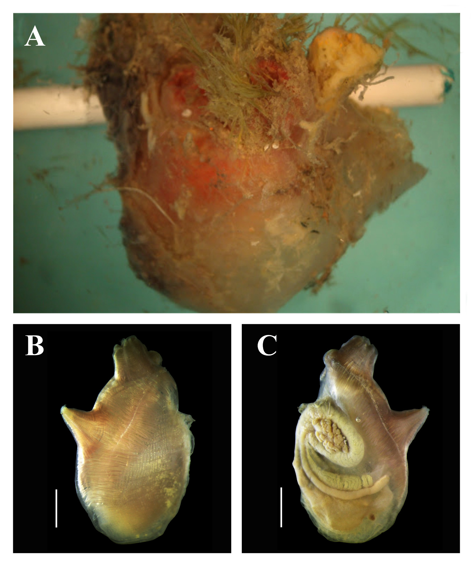
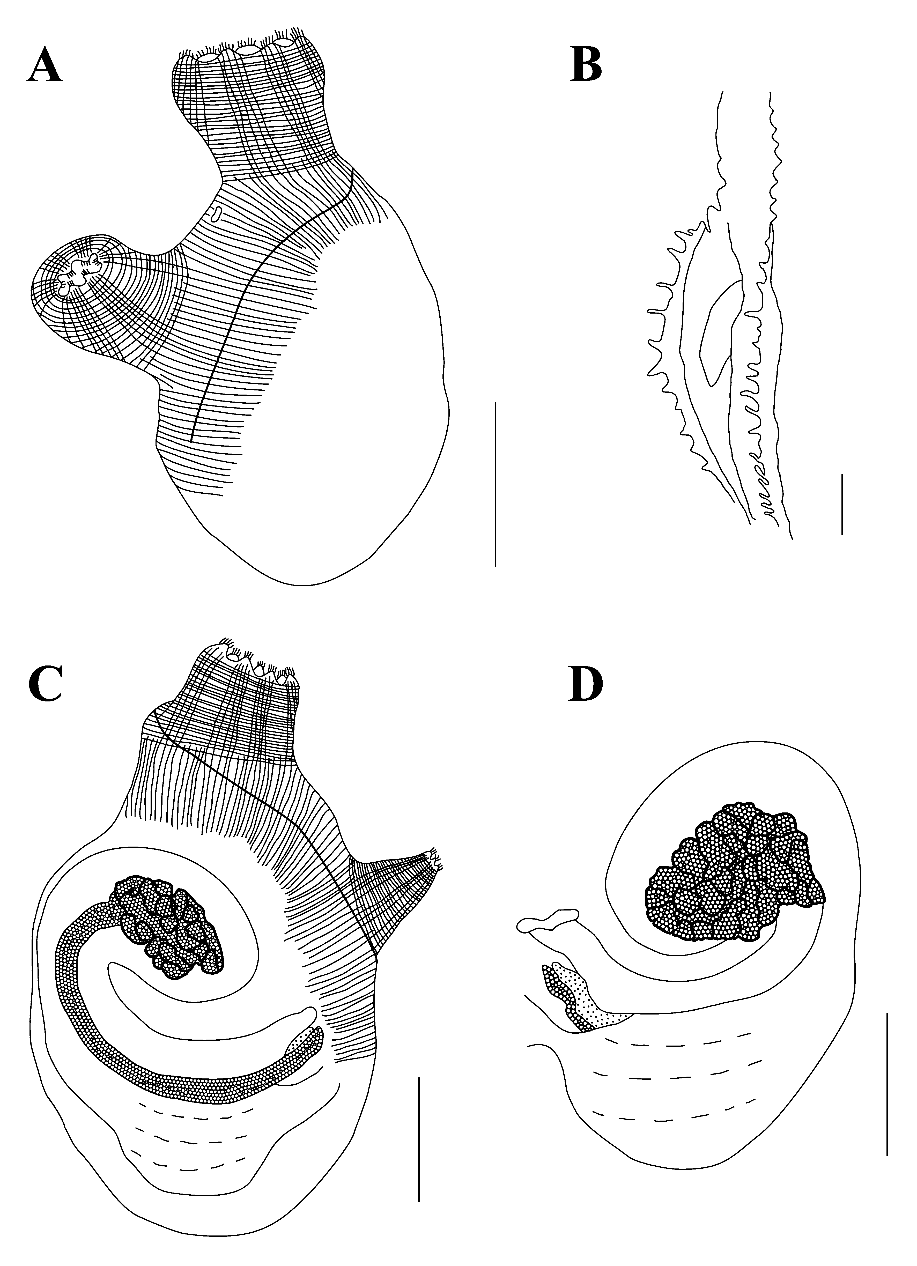
( Figs. 5–6 View FIGURE 5 View FIGURE 6 )
Material examined: DZUP ASC 55 – 2 ind.; STRI dock, Panama City; 1.5 m, inside bricks; 07/xii/2008; col.: R.M. Rocha. DZUP ASC 56 – 2 ind.; Marina Flamenco , Panama City; 1.5m, inside bricks; 04/i/2009; col.: R.M. Rocha. DZUP ASC 57 – 2 ind.; Isla Canales de Afuera; 5.0 m, under rocks; 13/i/2009; col.: R.M. Rocha.
Individuals are attached to the substrate by the left side of the body; there are no incrustations or epibionts. The color of living animals is red or yellow. The tunic is cartilaginous ( 0.6–1.5 mm thick), translucent and with numerous conic projections around the siphons; it may also have elongated projections on the ventral margin of the body.
The largest specimen measured 3.2 cm total length. The body is oblong, 1.3–3.2 cm long (including the oral siphon) and 0.9–1.2 cm wide, without tunic. Siphons are short ( 0.3–0.7 cm each one), the oral siphon is apical with eight lobes, while the atrial is displaced posteriorly 0.5–1.2 cm from the ring of tentacles and has 5–6 lobes. Lobes in both siphons have indented margins. The nervous ganglion is closer to the oral siphon. On the right side of the body there is a fold in the antero-dorsal region; in some individuals it is also present on the left side.
On both sides, the body wall musculature is formed by many short strong fibers ( 0.1–0.25 mm thick) perpendicular to the dorsal margin, forming a compact layer of muscles. These fibers extend posteriorly, but do not reach the ventral margin of the body. The longitudinal fibers in the siphons are organized in bands (one to each lobe).
There are 41–88 oral tentacles, of three sizes; the longest is 1.25–2.5 mm long. The prepharyngeal groove is double, with smooth margins, situated 0.3–0.5 mm from the ring of tentacles. The prepharyngeal area is usually smooth, but may have large papillae. The small peritubercular area forms a V and the dorsal tubercle aperture is Ushaped, with or without enrolled ends. The dorsal lamina is double anteriorly, with small finger like projections formed by the extremity of the left transverse vessels and other projections between them. It passes by the esophageal aperture on its left to the end of the pharynx, close to the stomach. There are no papillae on the right side of the dorsal lamina at the level of the esophageal aperture. There is a row of long languets on the right side of the esophageal aperture, formed by the elongation of the right transverse vessels of the pharynx. The pharynx has 26–32 longitudinal vessels on the right side, 24–31 on the left side and 33–73 transverse vessels. It is strongly pleated and there are 6–9 stigmata per mesh; the primary papillae are bi or trilobed. In some parts of the pharynx parastigmatic vessels are found, but secondary papillae are absent.
The alimentary canal is large, occupying more than half of the left side of the body. The stomach is rounded and wide, with 5–6 internal longitudinal folds. The isodiametric intestine forms two loops and the bilobed anus is located approximately 4.0– 10.5 mm from the oral tentacles. Small renal vesicles ( 0.04–0.08 mm diameter) cover the digestive tract. The peritoneum that covers the stomach and ascending portion of the intestine has irregular papillae, but on the rectum there are conical projections.
The ovary is compact, included in the primary intestinal loop, while the testis is ramified, with elongated follicles. Both gonads are visible from the outside and atrial cavity. The gonoducts open close to anus aperture, just posterior to it.
Remarks. The dissected specimens are in agreement with previous descriptions of A. liberata collected in Australia and Solomon Is.: small size (up to 2.0 cm total length), tunic with conical projections around the siphons, siphon lobes with indented margins, body wall with a fold on the right side, corporal musculature restricted to perpendicular fibers in the dorsal margin, longitudinal musculature in the siphons organized in bands, 6–8 stigmata per mesh and isodiametric intestine (Kott 1985; Nishikawa 1986). Nevertheless, there is some doubt about the identification because of differences in the descriptions and distribution. Living specimens are maroon-purple color and have 30–50 oral tentacles (Kott 1985; Nishikawa 1986), the ovary is ramified on the secondary intestinal loop in Australian specimens (Kott 1985), and there is a large geographical distance between previous records and this one.
Similar to A. liberata , Ascidia incrassata Heller, 1878 has projections on the margin of the siphonal lobes, body musculature on the right side formed by short fibers perpendicular to the dorsal margin, indented lamina on the right side of the esophageal aperture and the cauliflower-shaped ovary restricted to the primary intestinal loop. A. incrassata may be distinguished from the present specimens of A. cf. liberata by the large size (up to 10.0 cm total length and tunic 2.0 cm thick); absence of conical projections on the tunic, close to the siphons; body musculature only in the anterior region of the body (not posterior to the atrial siphon); and 7–12 stigmata per mesh in the pharynx (Monniot et al. 2001).
Recently, Carman et al. (2011) reported the presence of A. incrassata near the Pacific entrance of the Panama Canal. However, this identification should be corrected because the specimens better correspond to A. cf. liberata .
No known copyright restrictions apply. See Agosti, D., Egloff, W., 2009. Taxonomic information exchange and copyright: the Plazi approach. BMC Research Notes 2009, 2:53 for further explanation.
|
Kingdom |
|
|
Phylum |
|
|
SubPhylum |
Tunicata |
|
Class |
|
|
Order |
|
|
Family |
|
|
Genus |