
Cycloporus gabriellae Marcus 1952
|
publication ID |
https://doi.org/ 10.11646/zootaxa.3873.5.3 |
|
publication LSID |
lsid:zoobank.org:pub:687DC4E0-9B78-4AF0-9DD2-8B868E3B8EB5 |
|
DOI |
https://doi.org/10.5281/zenodo.6143930 |
|
persistent identifier |
https://treatment.plazi.org/id/927D87F1-FFDE-320E-FF78-7C8F68F8FD92 |
|
treatment provided by |
Plazi |
|
scientific name |
Cycloporus gabriellae Marcus 1952 |
| status |
|
Cycloporus gabriellae Marcus 1952 View in CoL
( Fig. 16 View FIGURE 16 )
Material examined. Two specimens (MNRJ-PLAT 63, 5 x 4 mm and 5 x 5 mm), one as sagittal sections of reproductive structures (5 slides). Three specimens (MNRJ-PLAT 64, 6 x 4 mm, 9 x 4 mm and 7 x 5 mm) one as sagittal sections of reproductive structures (10 slides), all collected 0 8.12.2007. Two specimens (MNRJ-PLAT 65, 5 x 3 mm and 7 x 5.5 mm), one as sagittal sections of reproductive structures (8 slides), collected 20.04.2008. One specimen (MNRJ-PLAT 66, 7 x 4 mm) as sagittal sections of reproductive structures (5 slides), collected 13.12.2008. Two specimens (MNRJ-PLAT 67, 7.5 x 5 mm, 6.7 x 4 mm), the larger as sagittal sections of reproductive structures (8 slides), collected 16.10.2009. All specimens collected at Praia das Conchas, Cabo Frio, Brasil, under rocks (22°52'33.05"S, 41°58'39.27"W) and preserved in ethanol 70%.
Distribution. Originaly described from Ilha de São Sebastião, São Paulo ( Marcus 1950), it was later reported from Curaçao and Antigua ( Marcus & Marcus 1968).
Diagnosis. Translucid body; color determined by intestinal content. Nine pairs of lateral branches of the main intestine, one anterior pair. Five to six pairs of uterine vesicles.
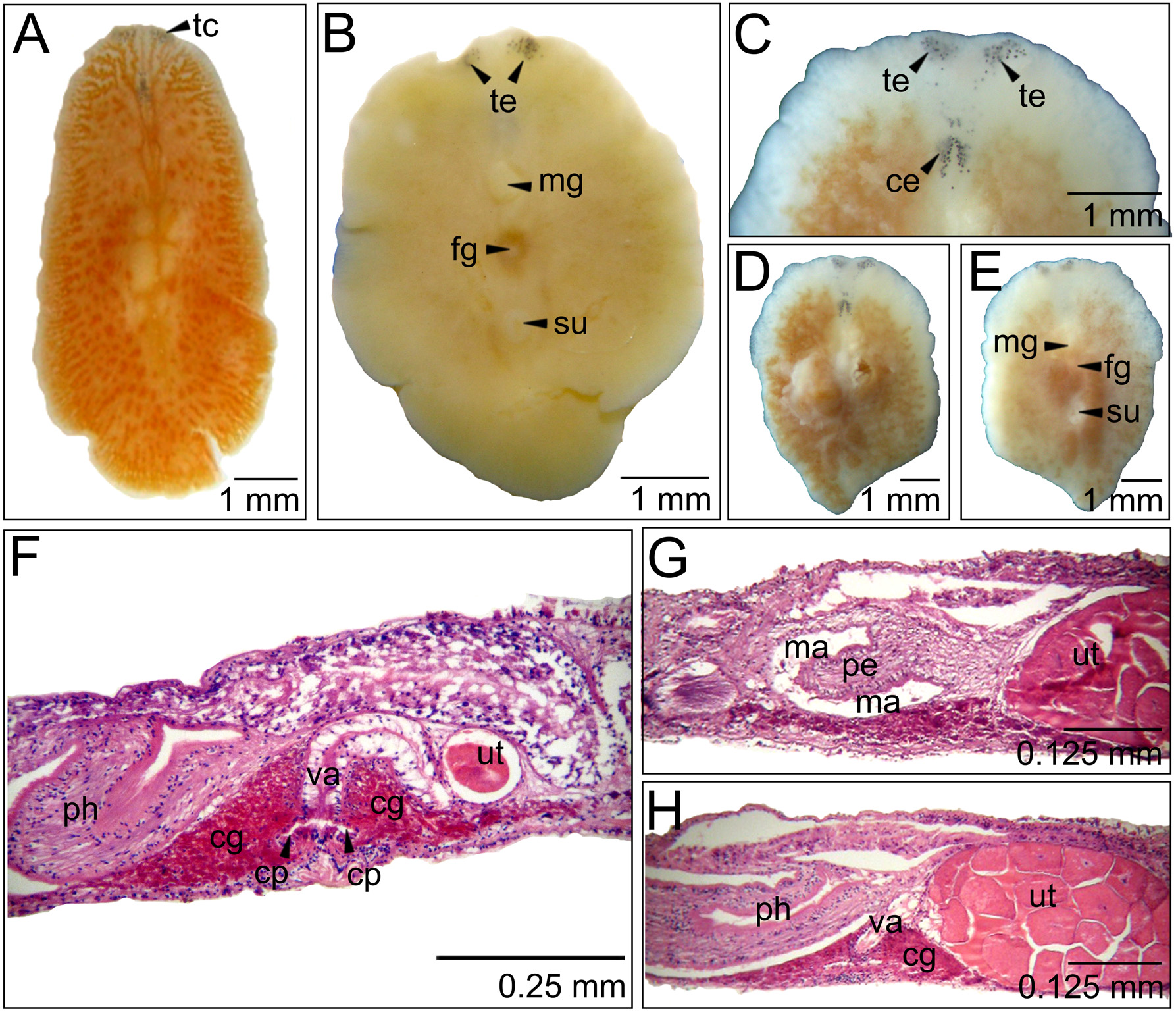
Description. Color: Translucid body, color determined by intestine content. Our specimens presented orange coloration when alive ( Fig. 16 View FIGURE 16 A). Intestine vesicles characteristic from the genus present at the body margin, white colored. In some specimens these vesicles are very abundant forming a conspicuous white band.
Form: Oval.
Tentacles: Short projections of the body margin ( Fig. 16 View FIGURE 16 A, B and C).
Eyespots: Tentacular eyespots distributed in ventral and dorsal groups, located at the tentacle margin. Ventral groups ( Fig. 16 View FIGURE 16 B and C) contain 13–30 eyespots and dorsal groups ( Fig. 16 View FIGURE 16 C and D) 25–45 eyespots. Cerebral eyespots, in two elongated groups, which become thinner towards the margin ( Fig. 16 View FIGURE 16 C), occupying 0.9 mm length, with 27–43 eyespots present, at 0.5–0.8 mm from the anterior margin. Most specimens presented around 35 eyespots in each group. Some eyespots are found scattered beyond the groups, toward the anterior margin, in a region that reaches 0.7 mm length, between tentacular and cerebral groups ( Fig. 16 View FIGURE 16 C).
Digestive system: Short pharynx, thick and tubular. Measures from 0.8 to 1.1 mm. Main intestine reaches 1.5 to 2 mm of the body. Nine lateral ramifications and one anterior present in the main intestine. Terminal intestine vesicles located throughout the body margin; occupy 0.1 mm of the margin.
Epidermis and body wall: Smooth dorsal surface. Dorsal epidermis (19 µm) a little thicker than the ventral epidermis (14 µm). Ciliated epidermis with rhabdites both dorsally and ventrally ( Fig. 16 View FIGURE 16 F). Epidermic glands also present. Voluminous sucker measures 0.25–0.4 mm. Muscular circular fibers constitute the external muscular layer and diagonal fibers the internal layer. Muscular layers thin in both body surfaces, a little thicker dorsally (11 µm) than ventrally (10 µm).
Gonopores: Male gonopore at 1.9 mm from the anterior margin, female pore at 2.2 mm ( Figure 16 View FIGURE 16 B and E). Female gonopore measures 0.2 to 0.25 and male pore measures 0.25 to 0.3 mm.
Male reproductive system: Testicles ventrally to main intestine. Male structures turned forwards. Seminal vesicle rounded with 270 µm diameter. Prostatic vesicle with 170 µm diameter. Male atrium measures 200 µm and penis papilla with 182 µm.
Female reproductive system: Ovaries dorsally to main intestine. Five to six pairs of uterine vesicles disposed at each side of the median line. Cement glands occupy only the region immediately beside the vagina and are found densely disposed. Vagina curved backwards with changed disposition, depending on the uteri content. Almost empty ( Fig. 16 View FIGURE 16 F) or full of oocytes ( Fig. 16 View FIGURE 16 G and H). Vagina measure 120–230 µm, oocytes measure 70.1–75 µm. Cement pouch, short and lightly curved backwards, measuring 49 µm. Female atrium measure 47 µm.
Taxonomic remarks. Cycloporus gabriellae is the only species of the genus reported from Brazil ( Marcus 1950, 1952). Our specimens present measures (from 5 x 3 mm to 7.5 x 5 mm) similar to the originally described (5 x 4 mm and 9 x 4 mm) by Marcus (1950) and smaller than those described by Marcus & Marcus (1968) (10 mm). However, they are in accordance with characteristics described in both works. Cycloporus is not a speciose genus. Until 2002 only six species were known around the world. Newman & Cannon (2002) increased this number to 14. Among these only C. gabriellae , C. australis and C. variegatus present nine main intestine lateral branches ( Newman & Cannon 2002). However, C. australis and C. variegatus present a higher number of cerebral tentacular eyespots and yellow dots in the margin, absent in C. gabriellae . Besides, Cycloporus variegatus present a median line, also absent in C. gabriellae ( Marcus 1950, 1952).
No known copyright restrictions apply. See Agosti, D., Egloff, W., 2009. Taxonomic information exchange and copyright: the Plazi approach. BMC Research Notes 2009, 2:53 for further explanation.
|
Kingdom |
|
|
Phylum |
|
|
Order |
|
|
Family |
|
|
Genus |