
Collaria Provancher, 1872
|
publication ID |
https://doi.org/ 10.11646/zootaxa.4138.2.1 |
|
publication LSID |
lsid:zoobank.org:pub:4E30E6F8-8950-4FC2-A733-555A3A16BB1F |
|
DOI |
https://doi.org/10.5281/zenodo.6075084 |
|
persistent identifier |
https://treatment.plazi.org/id/937187CE-FFB7-FFAF-FF78-F94C8EA862F3 |
|
treatment provided by |
Plazi |
|
scientific name |
Collaria Provancher, 1872 |
| status |
|
Genus Collaria Provancher, 1872 View in CoL
Type species. Collaria meilleurii Provancher, 1872 (Lectotype designated by Kelton, 1968).
Collaria Provancher, 1872: 79 View in CoL [n.gen.]; Carvalho 1959: 284 [catalog]; Schwartz 2008: 1179 [syn., diag., morph., phylogenetic relationships], Schuh 2002–2014 [catalog].
Trachelomiris Reuter, 1876: 61 [n. gen.] (syn. by Reuter 1905: 47); Carvalho 1959: 284 [catalog]; Schuh 2002–2014 [catalog].
Nabidea Uhler, 1878: 397 [n. gen.] (syn. by Uhler 1887: 230); Carvalho 1959: 284 [catalog].
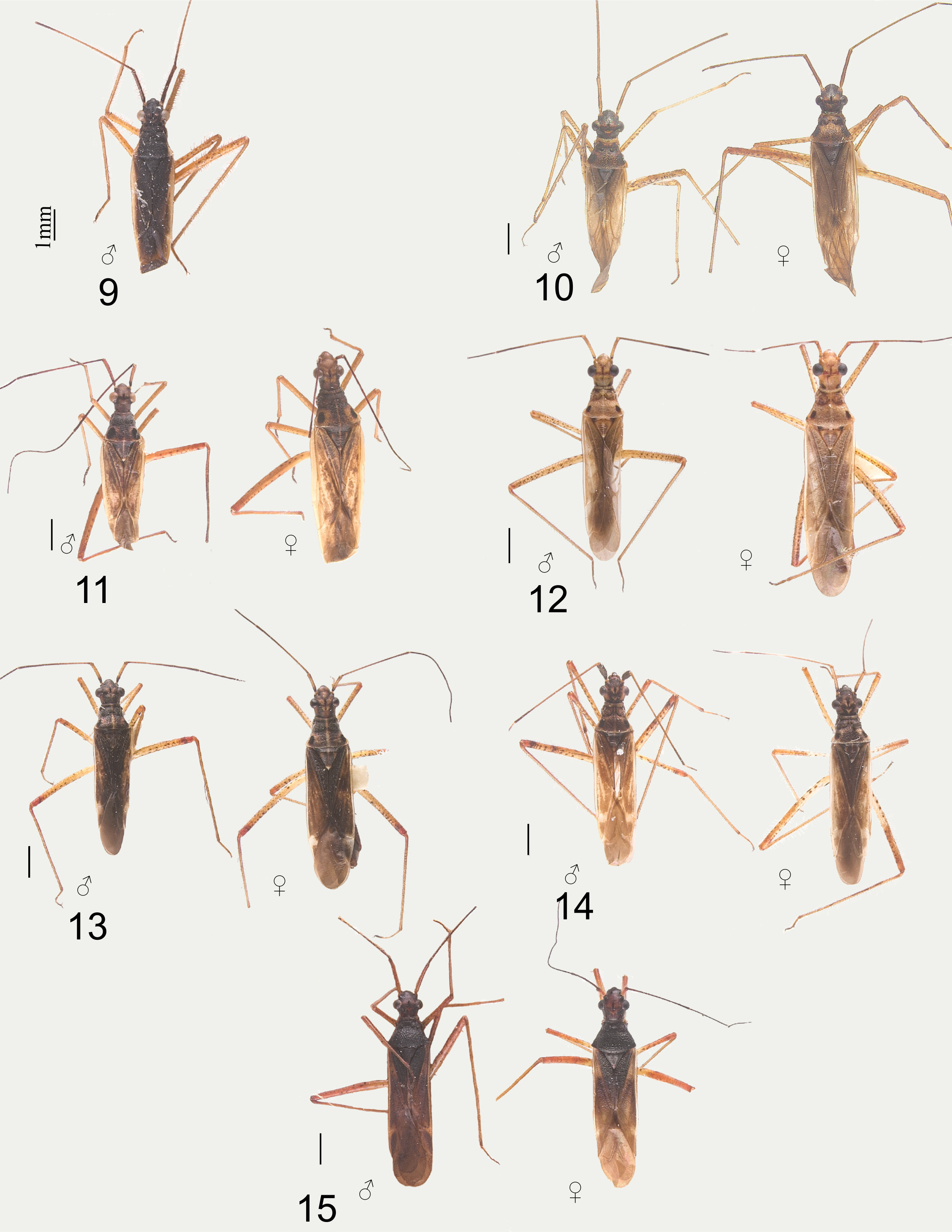
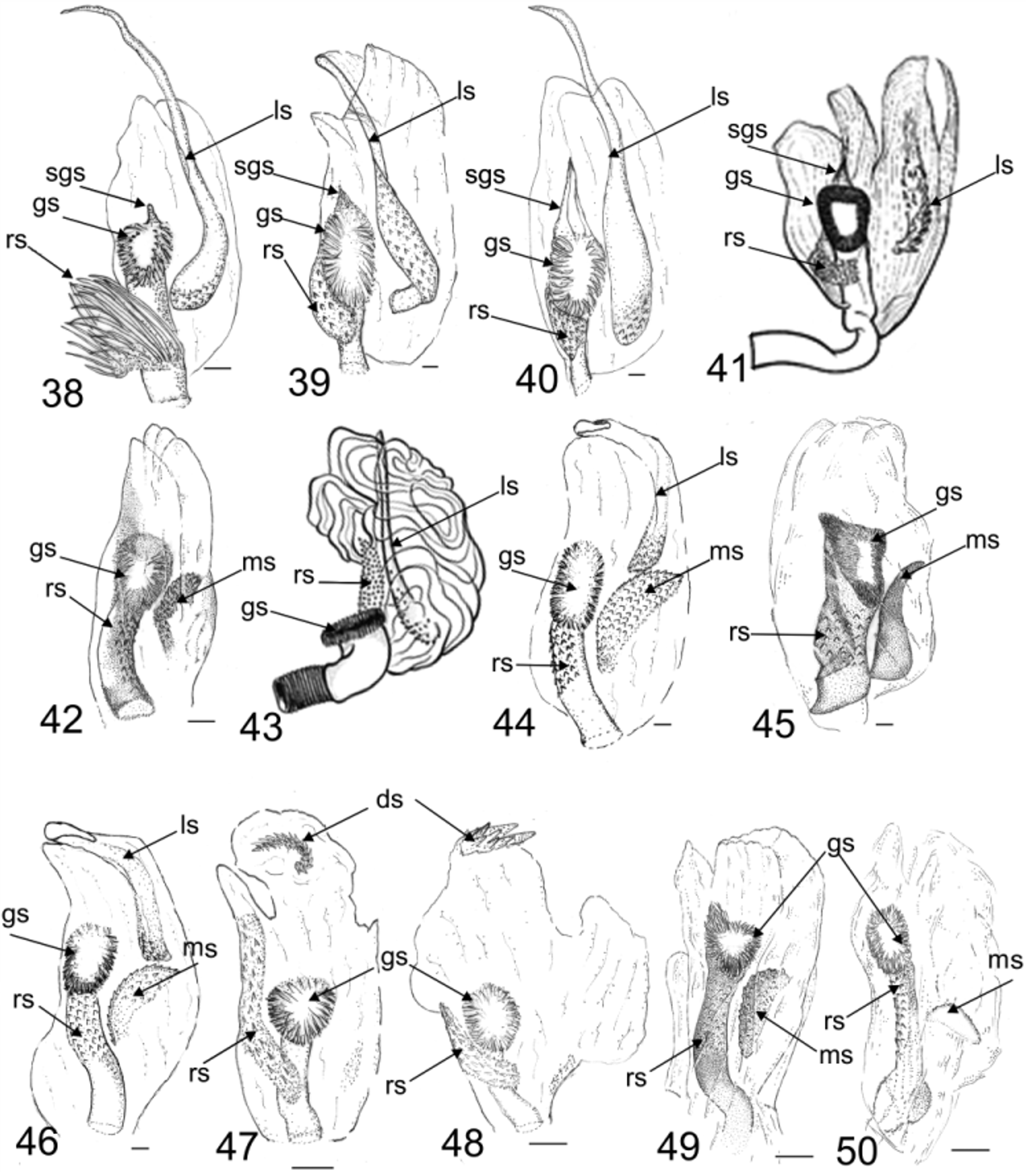
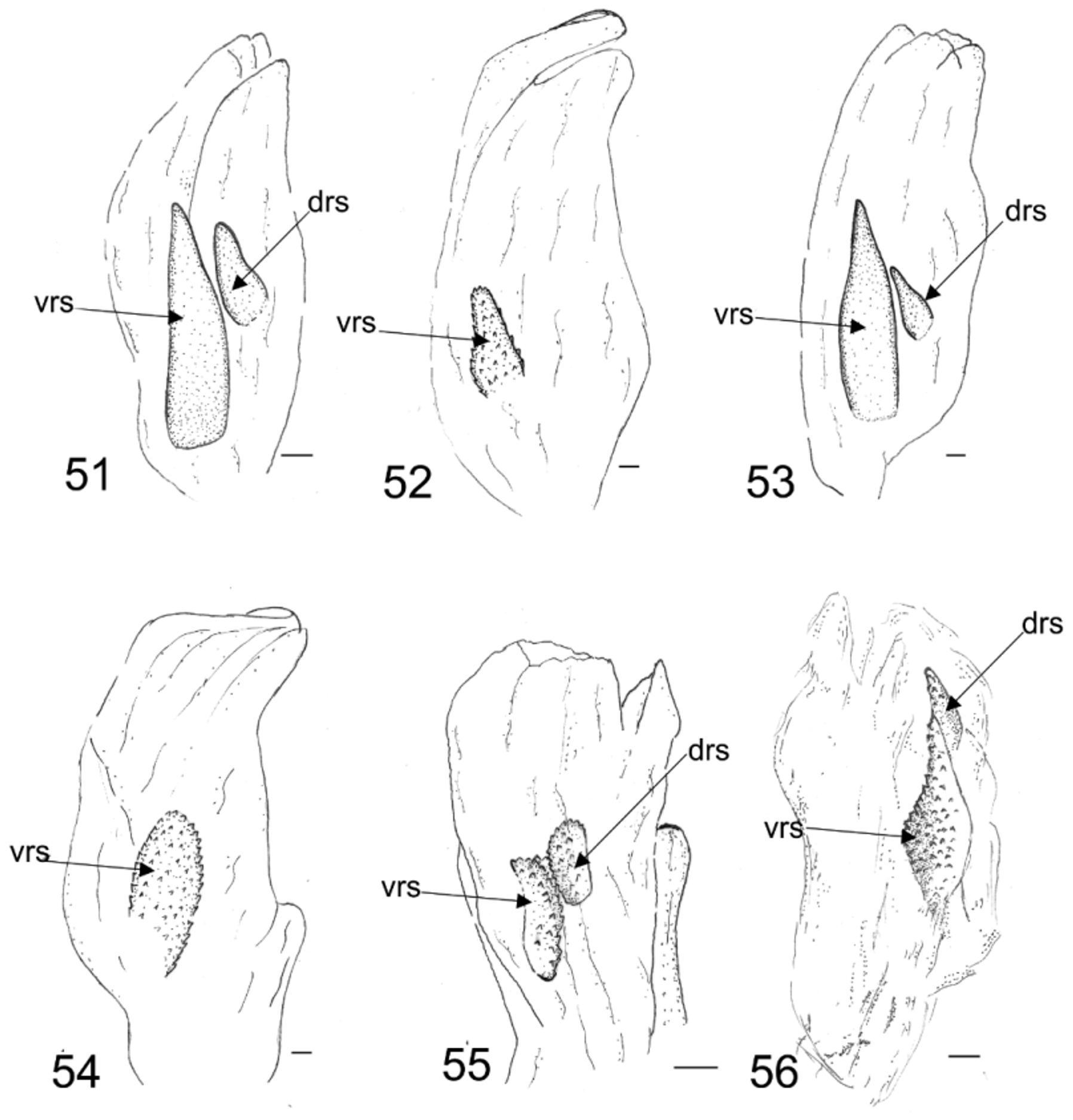
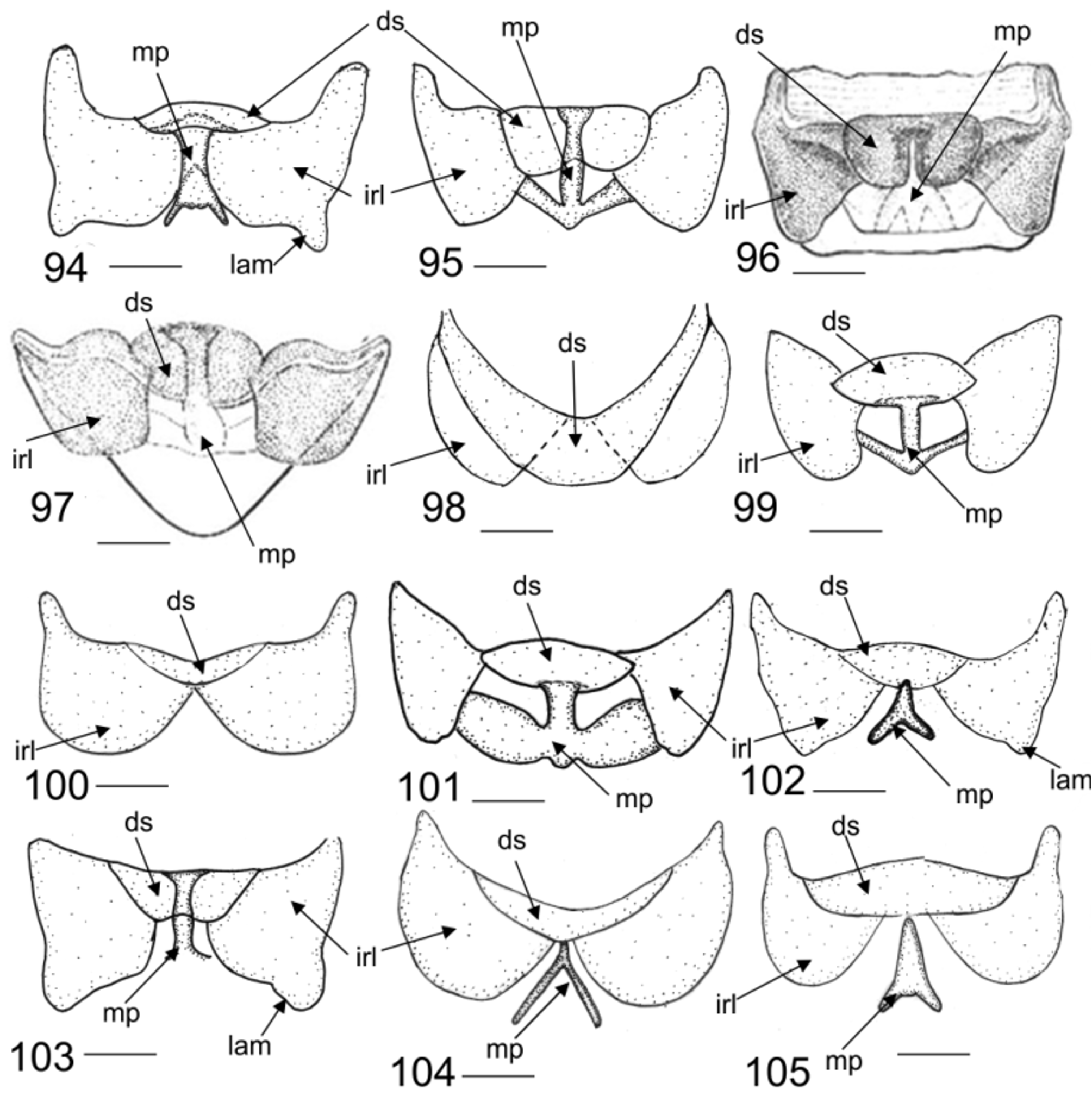
Diagnosis. Recognized by the broad frons, smoothly merging with clypeus; mandibular plate produced and strongly rounded; eye removed from pronotum by distance equal to twice distal width of antennal segment I ( Figs. 1–15 View FIGURES 1 – 8 View FIGURES 9 – 15 ), with ‘‘neck’’, sulcate vertex, anteocular portion in dorsal view shorter or equal than half of total length of the head, pronotal anterior lobe narrowed, proepisternum strongly rounded, hemelytra weakly pilose, finely punctate, male genitalia with endosoma usually with left and right lateral endosomal sclerites ( Figs. 38–58 View FIGURES 38 – 50 View FIGURES 51 – 56 View FIGURES 57 – 61 ) and female with well-developed dorsal structure, interramal lobes of posterior wall triangular or rounded, and medial process sclerotized ( Figs. 62–105 View FIGURES 62 – 71 View FIGURES 72 – 81 View FIGURES 82 – 93 View FIGURES 94 – 105 ).
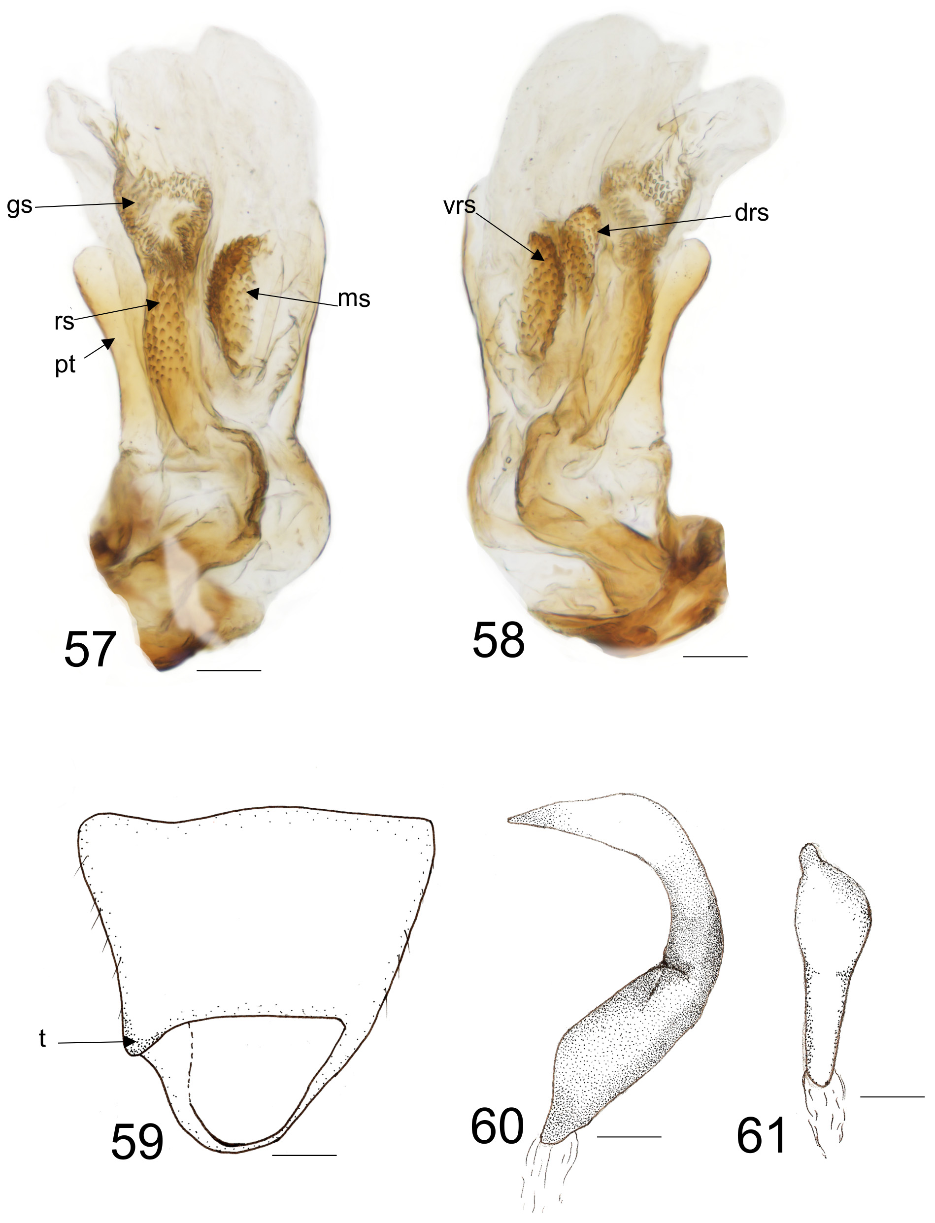
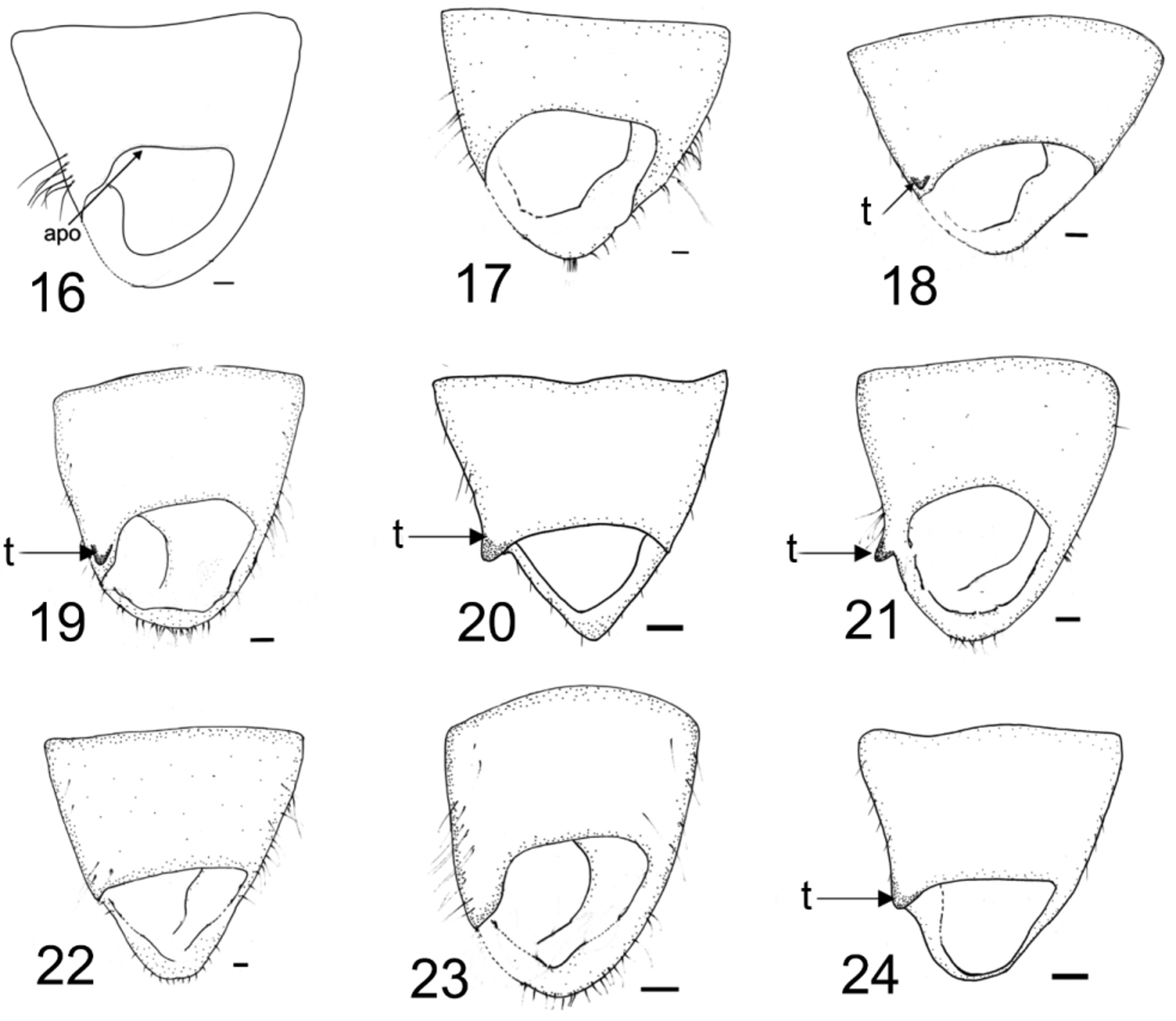
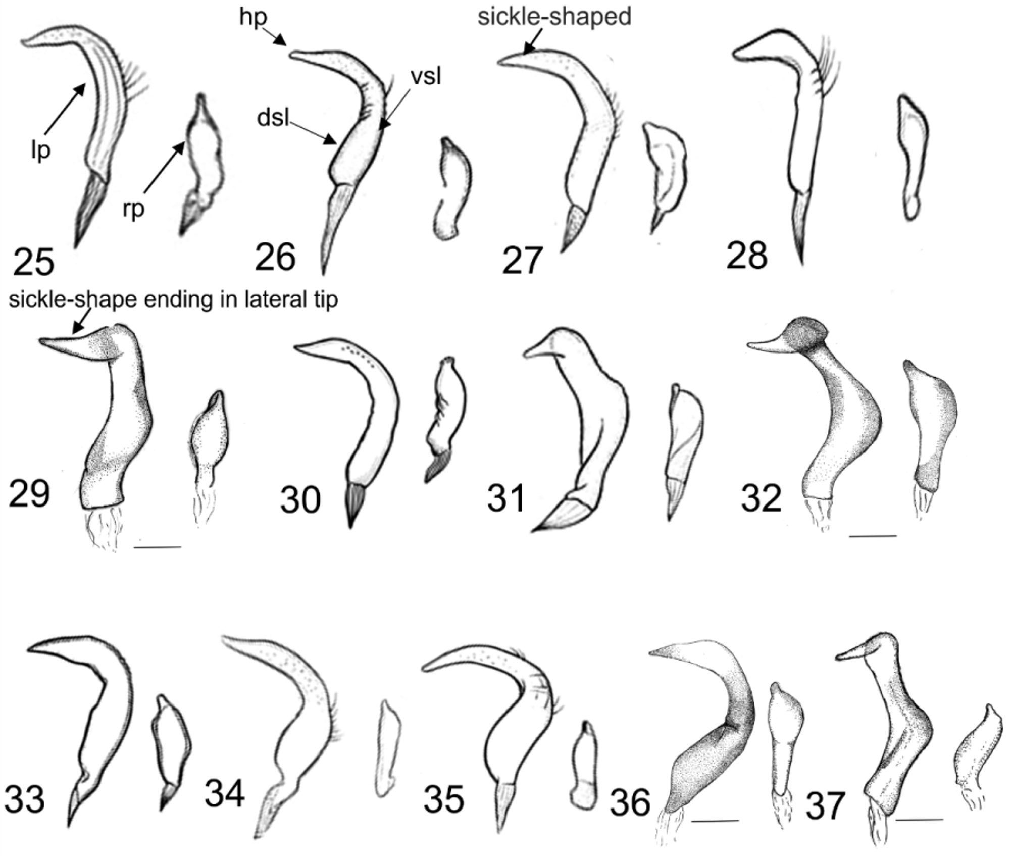
Redescription. Male: Macropterous, medium sized (5.44–7.37 Table 1). COLORATION: Brown to black, with brownish or yellowish areas ( Figs. 1–15 View FIGURES 1 – 8 View FIGURES 9 – 15 ). Head: Uniformly colored with black Y-shaped marking extending from longitudinal sulcus to frons, transverse pale spot (V-shaped) behind to longitudinal sulcus or paired transverse markings lateral to longitudinal sulcus; first antennal segment usually pale brown, remaining segments brown; labial segment IV apically dark brown; clypeus in dorsal view uniformly colored or with small black spot; mandibular, maxillary plate, and buccula uniformly colored or with black spots. Pronotum: With two dark spots on humeral region; uniformly colored or with pale humeral angles; scutellum brown to black; legs pale yellowish to brown; femur pale yellowish with rounded brown spots, tibia pale brown, and tarsus brown. Hemelytra: Brownish to dark brown with pale spots. Abdomen: Brown or black. VESTITURE AND STRUCTURE: Body with short to long, sparsely distributed setae. Head: Slightly wider than long, with sparse, long, semi-erect pilosity; short longitudinal sulcus; mandibular plate produced and rounded; eyes ovoid, located medially on head, removed from pronotum by a distance about length of an eye; antenna with short or long erect setae on segments, sometimes with short bristles in segment I; segment I same width of segment II, two to four times width of II, remaining segments thin and cylindrical; antennal sockets not reaching mandibular-maxillary plate suture; clypeus smooth and shiny; buccula short, not reaching anterior margin of eyes; labium reaching posterior coxae, with sparse semi erect, golden pilosity. Thorax: Collar with impressed sulcus; pronotum with lateral constriction and transversal shallow sulcus or without clearly divided areas; pronotal anterior lobe abruptly narrowed or gradually narrowed, lateral margin of pronotum rounded or carinate, with sparse or abundant pilosity; calli well delimited, convex and separated, reaching lateral margin of pronotum; scutellum triangular, flat; proepisternum visible in dorsal view, rounded-convex. Hemelytra: Smooth with short, sparse, erect setae. Legs: With erect or sub erect setae longer than width of each segment, hind tibia with microtrichia apically. Abdomen: With semi-erect setae. GENITALIA: Pygophore triangular, apex slightly rounded or triangular ( Figs.16–24 View FIGURES 16 – 24 ), tubercle on left lateroposterior margin of pygophore ( Figs. 18–21 View FIGURES 16 – 24 , t) or without a pointed expansion ( Figs. 22–23 View FIGURES 16 – 24 ); left paramere sickle-shaped ( Fig. 27 View FIGURES 25 – 37 ) or sickle-shaped ending in lateral tip ( Fig. 29 View FIGURES 25 – 37 ), with ventral (vsl) and dorsal (dsl) margins of sensory lobe nearly straight or clearly convex; hypophysis (hp) gradually acuminate from sensory lobe to apex; right paramere with basal sensory lobe bulbous, apex of hypophysis gradually acuminated ( Fig. 25–37 View FIGURES 25 – 37 ); phallotheca (pt) as in Fig. 57 View FIGURES 57 – 61 ; endosoma with various left and right lateral lobal sclerites: long sclerite (ls) strongly tapered towards apex and microtrichia in basal and medial region ( Figs. 38–40, 43, 44, 46 View FIGURES 38 – 50 ); distal sclerite (ds) elongated, C-shaped or combshaped ( Figs. 41, 47, 48 View FIGURES 38 – 50 ); medial left sclerite (ms) oval, semicircular or fusiform (with basal region wider) with trichia or smooth ( Figs. 42, 44, 45, 46, 49, 50 View FIGURES 38 – 50 ); ventral right sclerite (vrs) elongated, glabrous, fusiform or elliptical; dorsal right sclerite (drs) fusiform with apex broad ( Figs. 51–56 View FIGURES 51 – 56 ); ribbon like sclerite (rs) brush-shaped, with an expanded lobe extending beyond the distal margin of secondary gonopore, or armed only with a short lobe ( Figs. 38–50 View FIGURES 38 – 50 ); dorsal margin of secondary gonopore sometimes with sclerite (sclerite of secondary gonopore (sgs)).
Female: Similar to male in coloration and structure, but usually longer (5.98 –7.06, Table 1), usually macropterous, sometimes brachypterous. GENITALIA: First gonapophysis with apical grooved region (agrfg) weakly or strongly sclerotized, broad or acute ( Figs. 62–71 View FIGURES 62 – 71 ); second gonapophysis apically triangular, smooth or striated with teeth present or absent ( Figs. 72–81 View FIGURES 72 – 81 ); dorsal labiate plate with one or two small sclerites caudal to sclerotized rings ( Figs. 82–93 View FIGURES 82 – 93 ); posterior wall with triangular, rounded or sub quadrate interramal lobes, sometimes with a projection in lateroapical margin ( Figs. 94–105 View FIGURES 94 – 105 ); dorsal structure small, medium-sized (covering half of interramal lobes), or large (as large as interramal lobes) ( Figs. 94–105 View FIGURES 94 – 105 ); medial or sigmoid process strongly sclerotized, with inverted Y-shape, arrow-shaped or I-shape ( Figs. 94–105 View FIGURES 94 – 105 ).
Geographic distribution. Widely distributed in the Neotropical, Nearctic, and Afrotropical regions.
Plant associations. Eleven of the 15 Collaria species lack host-plant data, however the placement of the genus in the predominately monocot feeding tribe Stenodemini , indicates that the known, but limited information for species of Collaria , is with Poaceae genera ( Achnatherum , Andropogon , Avena , Brachiaria, Calamagostris , Digitaria , Eriochloa, Eulisine , Oryza , Panicum , Pennisetum , Triticum , Setaria , Sorghum , Zea ). There are also associations with some Fabaceae (e.g., Phaseolus ) ( Carvalho & Fontes 1981; Martinez & Barreto 1998; Hérnandez & Henry 2010; Schuh 2002–2014), a phenomenon that is intriguing. Other plant associations as those found here with Asteraceae ( Solidago canadensis ), Cyperaceae (Carex) , and Rosaceae (Rubus) are probably not true host plant associations, but accidental records. According to Cassis & Schuh (2012), clarification of host association issues might be improved with better host plant documentation derived from adequate fieldwork and a better understanding of the detailed biology of mirid species.
Discussion. The large postocular region of the head with the ovoid eyes, located in the medial portion of the head easily allows for the recognition of this genus. Collaria was hypothesized to be related to the Afrotropical genus Nabidomiris Poppius, 1914 , but differs from it by having the dorsal portion of clypeus not produced and by the presence sometimes of brachypterous females (e.g., Collaria villiersi ) (Schwartz 2008). Furthermore, Schwartz (2008) maintained that the male genitalia of Collaria have a complex endosoma. The endosoma of Collaria spp. usually has seven sclerites that differ in form, and the female genitalia have well developed dorsal structure, interramal lobes of posterior wall triangular or rounded, and medial or sigmoid process sclerotized as described below.


No known copyright restrictions apply. See Agosti, D., Egloff, W., 2009. Taxonomic information exchange and copyright: the Plazi approach. BMC Research Notes 2009, 2:53 for further explanation.
|
Kingdom |
|
|
Phylum |
|
|
Class |
|
|
Order |
|
|
Family |
Collaria Provancher, 1872
| Morales, Irina, Ferreira, Paulo S. F. & Forero, Dimitri 2016 |
Collaria
| Carvalho 1959: 284 |
| Provancher 1872: 79 |