
Pseudonannolene ambuatinga Iniesta & Ferreira, 2013
|
publication ID |
https://doi.org/ 10.11646/zootaxa.3702.4.3 |
|
publication LSID |
lsid:zoobank.org:pub:BFF2A19A-F6B9-4902-801F-7919326B7CAD |
|
DOI |
https://doi.org/10.5281/zenodo.6161498 |
|
persistent identifier |
https://treatment.plazi.org/id/9469F014-FFE7-8406-FF14-F9B2ED2EEDA6 |
|
treatment provided by |
Plazi |
|
scientific name |
Pseudonannolene ambuatinga Iniesta & Ferreira, 2013 |
| status |
|
Pseudonannolene ambuatinga Iniesta & Ferreira, 2013 View in CoL
( Figs. 2–6 View FIGURE 2. P View FIGURE 3. P View FIGURE 4. P View FIGURE 5. P View FIGURE 6. P )
Material examined: Holotype: 1 Male ( ISLA 2267) from Gruta Loca d’água de baixo, Pains/MG, Brazil, 28/I/ 2009.
Paratypes: 1 Male (fragmented, ISLA 2272) from Gruta Loca d’água de baixo, Pains/MG, Brazil, 28/I/2009; 2 Males (fragmented, ISLA 2273, ISLA 2275) from Gruta do Éden, Pains/MG, Brazil, 15/III/2012; 1 Female ( ISLA 2274) from Gruta do Éden, Pains/MG, Brazil, 15/III/2012; 4 Female (fragmented, ISLA 2268, ISLA 2269, ISLA 2270, ISLA 2271) from Gruta Loca d’água de baixo, Pains/MG, Brazil, 28/I/2009; 4 females ( ISLA 2276, ISLA 2277, ISLA 2278, ISLA 2279) from Gruta do Éden, Pains/MG, Brazil, 15/III/2012.
Etimology. Ambuatinga is formed by a combination of words: ‘‘ Ambua,’’ means ‘‘millipede’’ and ‘‘ tinga’’ means ‘‘white’’, both words coming from the Tupi-Grarani (Brazilian Indian languages). Therefore, ambuatinga means ‘‘white millipede.” It is to be treated as a noun in apposition.
Diagnosis. Body and eyes depigmented. Eyes with 27-33 ocelli. Basal section of gonopod 0.75 times longer than width and distal section 0.5 times longer than width. Solenomere rhomboid, spine directed laterally.
Measurements: Length from 40 up to 44 mm; maximum midbody diameter between 2.40 to 3.36 mm; body rings ranging between 61 to 66; length of antennae ranging from 2.5 to 2.89 mm (relation to diameter ranging 0.86 to 1.04); length of legs 1.60 to 2.11 mm (relation to diameter ranging 0.67 to 0.63); length of tarsal claw 0.1 to 0.14 mm (relation to diameter of 0.04).
Color: Whitish.
Description of Adults. Head ( Fig. 2 View FIGURE 2. P ): Head glabrous and depigmented. Three small labral teeth, a row of 15 labral setae and above a row of 6 supralabral setae. Mandibles depigmented, glabrous and with 2 external teeth, 4 internal teeth and 10 pectinate lamellae (difficult to see). Eyes depigmented with 27 to 33 ocelli, arranged in 4-5 rows. Antennae depigmented and densely setose. First antennomere small, with setae exclusively positioned on the distal edge. Second and third antennomeres of similar sizes. Fourth and fifth antennomeres shorter than third ( Fig. 3 View FIGURE 3. P A), the width in the fifth being longer than the fourth. Sixth antennomere longer and wider than the fourth and fifth and with four terminal sensory cones ( Fig. 5 View FIGURE 5. P C). Groups of basiconic sensilla on the edge of the fifth and sixth antennomeres ( Fig. 5 View FIGURE 5. P B).
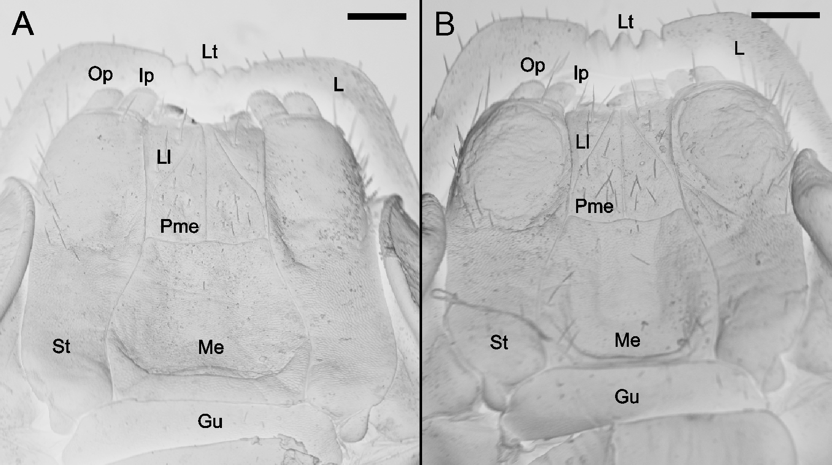
Gnathochilarium ( Fig. 4 View FIGURE 4. P A; B): Gula (Gu) with short setae. Mentum (Me) rounded with few setae visible. Males with a concavity deeper in Me than the female and Stipes (St) more rounded in distal region. St with basally and distally rounded lateral borders. Promentum (Pme) divided into two triangular parts separated by a midline suture. Laminae linguaies (Ll) triangular, entirely separated by Pme.
Trunk: Tergites and collum depigmented. Lateral region of rings with transverse striae present (striae variable in individuals). Anal shield and anal valve slightly pigmented.
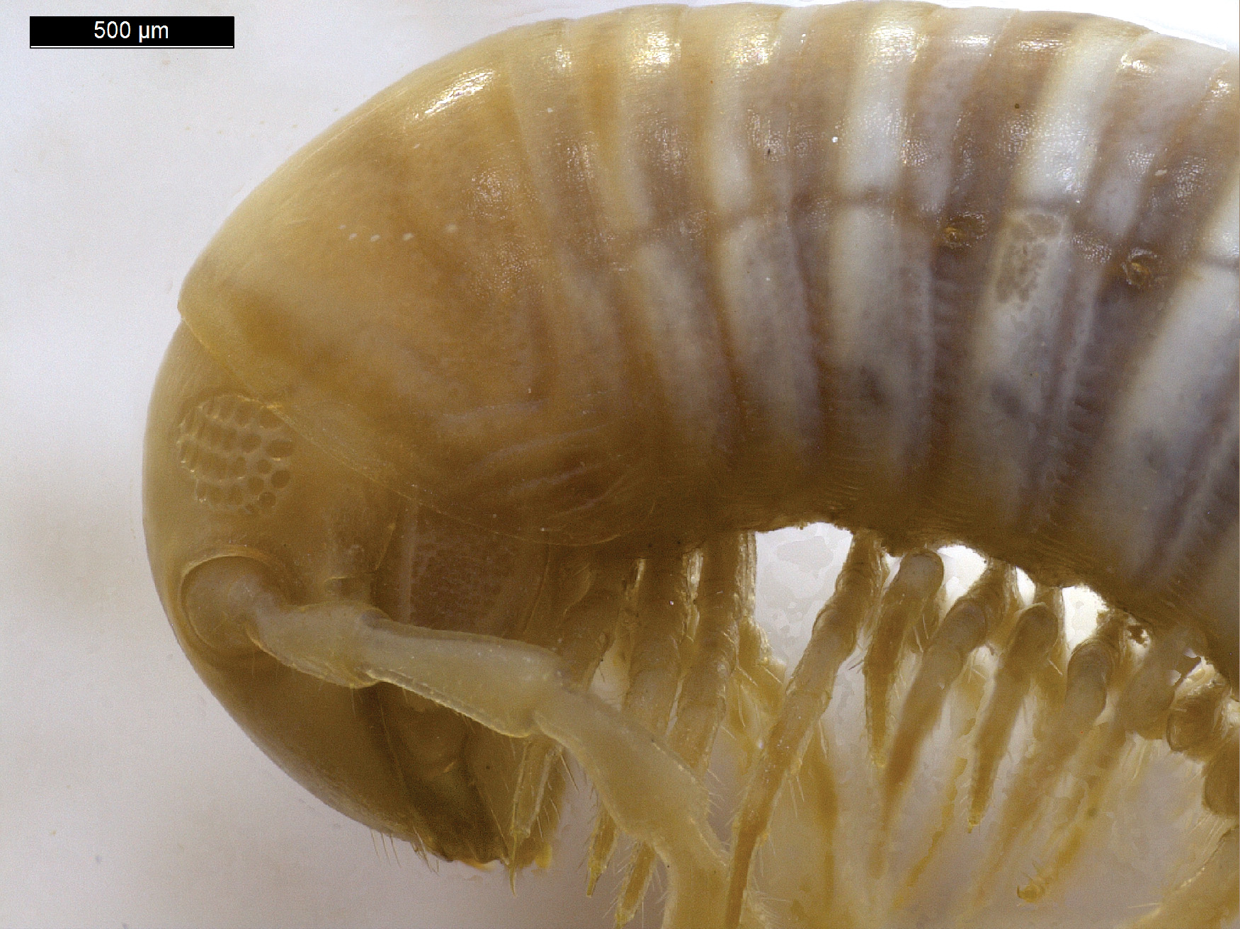
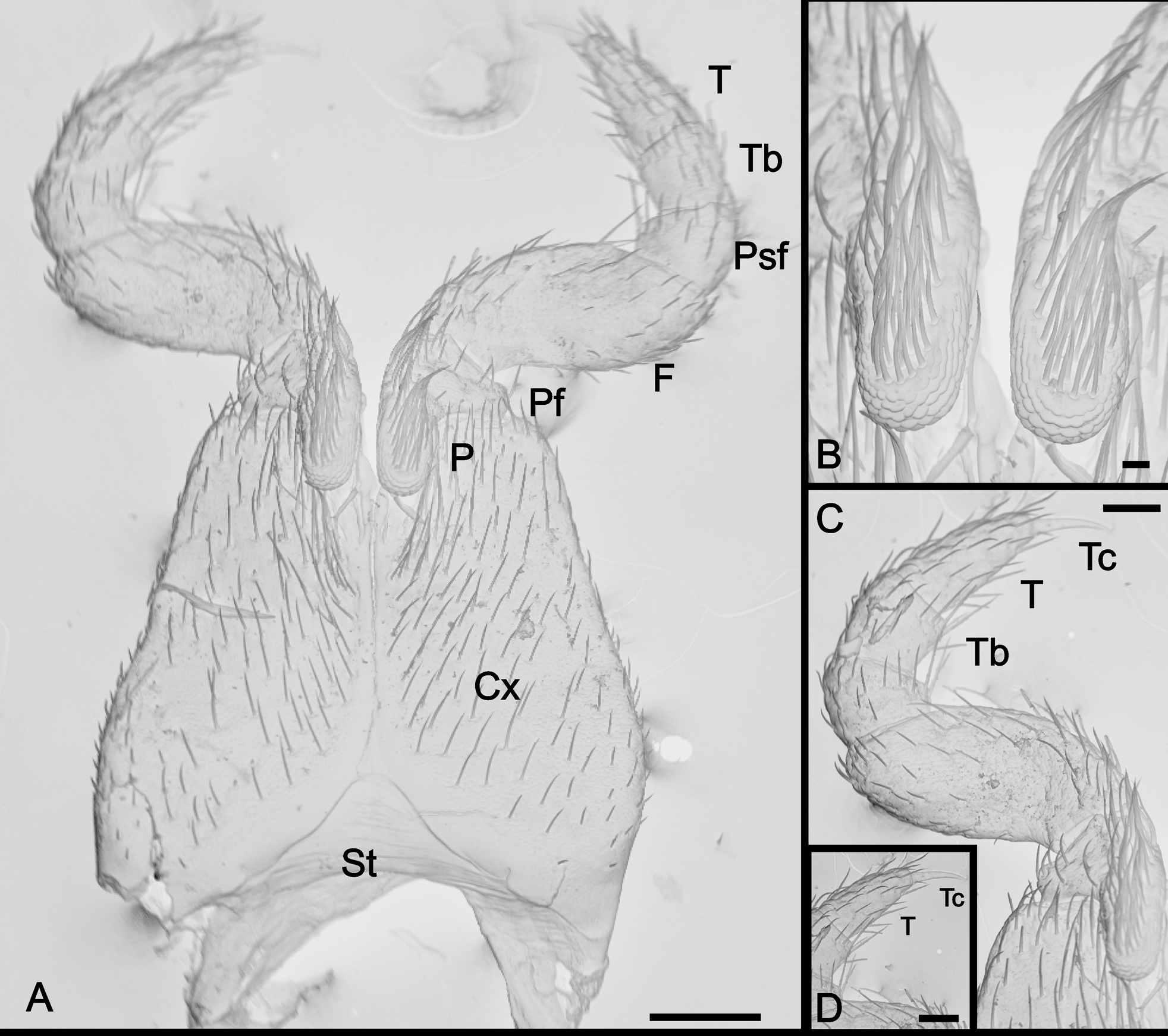
First male pair of legs ( Fig. 5 View FIGURE 5. P A): The first leg pair is modified and densely setose. Sternum (St) small and rounded. Coxae (Cx) larger than those of remaining legs and densely setose. Prefemur (Prf) with thin oral process parallel (P) to the coxa ( Fig. 5 View FIGURE 5. P B). Postfemur (Psf) and tibia (Tb) reduced ( Fig. 5 View FIGURE 5. P C). Femur (F) longer and wider than other podomeres. Terminal claw (Tc) not modified ( Fig. 5 View FIGURE 5. P D).
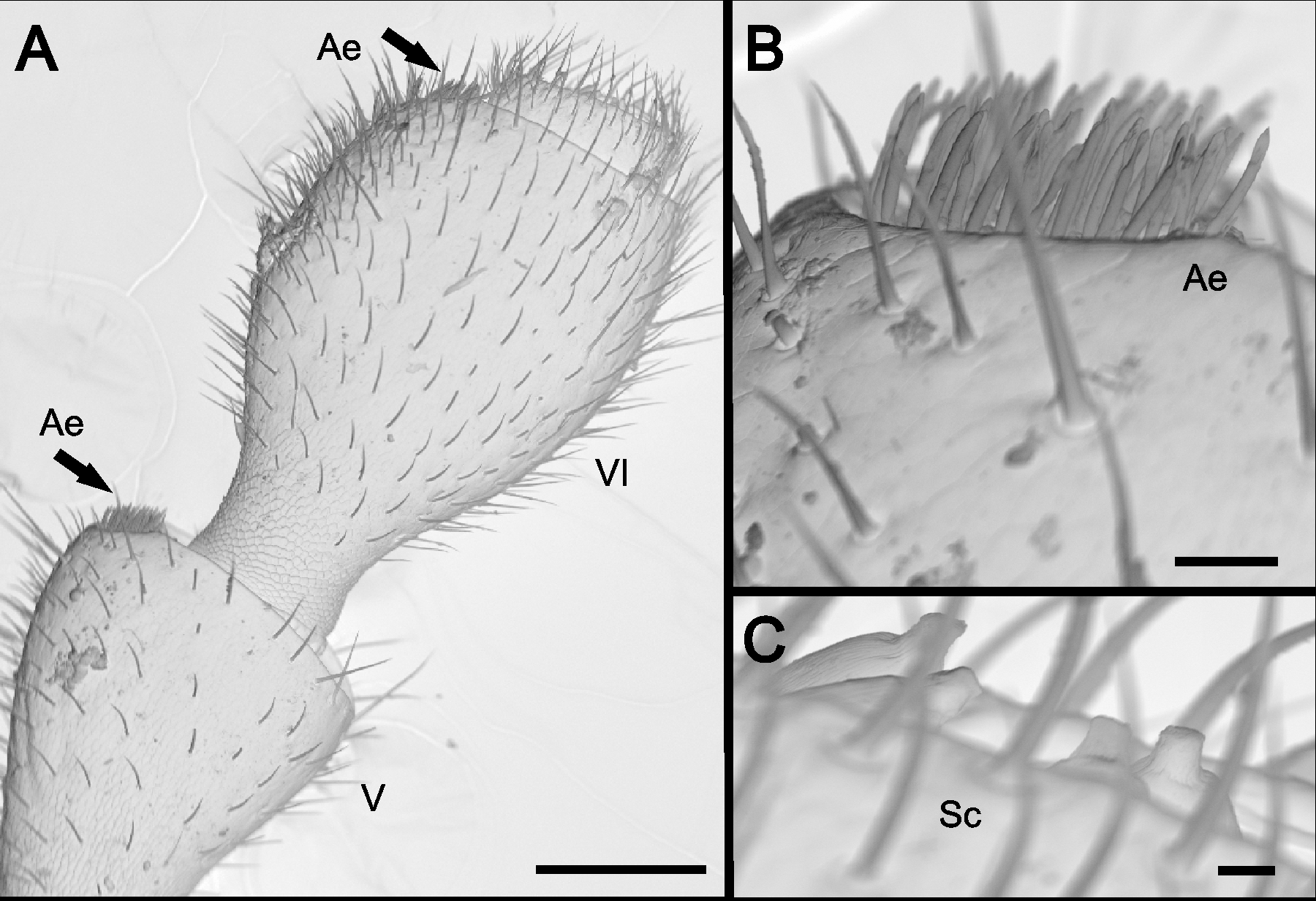
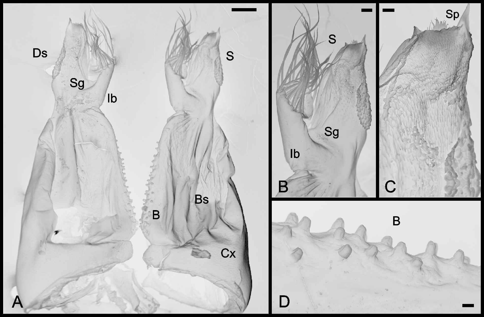
Gonopod ( Fig. 6 View FIGURE 6. P A): Coxae (Cx) reduced, glabrous and joined with basal section of telopodite. The basal section (Bs) of telopodite with surface more membranous than Cx. Shoulder absent. Length is about 0.75 times longer than width. Bs beset with basiconic sensilla (B) extending toward the internal margin of section ( Fig. 6 View FIGURE 6. P D).
Distal section (Ds) ( Fig. 6 View FIGURE 6. P B) sturdier and with length about 0.5 times longer than width. Ds divided into solenomere (S) ( Fig. 6 View FIGURE 6. P C) and an internal branch (Ib), separated by a seminal groove (Sg). Ib (coxosternal branch) like a shield of solenomere, densely setose. S rhomboid, with two different regions of squamous surface, a lateral surface more rounded and another spiniform in anteromedian region. Presence of a spine (Sp) in apex directed laterally.
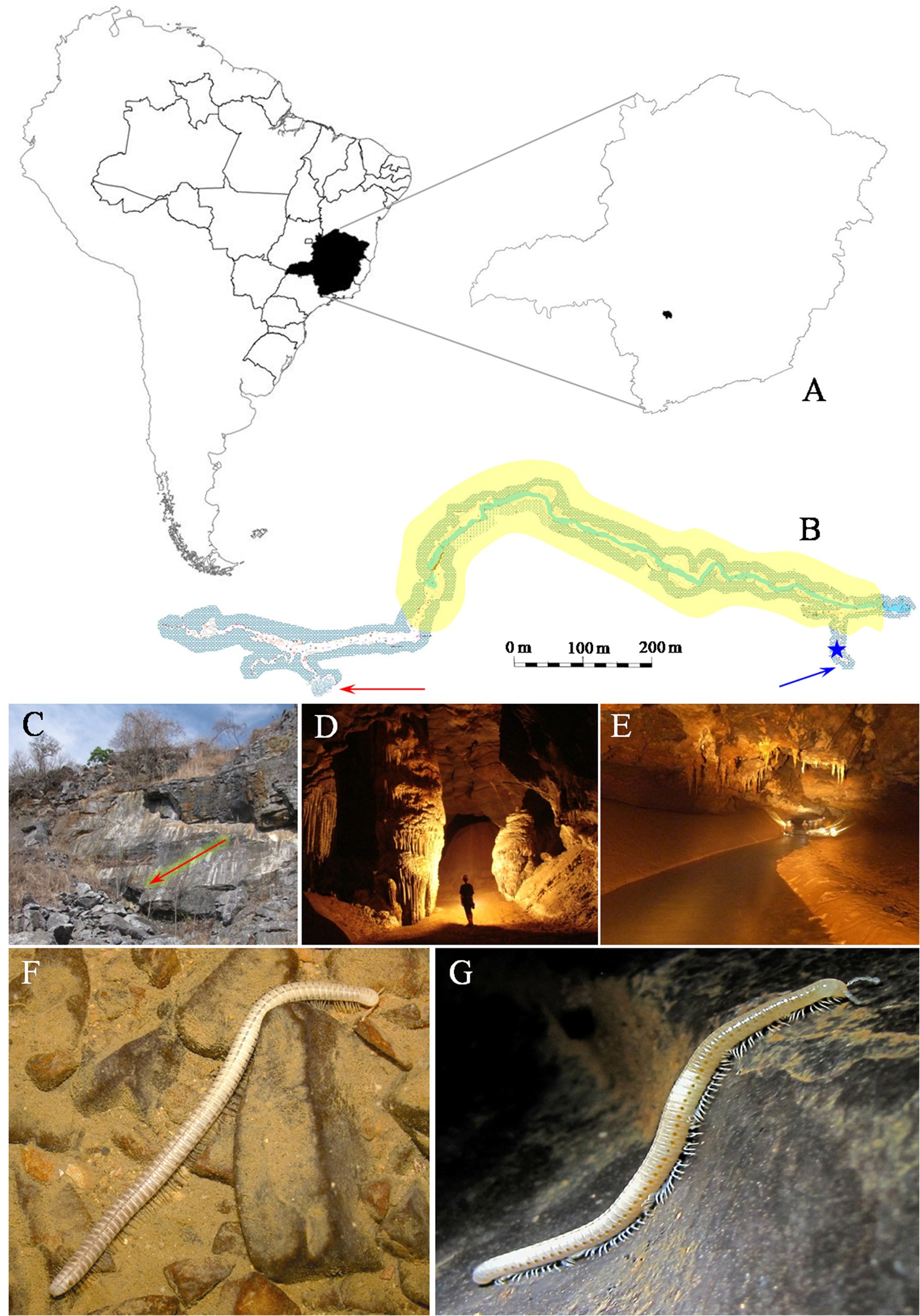
Remarks. Individuals of P.ambuatinga were observed in seven caves in the Pains region (Minas Gerais, Brazil), totaling 327 individuals, although few were collected for the study. In all caves, the organisms were always found in aphotic areas, far from entrances, also showing a clear preference for extremely humid locations. The largest populations were found in caves with underground rivers, such as the Gruta do Éden ( Fig. 1 View FIGURE 1 B, C, D, E) and the Loca d´água de baixo. Hundreds of individuals were observed in these caves. The organisms showed clear preference for plant remains that accumulated on the banks of rivers and for underground deposits of bat guano (especially that of the bat Desmodus rotundus , a hematophagous species). Furthermore, we observed completely submerged organisms, walking along the bottom of streams ( Fig. 1 View FIGURE 1 F), in the Gruta do Éden and Loca d´água de baixo. Some individuals were collected alive and reared in the laboratory. The terrarium was modified to contain two regions, one photic and the other aphotic (covered with aluminum foil and black plastic). Under these conditions, individuals exhibited clear preference for the aphotic area of the terrarium as expected, with some specimens burying themselves in sediments and remaining there.
No known copyright restrictions apply. See Agosti, D., Egloff, W., 2009. Taxonomic information exchange and copyright: the Plazi approach. BMC Research Notes 2009, 2:53 for further explanation.
|
Kingdom |
|
|
Phylum |
|
|
Class |
|
|
Order |
|
|
Family |
|
|
Genus |