
Aceria tripuraensis, Menon & Joshi & Ramamurthy, 2014
|
publication ID |
https://doi.org/10.11646/zootaxa.3760.4.4 |
|
publication LSID |
lsid:zoobank.org:pub:1289E1D4-B912-4DFE-9D77-729B6FAC1368 |
|
DOI |
https://doi.org/10.5281/zenodo.5040993 |
|
persistent identifier |
https://treatment.plazi.org/id/955CBA73-FFDA-FFBB-FF61-F354FE857E86 |
|
treatment provided by |
Felipe |
|
scientific name |
Aceria tripuraensis |
| status |
sp. nov. |
Aceria tripuraensis n. sp.
( Figs.1–13 View FIGURE 1 View FIGURES 2–9 View FIGURES 10–13 )
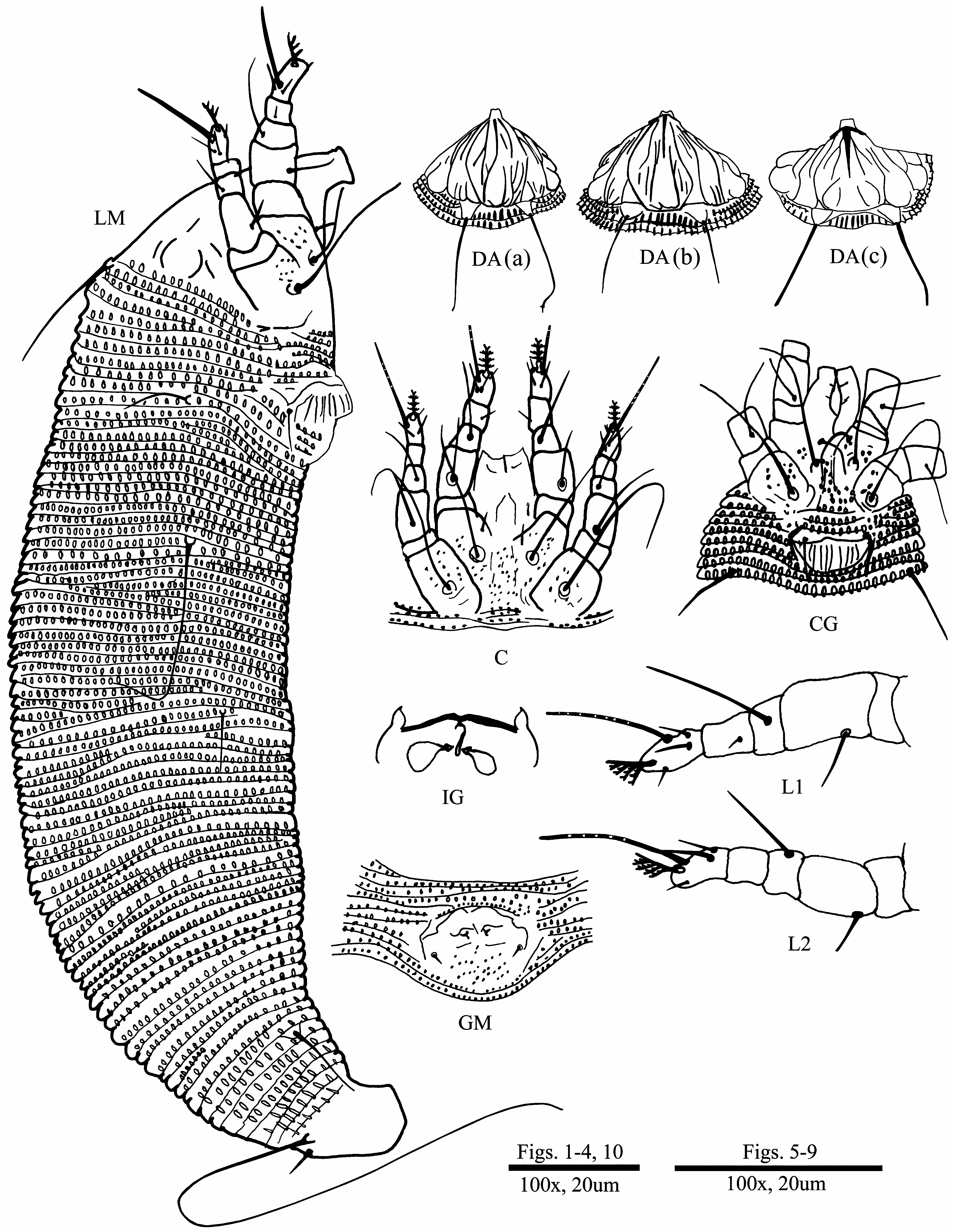
Diagnosis. Prodorsal shield with rounded lobes on postero-lateral margins and shield design comprised of one median, two admedian and four submedian lines. Solenidia on tarsus I and II, stout with transverse sculptures, at least 2.5x longer than respective empodia; empodia 4-rayed. Coxisternal plates microtuberculated. Female genital cover flap with longitudinal ridges. Opisthosomal setae ( d) long, almost 3.5x the length of setae ( c2), 2.9x the length of setae ( f) and 12x the length of the shortest setae ( e); setae ( h2) nearly 13.3x the length of setae ( h1). Live mites are transparent to white in colour.
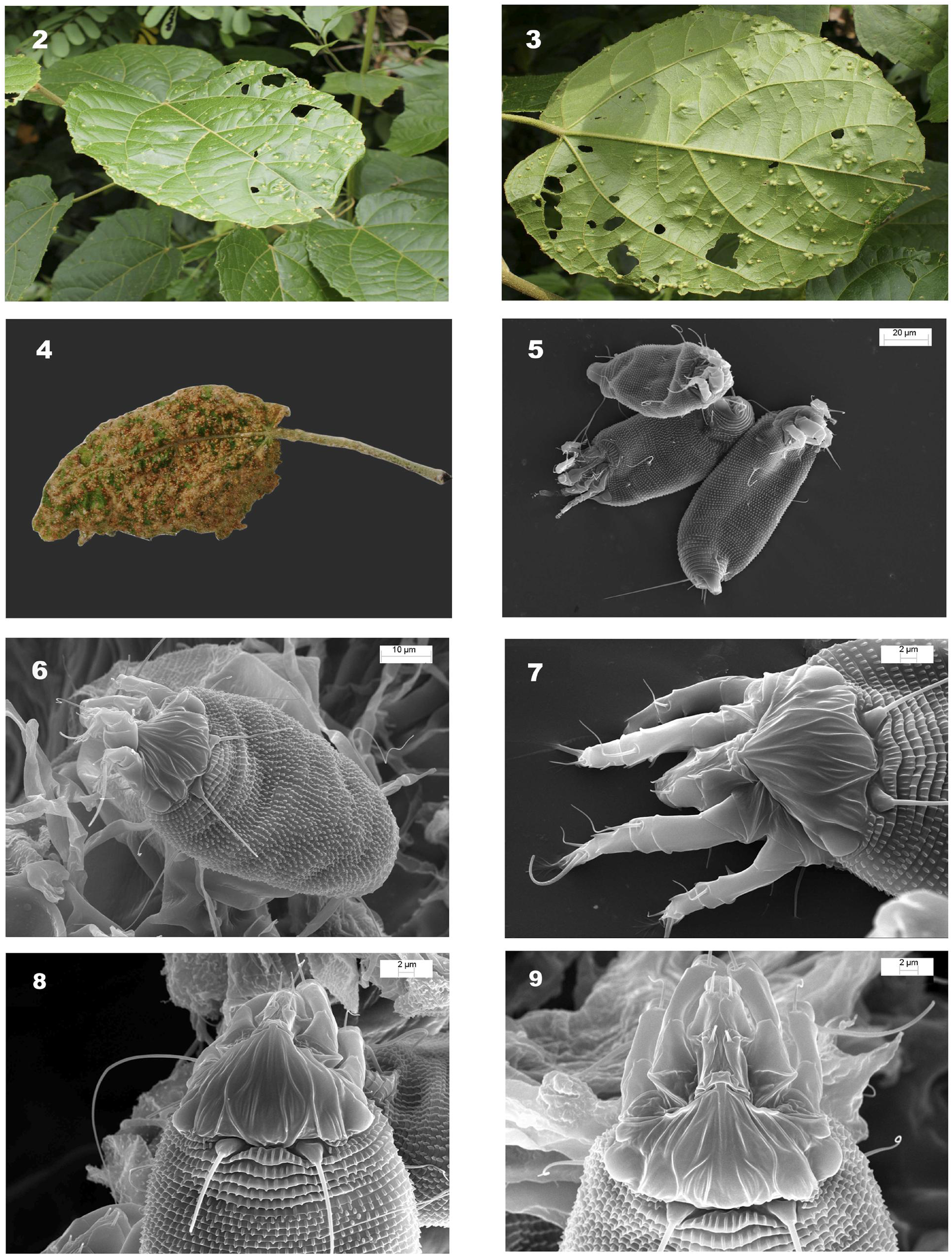
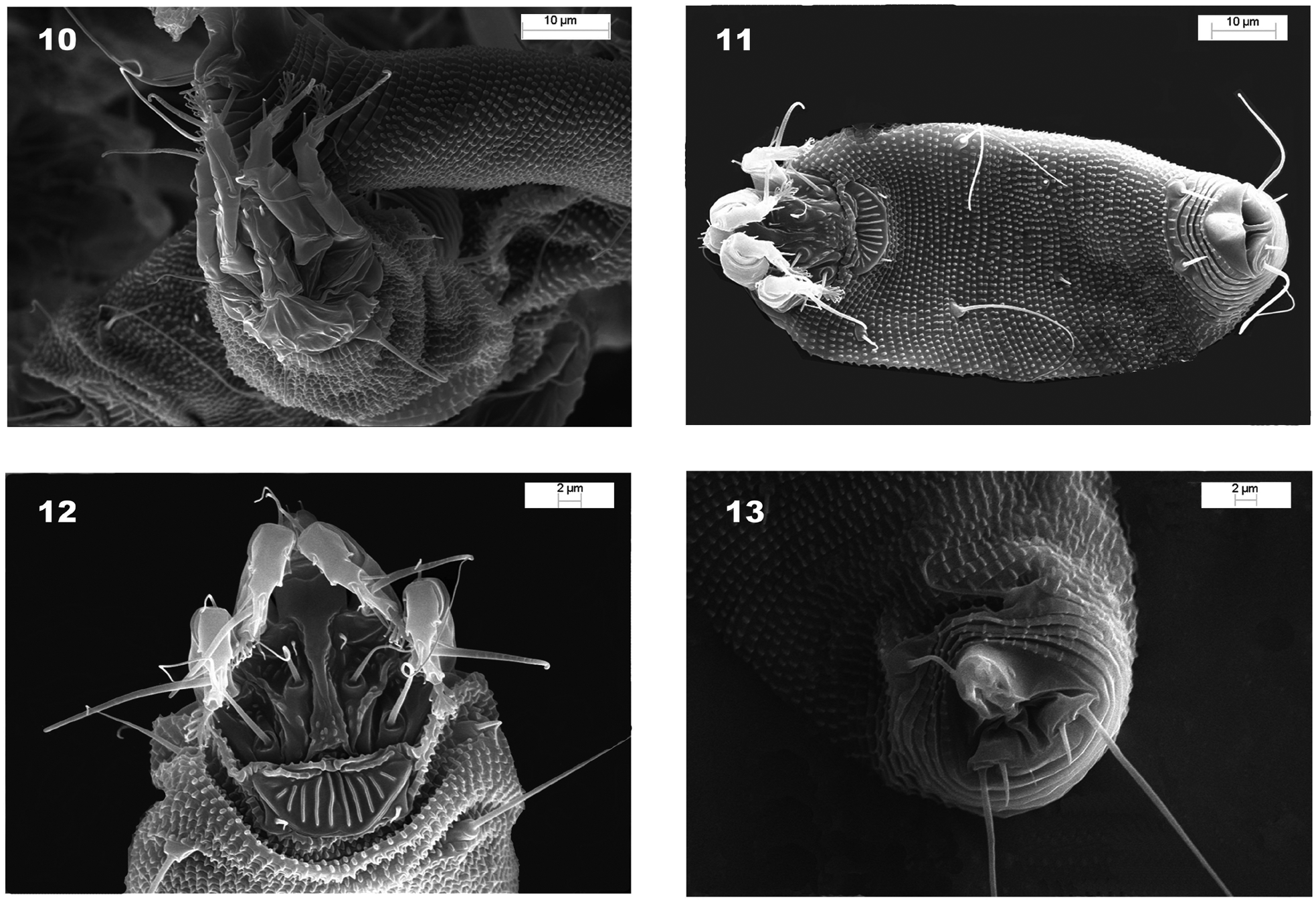
Description. FEMALE (n=10). Body worm-like 180, 156±20 (130–180), 41, 36±5 (30–43) wide; white in colour. Gnathosoma 12, 12± 2 (9–15) projecting downwards, pedipalp genual setae ( d) 2.5±0.5 (2–3), cheliceral stylets 15, 14±1 (12–15). Prodorsal shield broad at base, 19, 20±2 (17–23), 35, 30±3 (26–35) wide; frontal lobe partially buried into flexible cuticle of basal pedipalp; prodorsal shield (based on SEM images, Figs. 6–9 View FIGURES 2–9 ) with median line, prominently visible on anterior half of shield; admedian lines complete, extending outwards and bifurcating in middle of prodorsal shield; short lines present below bifurcation of admedian lines; first submedian lines complete, meeting posteriorly to form a vase-like structure, enclosing median and admedian lines completely and sometimes bifurcating near base of prodorsal shield; second submedian lines present only on anterior 1/3 of shield; third submedian lines complete, running obliquely and extending to base of prodorsal shield; fourth pair of submedian lines extending up to lobe structures as present laterally on prodorsal shield. Prodorsal shield (as examined under phase contrast; Fig. 1 View FIGURE 1 DA) characterized by many lines: straight, admedian line, vase-shaped; submedian lines enclosing various lines in middle and postero-lateral rounded lobes, prominently visible. Posterior margin of prodorsal shield with sulcus (furrow) at level of scapular setae. Scapular tubercles subcylindrical, arising from under posterior margin of prodorsal shield with two annuli lateral to each tubercle, 11, 13±1 (11–14) apart, directing scapular seta ( sc) divergently backwards; ( sc) 22, 23±1 (21–25), spanning 9, 11±2 (9–15) annuli. Legs. Leg I 27, 27±1 (26–28); trochanter 4, 4±1 (3–4), femur 10, 9±1 (8–10), basiventral femoral seta ( bv) 7, 6±1 (5–7); genu 4, 4±1 (4–5), antaxial genual seta ( l″) 14, 13±1 (12–15); tibia 5, 5±1 (4–6), paraxial tibial seta ( l′) 2, 2±0 (2); tarsus 7, 6±1 (5–7), tarsal solenidion ( ω) 12, 13±1 (12–14), rod-like, without knob, but with very faint transverse sculpturing over entire length, empodium 6, 5±1 (5–6), simple, 4-rayed, paraxial fastigial seta ( ft′) 3, 4±1 (4–5), antaxial fastigial seta ( ft″) 4, 5±1 (4–6), unguinal seta ( u′) 3, 2±1 (2–3). Leg II 24, 24±1 (23–25); trochanter 3, 3±1 (3–4); femur 9, 9±1 (8–10); basiventral femoral seta ( bv) 6, 7±1 (6–9); genu 4, 4±1 (3–4), antaxial genual seta ( l″) 8, 7±1 (5–9); tibia 5, 4±1 (3–5); tarsus 6, 5±1 (6–7), tarsal solenidion () 15, 16±1 (15–17), rod-like, without knob, but with very faint transverse sculpturing over entire length (visible in SEM micrographs; Figs. 10 & 13 View FIGURES 10–13 ), tarsal empodium 5, 5±1 (4–6), simple, 4 rayed, paraxial fastigial seta ( ft′) 3, 3±1 (3–4), antaxial fastigial seta ( ft″) 4, 4±1 (3–5), unguinal seta ( u′) 3, 2±1 (2–3). Coxal area granular, sternal line present, anterolateral setae on coxisternum I ( 1b) 5, 5±1 (4–5), 7, 7±1 (6–8) apart; proximal setae on coxisternum I ( 1a) 25, 22±3 (20–26), 8, 7±1 (6–8) apart; proximal setae on coxisternum II ( 2a) 37, 35±2 (30–36), 16, 16±1 (16–17) apart. Coxisternal area with 4–5 microtuberculated annuli. Genitalia 10, 7±1 (5–9) long, 16, 16±1 (15–16) wide; epigynium with 10–12 longitudinal ridges; central ridge longer than lateral ridges; internal female genitalia with foreshortened anterior apodeme; proximal seta on coxisternum III ( 3a) 5, 6±1 (4–7). Opisthosoma with annuli subequal dorsoventrally. Opisthosomal seta ( c2) 14, 14±1 (13–15), on ventral annulus 9–10; opisthosomal seta ( d) 48, 49±2 (46–53), 31, 30±1 (30–31) apart, on ventral annulus 17, 18±1 (16–19); opisthosomal seta ( e) 4, 4±1 (4–5), 16, 16±1 (14–17) apart, on annulus 32, 33±2 (31–35); opisthosomal seta ( f) 17, 17±1 (15–18), 14, 13±1 (12–14) apart, on annulus 54 (5th annulus from rear), 56±2 (53–60). Number of dorsal annuli 65, 65±1 (63–68) with oval/elongated microtubercles; 2 annuli present laterally to each scapular tubercle; first annulus posterior to prodorsal shield broadened with elongated microtubercles; widely spaced microtubercles present on posterior 5–8 annuli, becoming reduced in size; last 5 to 8 annuli smooth dorsally in some females. Number of ventral annuli 59, 61±2 (58–65) also with oval microtubercles, becoming narrower, rib-like and closely spaced posterior to seta ( f). Opisthosomal seta ( h2) 74, 75±4 (70–81); opisthosomal seta ( h1) 6, 6±1 (4–7).
MALE (n=2). Similar to female, 137.5±10 (130–145), 47.5±0.7 (47–48) wide. Gnathosoma projecting downwards; pedipalp genual setae ( d) 2.5±0.5 (2–3); chelicerae 13±1.4 (12–14); rostrum 11±1.4 (10–12). Prodorsal shield 20.5±0.7 (20–21) long, 27±1.4 (26–28) wide; dorsal tubercles near rear shield margin 15.5±0.7 (15–16) apart, directing scapular seta ( sc) divergently backwards; ( sc) 18.5±0.7 (18–19), spanning 11–12 annuli. Legs. Leg I 25; femur 9.5±0.7 (9–10), basiventral femoral seta ( bv) 7±1.4 (6–8); genu 3.5±0.7 (3–4), antaxial genual seta ( l″) 13; tibia 4.5±0.7 (4–5), paraxial tibial seta ( l′) 2; tarsus 5.5±0.7 (5–6), tarsal solenidion ( ω) 13±2.8 (11–15), rod-like, without knob, but with very faint transverse sculpturing along entire length, empodium 5.5±0.7 (5–6), 4 rayed, paraxial fastigial seta ( ft′) 4, antaxial fastigial seta ( ft″) 5, unguinal seta ( u′) 2. Leg II 23; femur 9.5±0.7 (9–10); basiventral femoral seta ( bv) 5.5±0.7 (5–6); genu 3, antaxial genual seta ( l″) 9.5±0.7 (9–10); tibia 4, tarsus 4.5±0.7, tarsal solenidion ( ω) 15, not knobbed, rod-like, but with very faint transverse sculpturing along entire length, empodium 5, 4-rayed, paraxial fastigial seta ( ft′) 3, antaxial fastigial seta ( ft″) 5, unguinal seta ( u′) 2. Anterolateral setae on coxisternum I ( 1b) 4±1.4 (3–5), 8±1.4 (7–9) apart; proximal setae on coxisternum I ( 1a) 16.5±2.1 (15–18), 8.5±0.7 (8–9) apart and proximal setae on coxisternum II ( 2a) 29±1.4 (28–30), 17±2.8 (15–19) apart. Genitalia 17 wide, 9.5±0.7 (9–10), genital seta ( 3a) 10±1.1 (9–12). Opisthosoma. Opisthosomal seta ( c2) 14±1.4 (13–15) on annulus 9–10; opisthosomal seta ( d) 42.5±3.5 (40–45), 34±2.8 (32–36) apart on annulus 21.5±0.7 (21–22); opisthosomal seta ( e) 3.5±0.7 (3–4), 21 apart, on annulus 36.5±0.7 (36–37); opisthosomal seta ( f) 15.5±2.1 (14–17), 13.5±0.7 (13–14) apart, on annulus 61.5±0.7 (61–62). Number of dorsal annuli 66.5±0.7 (66– 67), microtuberculated; number of ventral annuli 68±2.8 (66–70), microtuberculated. Opisthosomal seta ( h2) 49±1.4 (48–50); opisthosomal seta ( h1) 5±1.4 (4–6).
NYMPH. Not found.
LARVA (n=5). Body 100 (in all specimens measured), 36.6±4.7 (30–40) wide. Gnathosoma projecting downwards; chelicerae 12.2±0.8 (11–13); gnathosoma 11.2±1.3 (10–13). Prodorsal shield 20.4±3.2 (15–23) long, 20 wide; dorsal tubercles near rear shield margin directing scapular seta ( sc) divergently backwards; ( sc) 4.4±0.5 (4–5), spanning 5–6 annuli. Legs. Leg I 13; femur 5, basiventral femoral seta ( bv) 2.6±0.5 (2–3); genu 2, antaxial genual seta ( l″) 2.6±0.5 (2–3); tibia 2, paraxial tibial seta ( l′) 2; tarsus 4, tarsal solenidion ( ω) 5, not knobbed, transverse sculpturing not visible, empodium 4, 3-rayed, paraxial fastigial seta ( ft′) 2, antaxial fastigial seta ( ft″) 2, unguinal seta ( u′) not visible. Leg II 13; femur 4.6±0.5 (4–5); basiventral femoral seta ( bv) 2.4±0.5 (2–3); genu 2, antaxial genual seta ( l″) 2.4±0.5 (2–3); tibia 2; tarsus 3.6±0.5 (3–4), tarsal solenidion ( ω) 5.4±0.5 (5–6), not knobbed, transverse sculpturing not visible,empodium 3, 3-rayed, paraxial fastigial seta ( ft′) 2, antaxial fastigial seta ( ft″) 2, unguinal seta ( u′) not visible. Anterolateral setae on coxisternum I ( 1b) not seen; proximal setae on coxisternum I ( 1a) 5±1 (4–6), 7 apart and proximal setae on coxisternum II ( 2a) 10±2 (8–12), 15 apart. Genitalia not formed. Opisthosomal seta ( c2) 3.6±0.8 (3–5) on annulus 7–10; opisthosomal seta ( d) 9±1.2 (7–10), 24 apart, on annulus 15–20; opisthosomal seta ( e) not seen; opisthosomal seta ( f) 9.8±1.4 (8–12), 11 apart, on annulus 36– 42. Number of dorsal annuli 61.4±2 (59–64), microtuberculated; number of ventral annuli 41.4±3.7 (36–45), microtuberculated. Caudal seta ( h2) 17.8±2.2 (15–20); accessory seta ( h1) 3±1.2 (2–6).
Type material. Holotype female, 20 female paratypes on 20 microscope slides; GoogleMaps 2 male paratypes on 2 slides; 5 larva on 3 slides deposited in NPC, India with registration number: 1791–1810/13; GoogleMaps 2 female paratypes on 2 microscope slides deposited in NMNH, SEL, USDA with transaction number: 206557. All ex Hibiscus macrophyllus Roxb. ex Hornem. (Malvaceae) , locality: Ishaan Chandranagar, Agartala , Tripura ( 23°45'55"N 91°14'33"E), collected by V. V. Ramamurthy on 20 August 2011. GoogleMaps
Etymology. The specific designation tripuraensis is derived from the name of the north-eastern state of India, ‘Tripura’, from where the type host plant was collected.
Host plant. Hibiscus macrophyllus Roxb. ex Hornem. (Malvaceae) .
Relation to the host plant. This mite causes bud galls with domes on the lower leaf surfaces. The leaves appear bronzed, with reddish coloured pockmarks on the dorsal surface ( Figs. 2–4 View FIGURES 2–9 ).
Remarks. This new species is distinct among Aceria spp. having 4-rayed empodia and reported from India in the presence of prominent lobes on the postero-lateral margins of the prodorsal shield. In addition to this character, the new species is distinct among the species of Aceria that are specific to the host plants of the family Malvaceae in its characteristic prodorsal shield design and legs I and II with very long solenidia with faint transverse sculptures.
No known copyright restrictions apply. See Agosti, D., Egloff, W., 2009. Taxonomic information exchange and copyright: the Plazi approach. BMC Research Notes 2009, 2:53 for further explanation.
|
Kingdom |
|
|
Phylum |
|
|
Class |
|
|
Order |
|
|
Family |
|
|
Genus |