
Rakaia Hirst 1925
|
publication ID |
https://doi.org/ 10.11646/zootaxa.133.1.1 |
|
publication LSID |
lsid:zoobank.org:pub:175812D4-7628-441E-8A7A-EE9EBC2E1FDD |
|
DOI |
https://doi.org/10.5281/zenodo.5093221 |
|
persistent identifier |
https://treatment.plazi.org/id/97070A43-FF97-5A2A-5708-F9FAB9DCADEB |
|
treatment provided by |
Felipe |
|
scientific name |
Rakaia Hirst 1925 |
| status |
|
Type species: Rakaia antipodiana Hirst 1925 View in CoL .
Species account and distribution: 23 valid species (including the new species described here) are known, with 3 occurring in Queensland ( Australia) and the rest in New Zealand. Six of these species comprise two subspecies each.
Rakaia macra View in CoL sp. nov.
( Figs. 1-30 View FIGURES 1-2 View FIGURE 3 View FIGURES 45 View FIGURES 67 View FIGURES 811 View FIGURES 1216 View FIGURES 1722 View FIGURES 2324 View FIGURES 2528 View FIGURES 2930 )
Types. Holotype: male from Waipori (Otago, South Island of New Zealand), 18 March 1977, R. Forster leg., deposited in OMNZ . Paratypes: 5 males and 6 females, same collecting data as holotype, deposited in OMNZ ; 6 males and 6 females, same collecting data as holotype, deposited in MONZ ; 6 males and 6 females, same collecting data as holotype, deposited in MCZ 48184-48195 View Materials (including 2 male and 2 females mounted on SEM stubs).
Other material examined. 39 males and 36 females from Waipori (Otago, South Island, of New Zealand), 18 March 1977, R. Forster leg., deposited in OMNZ ; 16 males, 18 females and 6 juveniles from Waipori (Otago, South Island of New Zealand), 21 March 1977, R. Forster leg., deposited in OMNZ ; 19 males, 19 females, and 10 juveniles from Waipori (Otago, South Island of New Zealand), 20 September 1977, R. Forster leg., deposited in OMNZ .
Etymology. Latin adjective: macra = lean, skinny. The specific epithet refers to the general appearance of the animal’s body, particularly in lateral view.
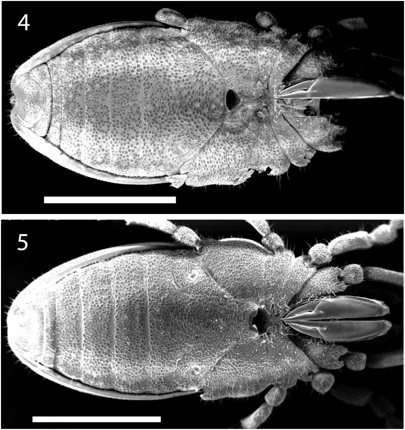
Diagnosis: Pettalid with dorsal scutum flat ( Figs 6, 7 View FIGURES 67 ). Trochanter of palp with conspicuous ventral process ( Fig. 11 View FIGURES 811 ); chelicerae with distinct outer lateral ridge on the surface of the second article ( Figs. 4, 5 View FIGURES 45 , 8 View FIGURES 811 ), lacking a ventral process on the first article ( Fig. 8 View FIGURES 811 ); dentition of the mobile digit of chelicerae uniform ( Fig. 9 View FIGURES 811 ).
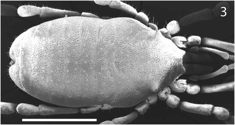
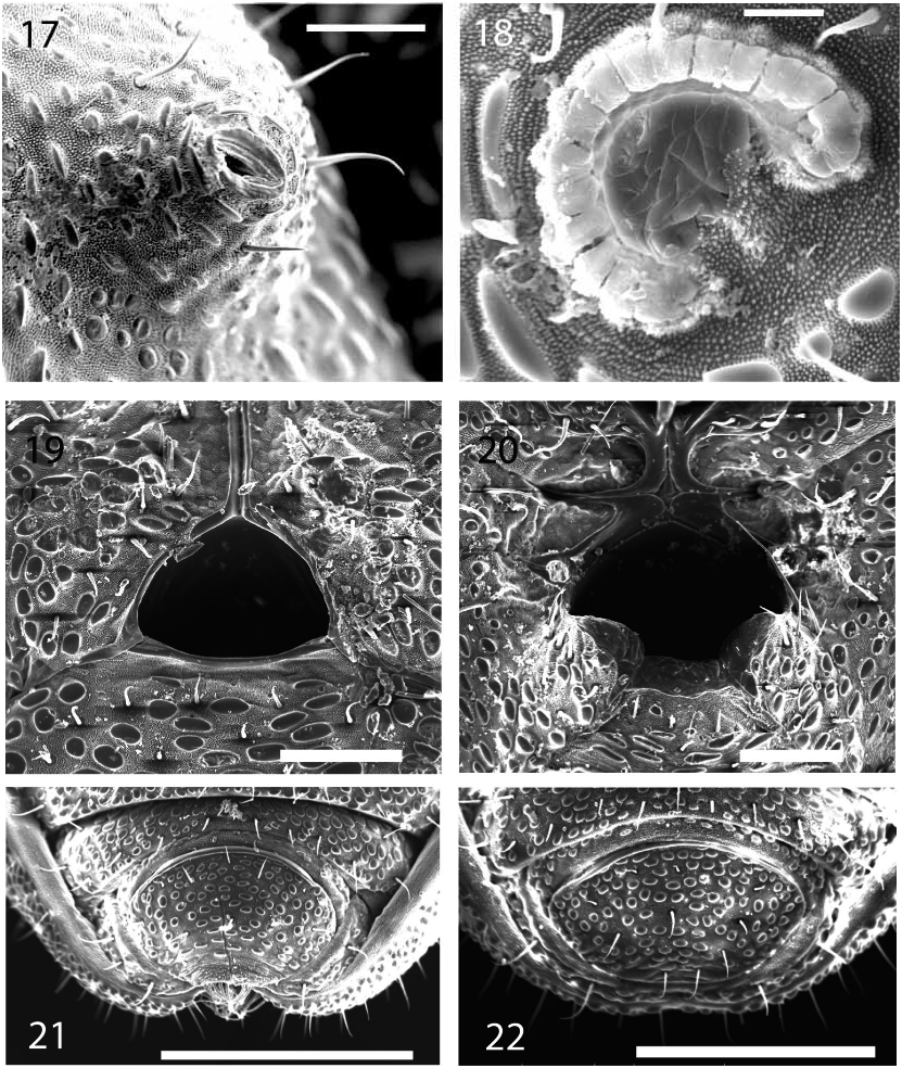
Description: Total length of male holotype (female paratype measurements in parentheses) 2.25 (2.60), width across ozophores 0.855 (0.769), greatest width 1.19 (1.29); length/width ratio 1.89 (2.02). Body brownyellow (in alcohol) ( Figs. 1, 2 View FIGURES 1-2 ) with most of the dorsal surface and legs showing a tuberculategranulate microstructure. Anterior margin of dorsal scutum with two projections delimiting the insertion of the chelicerae ( Figs. 1 View FIGURES 1-2 , 3 View FIGURE 3 ); prosomal region trapezoidal. Eyes absent. Ozophores conical and positioned dorsally, somewhere between types 2 and 3 of Juberthie (1970) ( Figs. 3 View FIGURE 3 , 17 View FIGURES 1722 ). Transverse prosomal sulcus distinct but not conspicuous ( Fig. 3 View FIGURE 3 ). Transverse opisthosomal sulci distinct by lacking granulation, longitudinal opisthosomal sulcus absent ( Figs. 1 View FIGURES 1-2 , 3 View FIGURE 3 ). Dorsal scutum flat ( Figs. 6, 7 View FIGURES 67 ); opisthosomal region reaching its maximum width at segment II.
Coxae of legs I and II mobile; coxae of remaining legs fused. Ventral prosomal complex of male with coxae II, III, and IV meeting in the midline ( Fig. 4 View FIGURES 45 ). Male gonostome small, subtriangular, slightly wider than long, bordered on posterior margin by the first opisthosomal sternite ( Fig. 19 View FIGURES 1722 ); male gonostome shorter than length of seam of contact of left and right coxae IV parallel to midline. Ventral prosomal complex of female with only coxae II and III meeting in the midline ( Fig. 20 View FIGURES 1722 ). Female gonostome roughly round in shape, with the edges of coxae of leg IV and first opisthosomal sternite forming a partial “tube” at the posterior margin of the opening ( Figs. 5 View FIGURES 45 , 20 View FIGURES 1722 ).
Spiracles Cshaped (sensu Giribet and Boyer 2002), with one of the edges recurving internally as found in the “open circle” type ( Fig. 18 View FIGURES 1722 ). Sternal opisthosomal region without modifications or glandular pores ( Figs. 2 View FIGURES 1-2 , 4, 5 View FIGURES 45 ). Anal region with sternites 8 and 9 and tergite IX free, not forming a corona analis ( Figs. 21 View FIGURES 1722 , 23 View FIGURES 2324 ). Area of contact of tergite IX and sternite 9 of “pettalid” type ( Giribet and Boyer 2002) in which tergite IX laterally covers sternite 9 and clearly meets sternite 8. Anal plate of male without longitudinal carina; posterior margin of anal plate slightly concave, bearing a scopula ( Fig. 23 View FIGURES 2324 ). Scopulae present on each lobe of tergite VIII ( Fig 24 View FIGURES 2324 ). Cuticle with granular surface in all ventral areas including coxae and anal plate. Anal glandular pore not observed in males.
Chelicerae short, extremely robust, with very few long setae. Proximal article with hooklike dorsal crest (“dorsal ridge” of Hansen and Sørensen 1904 and Forster 1948; “dorsal transverse crest” of Juberthie 1970), without ventral process, ornamented over almost its entire surface from the dorsal ridge to the second article ( Fig. 8 View FIGURES 811 ). Chelicerae of the protruding type described by Giribet (in press). Second article extremely wide, with a distinct longitudinal ridge on the outer lateral surface running towards the base of the mobile digit ( Figs. 5 View FIGURES 45 , 8 View FIGURES 811 ). Mobile digit with 12 uniform denticles of the same type ( Fig. 9 View FIGURES 811 ). Proximal cheliceral article of male paratype 0.697 long, 0.303 wide; second article 0.947 long, 0.273 wide; mobile digit 0.121 long, 0.083 wide, 13% of second article length.
Palp ( Fig. 10 View FIGURES 811 ) with a sharp, spiny ventral process on the trochanter ( Fig. 11 View FIGURES 811 ). Measurements of palpal articles in male paratype, length/width (L/W ratio), from trochanter to tarsus: 0.275 / 0.129 (2.13); 0.422 / 0.100 (4.22); 0.311 / 0.111 (2.80); 0.333 / 0.222 (1.5). Palpal claw 0.041 long.
Legs with all claws smooth, without ventral dentition or lateral pegs ( Figs. 1216 View FIGURES 1216 ). Surface of most articles clearly ornamented with granules, including metatarsi II and III; metatarsus of legs I and II with ornamentation only in the proximal half; tarsi without granules. Ventral side of tarsus I with a concentration of short sensory hairs occupying about half of the total tarsal length, not forming a distinct solea ( Fig. 12 View FIGURES 1216 ). Tarsus IV of male entire, carrying a short, thick adenostyle projecting upward and slightly distally ( Fig. 15 View FIGURES 1216 ). Leading edge of adenostyle base at about 40% of the tarsal length. Tarsus IV of the female without modifications ( Fig. 16 View FIGURES 1216 ).
Leg measurements of male paratype in µm [length/width (L/W ratio)]:
Tr Fe Pa Ti Mt Ta Total Leg I 224/210 (1.07) 605/171 (3.54) 468/171 (2.15) 447/171 (1.61) 289/145 (1.99) 500/184 (2.72) 2533 Leg II 132/140 (0.94) 421/132 (3.19) 228/140 (1.63) 281/140 (2.01) 211/105 (2.01) 360/123 (2.93) 1633 Leg III 226/170 (1.33) 396/151 (2.62) 255/151 (1.69) 321/179 (1/79) 245/132 (1.86) 415/142 (2.92) 1858 Leg IV 224/184 (1.22) 579/184 (3.15) 342/197 (1.74) 395/197 (2.01) 289/158 (1.83) 526/188 (2.80) 2355
Penis small, typical of pettalids ( Figs. 2528 View FIGURES 2528 ). Setal formula 3, 6, 3 or 4 (two penises examined). Ventral side ornamented with tiny denticles along distolateral and distal margins. Three ventral setae set back far from distal margin, their bases separated by distances roughly equal to setal diameter. Rounded distal margin of penis with 6 apical setae. Dorsal side of penis with a group of 3 or 4 long setae on each side, their broad bases separated by distances roughly equal to setal diameter and arranged in a V. Gonopore complex with two movable fingers (m.f.) in the shape of pronounced hooks, with denticles at their bases ( Figs. 26, 28 View FIGURES 2528 ).
Ovipositor ( Figs. 29, 30 View FIGURES 2930 ) wide, composed of two apical lobes and 29 circular articles, each of the latter furnished with eight setae. Apical lobes carrying several setae (increasing in length towards the tip); a long terminal seta rising from a small socket at the end of each lobe. Sensitive processes carrying a long, multibranched seta on the distolateral side of each terminal lobe.
Variation: Range of measurements: male (n = 6), female (n = 6) in parentheses: dorsal scutum length 2.242.50 (2.482.64).
Distribution and habitat: Known only from Waipori, Otago, South Island of New Zealand. Collected by Ray Forster three times in 1977: March 18, March 21, and September 20. Although there is a scenic reserve at this site, no further information about these collections is available. A collection of specimens of an undescribed species is also known from this locality.
Remarks: Rakaia macra sp. nov. clearly belongs to the family Pettalidae because of the ozophore position and the modified anal region. However, the dentition of its chelicerae is unusual for this group; a homogeneous dentition has been previously reported only in the genera Purcellia Hansen & Sørensen 1904 and Parapurcellia Rosas Costa 1950 among the Pettalidae . Rakaia macra sp. nov. resembles other Rakaia species which, in ventral view, appear to have scopulae only on the posterior margin of the anal plate. These include Rakaia lindsayi Forster 1952 , R. magna Forster 1948 , R. media Forster 1948 , and R. solitaria Forster 1948 .
Rakaia macra sp. nov. is distinguished from all other congeners by the presence of an outer lateral ridge on the second cheliceral article ( Fig. 8 View FIGURES 811 ). Additionally, it is distinguished from R. lindsayi by the uniform dentition in the mobile cheliceral digit ( Fig. 9 View FIGURES 811 ), the presence of a ventral process on the trochanter of the pedipalp ( Fig. 11 View FIGURES 811 ), and the absence of a ventral process on the chelicerae ( Fig. 8 View FIGURES 811 ). R. macra sp. nov. is distinguished from R. magna by size; specimens of R. magna are 3.0 mm or more in length, while all specimens of R. macra are smaller than 2.70 mm in length. R. macra is distinguished from R. media by the shape of the male tarsus IV; R. media has a pyriform male tarsus IV (sensu Forster 1952), while the male tarsus IV of R. macra tends to be elongate (sensu Forster 1952). R. macra is distinguished from R. solitaria by elongate vs. pyriform male tarsus IV, and the shape of the adenostyle, which is long and slender in R. solitaria but stout in R. macra ( Fig. 15 View FIGURES 1216 ).
The penis of R. macra sp. nov. ( Figs. 2528 View FIGURES 2528 ) is very similar to that of several pettalids: R. daviesae Juberthie 1989 ( Juberthie 1989: figure 7), Austropurcellia scoparia Juberthie 1988 ( Juberthie 1988: figures 1013), Chileogovea jocasta Shear 1993 ( Shear 1993: figures 910), and Chileogovea oedipus Roewer 1961 ( Shear 1993: figures 1516), but setal formula differs among the various species. For example, there are 8 or 9 dorsal setae in R. daviesae but only 3 or 4 in R. macra sp. nov. The ventral setae of the penis are also found in different numbers in the two species: 2 in R. daviesae , but 3 in R. macra sp. nov. The mobile digit with two prominent hooks is present in all of the species listed above, but is absent in the pettalid species Purcellia illustrans Hansen & Sørensen 1904 ( Hansen and Sørensen 1904: Plate IV, figures 1o1q).
The ovipositors of Rakaia macra and Rakaia daviesae appear nearly identical ( Figs. View FIGURES 2930
2930, Juberthie 1989: figure 7) except in the number of circular articles: 26 in R. daviesae but 29 in R. macra sp. nov. The sensitive processes and general configuration of the distal end of the first article and lobes appears identical to that illustrated for Metasiro and described as typical of Chileogovea , Parapurcellia , Rakaia , and the majority of Siro species ( Fig. 30 View FIGURES 2930 , Juberthie 1970: figure 12c).


No known copyright restrictions apply. See Agosti, D., Egloff, W., 2009. Taxonomic information exchange and copyright: the Plazi approach. BMC Research Notes 2009, 2:53 for further explanation.