
Proteus
|
publication ID |
https://doi.org/ 10.1093/zoolinnean/zlac063 |
|
DOI |
https://doi.org/10.5281/zenodo.7695698 |
|
persistent identifier |
https://treatment.plazi.org/id/9750C307-FF84-4C37-FEFE-F510FD3CF9FA |
|
treatment provided by |
Plazi |
|
scientific name |
Proteus |
| status |
|
PROTEUS LAURENTI, 1768 View in CoL View at ENA
Species: Proteus anguinus * Laurenti, 1768 View in CoL View at ENA .
Otic–occipitum complex
In proteids, the prootic, opisthotic and exoccipital remain unfused in the adult; therefore, there is no otic–occipitum complex.
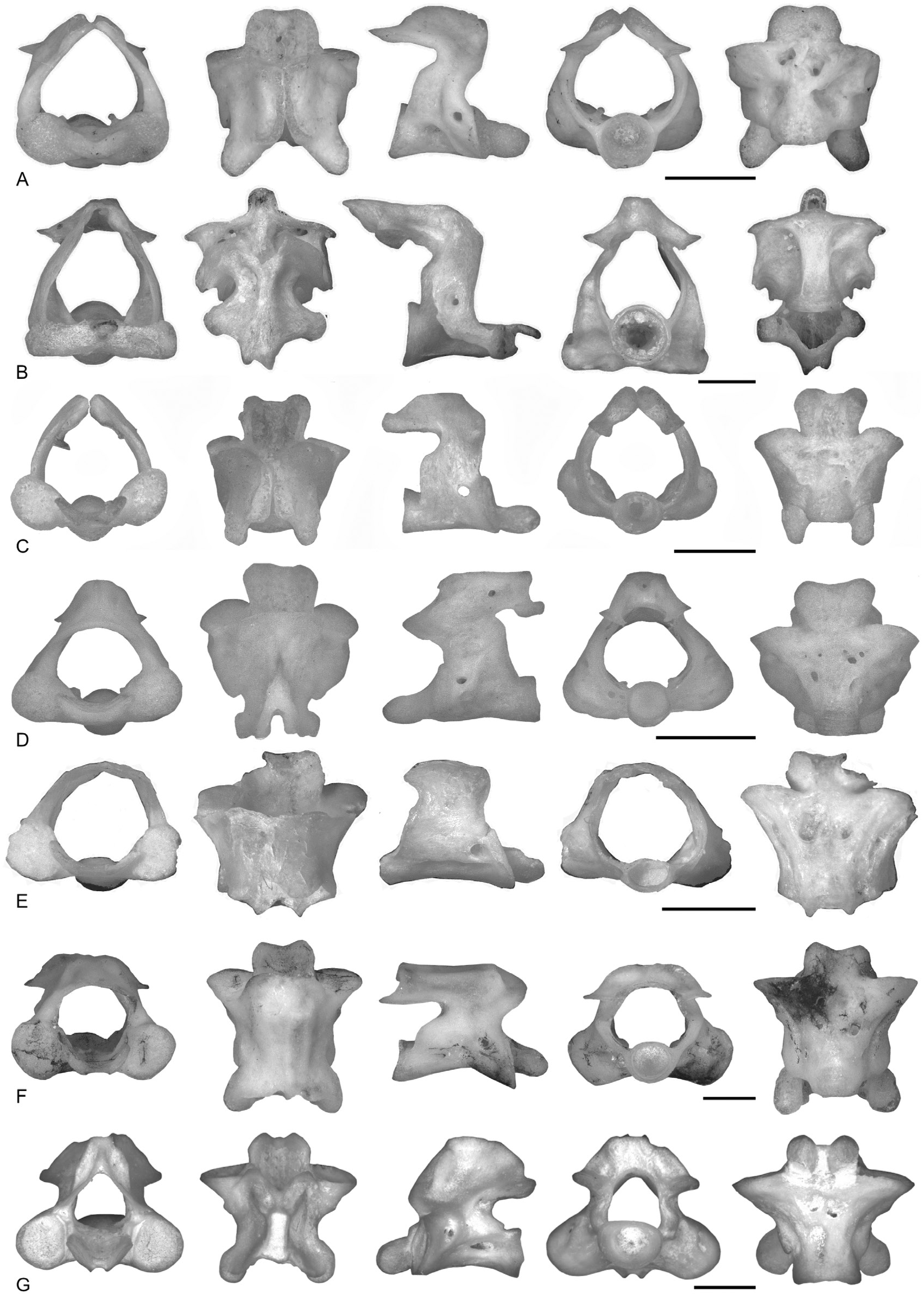
Atlas ( Fig. 6B View Figure 6 )
The neural canal is triangular in anterior view and is at least twice as high as each occipital joint.In posterior view, the neural canal is slightly wider than the circular cotyle. The occipital joints are elliptical, with the major axis being horizontal (or sub-horizontal), and exhibit strong dorsoventral compression in anterior view. They are confluent in the middle, with the flat and narrow odontoid process dorsal to them. The odontoid process is narrow and formed by a single, flat articular surface. The neural crest is low and hourglass-shaped in dorsal view. The secondary crests are absent. The neural spine is absent, and the posterodorsal area of the neural arch in dorsal view is forked and extends beyond the postzygapophyses. The lateral surface of the atlas bears only the foramen of the first spinal nerve. The incisura vertebralis cranialis is absent or high and narrow. In lateral view, the dorsal edge of the neural arch is sub-horizontal. The neural arch between the incisura caudalis and the cotyle is concave, inclined or sub-vertical. The maximum concavity of the incisura vertebralis caudalis is dorsal to the horizontal plane containing the maximum concavity of the incisura cranialis. The lateral crests extend more dorsally to the postzygapophyses without contacting them. The inferior crests are low or absent. In posterior view, the neural arch is dorsally convex (inverted V-shaped). The postzygapophyses are entirely more posteriorly extended than the cotyle in lateral view. In dorsal view, the neural arch dispalys an anteriorly V-shaped concavity and is forked posteriorly. The cotyle is not visible in dorsal view. The ventral surface is usually smooth.
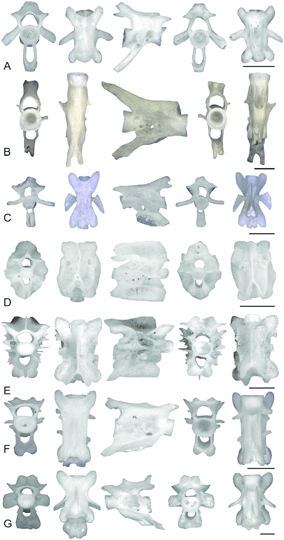
Precaudal vertebrae ( Fig. 9B View Figure 9 )
The precaudal vertebrae are amphicoelous. The neural canal is elliptical and slightly lower than the cotyle. The cotyles are circular or slightly elliptical, with the major axis being horizontal. In lateral view, the anterior edge of the neural arch between the anterior cotyle and the prezygapophyses is concave or vertical. The transverse processes are laminar, rectangular or triangular (proximally enlarging) in anterior view. They are orientated slightly posteriorly, and in lateral view they cover the posterior edge of the neural arch between the centrum and postzygapophyses entirely or in part. The diapophysis is cylindrical and proximally hollow, whereas the parapophysis is smaller and not hollow. In lateral view, most of the height of the vertebrae is formed by the centrum and the neural canal (only one-fifth of the height of the vertebra is formed by the neural arch dorsal to the postzygapophyses). In lateral view, the neural arch dorsal to the prezygapophyses is variably visible. The blade-like neural crest is absent or low, starting posterior to the anterior edge of the neural arch, and it broadens posteriorly in some cases. The neural spine is absent. The posterodorsal area of the neural arch in dorsal view is forked and projects posteriorly further than the postzygapophyses. The anterior and posterior zygapophyseal crests are absent. The ventral lamina is wide, triangular or trapezoidal in outline. The anterior ventral crests are anteriorly convex, whereas the posterior crests are posteriorly concave. The lateral surface of the vertebrae is smooth. The only foramen present is visible in anterior and lateral view, in the ventral half of the proximal edge of the transverse processes (at the base of parapophyses). The incisura vertebralis caudalis is not deep and is present only as a small posterior concavity (wider in the first precaudal vertebrae). The anterior edge of the neural arch between the centrum and postzygapophyses is slightly concave or convex. In posterior view, the neural arch is dorsally forked, with a deep incisura dorsalis. Half of the postzygapophyses extends posteriorly beyond the cotyle in lateral view. In dorsal view, the neural arch is anteriorly concave (U-shaped), and the incisura dorsalis is visible posteriorly in the forked neural arch. The edge of the anterior cotyle is visible in dorsal view, whereas the posterior cotyle is not visible. In lateral view, the ventral profile of the centrum is horizontal. The ventral surface bears two foramina and a subcentral keel.
Caudal vertebrae ( Fig. 12B View Figure 12 )
The caudal vertebrae are longer than high (height/length ratio <1). The neural canal is pentagonal or circular, whereas the haemal canal is triangular or circular. The neural canal is wider and lower than the haemal canal. The transverse processes are absent or transformed into horizontal laminae. The neural crest is absent or low. Zygapophyseal and ventral crests are absent. The lateral surface is smooth or with a single foramen visible at the base of the haemal arch. In lateral view, the anterior edge of the haemal arch is convex or anteriorly inclined, whereas the posteroventral edge of the haemal arch forms a sharp tip in lateral view. The haemal crest is either low or absent. The posterodorsal ends of the neural arches and posteroventral ends of the haemal arches are forked. The width between the prezygapophyses is greater than that between the postzygapophyses in the first caudal vertebrae; pre- and postzygapophyses are absent in the caudal vertebrae from the posterior half of the tail.

No known copyright restrictions apply. See Agosti, D., Egloff, W., 2009. Taxonomic information exchange and copyright: the Plazi approach. BMC Research Notes 2009, 2:53 for further explanation.