
Ceratophysella hermosa ( Wray, 1953 )
|
publication ID |
https://doi.org/10.11646/zootaxa.3918.3.1 |
|
publication LSID |
lsid:zoobank.org:pub:E69AC33B-2E8A-4914-B64F-C2DF918612BE |
|
DOI |
https://doi.org/10.5281/zenodo.5664739 |
|
persistent identifier |
https://treatment.plazi.org/id/985F8790-2F41-FFDA-97BE-FB36DC663756 |
|
treatment provided by |
Plazi |
|
scientific name |
Ceratophysella hermosa ( Wray, 1953 ) |
| status |
comb. nov. |
Ceratophysella hermosa ( Wray, 1953) new combination
Figs. 1A, 1 View FIGURE 1. A C, 2, 3
Mitchellania hermosa Wray 1953: 1 .
Hypogastrura ( Mitchellania) hermosa Christiansen & Bellinger 1980: 178 ; 1998: 186.
Specimens examined. Lectotype female, paralectotype female (labeled Mitchellia hermosa n. sp. n. gen.) (by present designation), USA, North Carolina, Yancey County, Mt. Mitchell, elev. 6,500 ft. ( 1,981 m), 5 October 1949, spruce “woodsmold,” D. L. Wray & E. D. Wray, colls.; 1 specimen, USA, Tennessee, Sevier County, elev. 5,800 ft. ( 1,768 m), 27 October 1959, T. P. Copeland, coll.; 1 specimen, USA, Tennessee, Sevier County, Great Smoky Mountains National Park, Goshen Prong, elev. 917 m, pitfall trap, 5–18 December 2001, I. Stocks, coll.
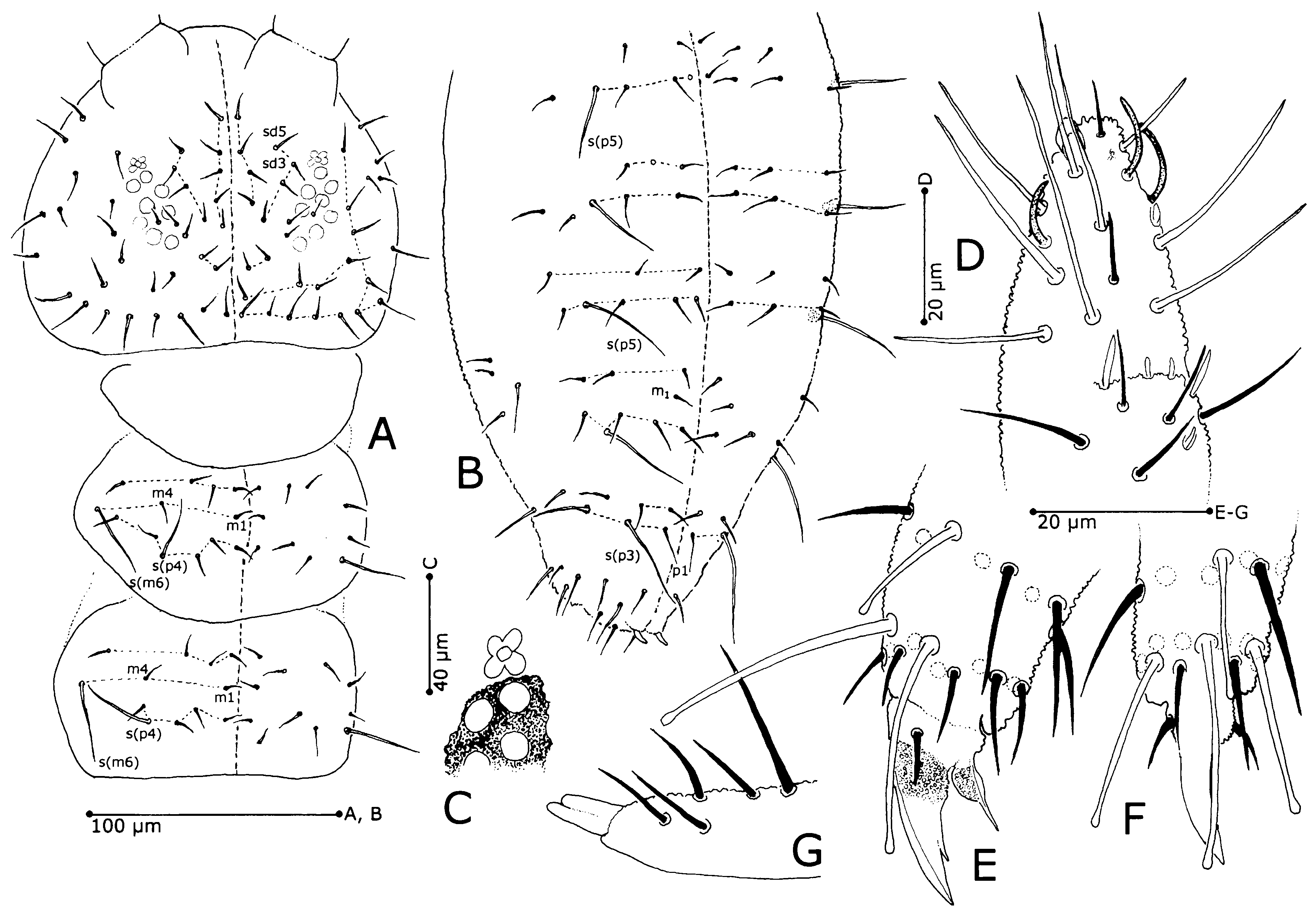
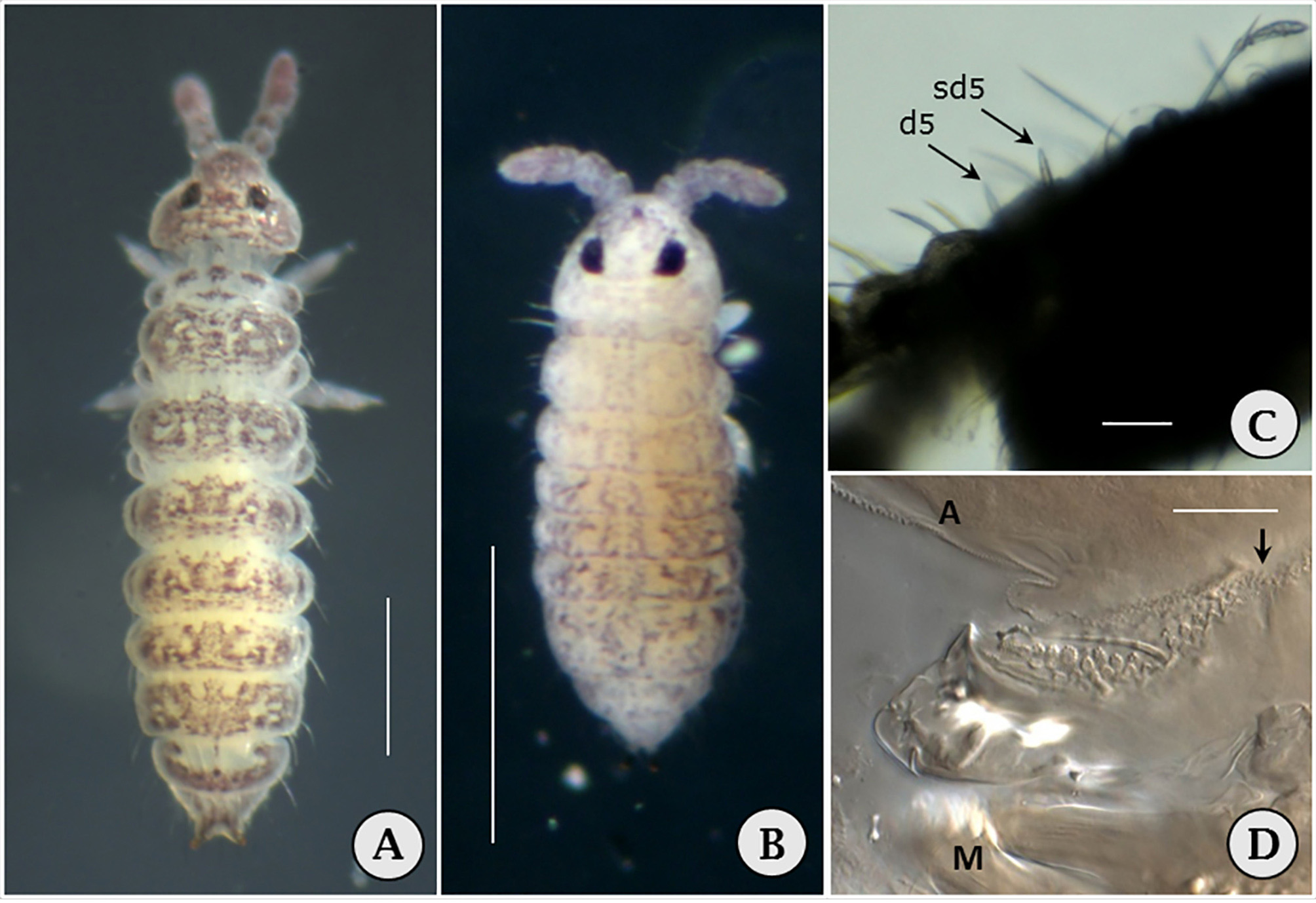
Redescription. Lectotype 1.97 mm [2.0 mm]. Color in ethanol violet-gray to dark gray in mosaic pattern on light background ( Fig. 1A View FIGURE 1. A ) [yellowish-white background with splotches and areas of purplish pigment]. Eye patches black, anal spines amber. Granulation coarse, Yosii’s ‘a’ number 18–24 between p2 setae on Abd. V (p1 setae absent); macrosetae and mesosetae strongly differentiated; macrosetae and longer mesosetae stout, coarsely serrate on one side. On head, setae d5 and sd5 modified as thick, pointed, erect spines ( Figs. 1 View FIGURE 1. A C, 2B, C); seta sd4 absent; setae d2, g3, p3 and v2 longer and stouter than other dorsal head setae. Lateral pronotal seta longer than inner setae ( Fig. 2 View FIGURE 2 A). On mesonotum and metanotum seta p2 a macroseta, most other setae mesosetae or microsetae; seta m2 present on Th. II and III. Dorsal meso- and metanotal sensilliform setae short, no longer than m-setae. Lateral sensilliform setae filiform on meso- and metanota, mesonotal microsensillum oval, just anterior to seta m6 ( Figs. 2 View FIGURE 2 D, E) Sensilliform setae of Abd. I–IV much shorter than neighboring p-setae. Setae p2, p4 and p6 on Abd. I–IV and setae p2 and p5 on Abd. V very long and stout macrochaetae. M-seta row nearly complete on Abd. I–IV, with setae m1, m4, m5 and either m2 or m3. Seta p1 absent from Abd. V. Plurichaetosis frequent, with setae a3, a4, and p3 sometimes doubled on thoracic and abdominal tergites.
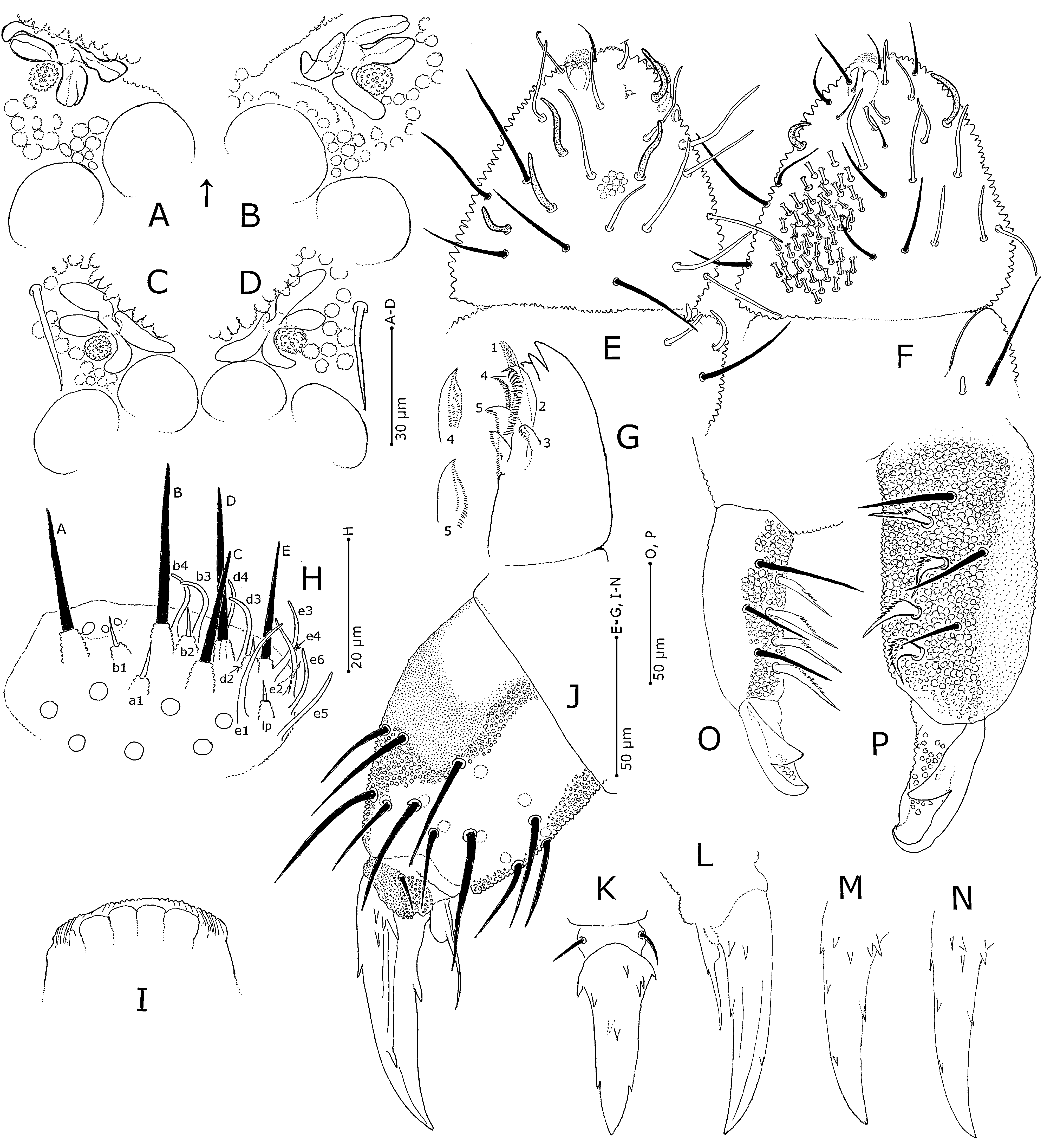
Antennal segment IV with finely granulated lobes surrounding aperture of trilobed apical vesicle, subapical spherical organite, short, peg-like microsensillum, and seven sensilla; two most lateral sensilla shorter than the five more dorsal sensilla ( Fig. 3 View FIGURE 3 E, F); most dorsal setae sensilliform, most ventral setae setiform; subapical hooked sensilliform seta present. Ventral sensory field with 50–60 short, thick, brush-tipped setae and two longer, more apical brush-tip setae ( Fig. 3 View FIGURE 3 F). Sense organ of Ant. III with two small oval to curved sensilla, two flanking sensilliform setae, and subventral microsensillum ( Figs. 3 View FIGURE 3 E, F). Eversible sac between Ant. III and IV apparently absent. Antennal segment I with 7 setae, Ant. II with 13 setae.
Ocelli 8+8. Postantennal organ width about 1.5× width of nearest ocellus, with four lobes and granulate accessory tubercle ( Figs. 3 View FIGURE 3 A–D); anterior lobes longer than posterior lobes; more medial anterior lobe frequently with small extension on more lateral end. Posterior lobes curved closely around accessory tubercle. Labrum with 5, 5, 4 setae, four large but weak lobes medially and small lobe on each side of larger lobes, and minutely crenate anterior margin becoming longitudinally grooved laterally ( Fig. 3 View FIGURE 3 I). On head of maxilla lamella 1 minutely serrate and extending just past teeth, lamella 4 exposed and reaching about as far as lamella 2 ( Fig. 5 View FIGURE 5 G). Outer lobe of maxilla with two sublobal hairs. Labium with 4 basomedial and 4 basolateral setae. Labial palpus with 7 proximal setae; all guard setae present except d1 and e7, and much shorter than sensilla A–E ( Fig. 3 View FIGURE 3 H); guard setae a1, b1, d2, e2 and lateral papilla lp composed of conical granulated base and spine-like apex; other guard setae cylindrical, sensilliform, rounded at tip.
Tibiotarsal cuticle strongly granulated distally and on ventral half proximally, very finely granulated dorsoproximally, all setae inserted in strongly granulated region; tibiotarsi I–III with 19, 19, 18 setae, respectively, proximal whorl on tibiotarsus III with 7 setae ( Fig. 3 View FIGURE 3 J). Clavate tenent hairs absent. Unguis with variable number of small basal and lateral teeth ( Figs. 3 View FIGURE 3 J–N); one or more pairs of entire or bifurcated basal lateral teeth and one pair of distal lateral teeth, one or two dorsal teeth at varying locations on the unguis, and strong inner tooth. Unguiculus with rounded lamella and apical filament extending just past inner ungual tooth.
Ventral tube with 4+4 setae. Tenaculum with 4+4 teeth, without setae. Length of dens about twice length of mucro; dens strongly tuberculate dorsally, with 7 setae ( Figs. 3 View FIGURE 3 O, P); three outer setae slender, four inner setae strongly expanded basally then flattened and tapering distally with prominent serrations. Mucro broad, with strong triangular lateral flap; floor of spoon-like part with strong granulation. Anal spines very long, tapering, 1.5× length of inner hind unguis, nearly 3 times longer than basal papillae ( Figs. 2 View FIGURE 2 A, B).
Remarks. Both of the original specimens were badly fragmented when the slides were received; therefore, 2 more recently collected specimens belonging to this species were used for additional study. The lectotype is the same specimen illustrated by Wray (1953), recognizable by the folded, twisted PAO ( Fig. 5 View FIGURE 5 B) that Wray interpreted as having six lobes. The illustration in Wray (1953) suggested to Christiansen & Bellinger (1980) a specimen in molt.
Ceratophysella hermosa will not key to itself in Christiansen & Bellinger (1998) because of Wray’s misinterpretation of the PAO. It differs from all other described Mitchellania -like Ceratophysella in having only 1+1 setae (p2) between sensilliform setae p3. Seta p1 is absent from Abd. V. The designation of the most medial pseta as p2 is based on its location between setae a1 and a3, as well as its identical appearance to the p2-setae on the other segments.
Skarżyński & Christiansen (2008) synonymized Mitchellania Wray, 1953 with Ceratophysella , but did not actually form the new combinations for species previously included in Mitchellania . Therefore, this action is taken here for C. hermosa ( Wray, 1953) new combination (= Mitchellania hermosa Wray, 1953 ). Ceratophysella hermosa is the type species of the genus Mitchellania Wray, 1953 . For the sake of taxonomic stability it is transferred to Ceratophysella due to the presence of a Ceratophysella -like mucro ( Skarżyński & Christiansen 2008).
Ceratophysella hermosa appears to be on the taxonomic fringe of its genus, particularly in the appearance of the labial palpus. In Ceratophysella jondavi redescribed in this paper and Ceratophysella spp. described elsewhere, the guard setae are longer than sensilla A–E, as illustrated by Fjellberg (1999). The guard setae of C. hermosa are much shorter than sensilla A–E. This distinctive labial palpus, presence of 7 proximal labial setae, the distinctive PAO with posterior lobes bent around the accessory tubercle, spine-like nature of head setae d5 and sd5, absence of head seta sd4, absence of seta p1 on Abd. V and very short sensilliform setae all suggest a separate line within Ceratophysella sensu lato. Molecular analysis of C. hermosa and other Ceratophysella spp. is needed to provide a firmer foundation for hypogastrurid springtails with a Ceratophysella -like mucro.

No known copyright restrictions apply. See Agosti, D., Egloff, W., 2009. Taxonomic information exchange and copyright: the Plazi approach. BMC Research Notes 2009, 2:53 for further explanation.
|
Kingdom |
|
|
Phylum |
|
|
Class |
|
|
Order |
|
|
Family |
|
|
Genus |
Ceratophysella hermosa ( Wray, 1953 )
| Bernard, Ernest C. 2015 |
Hypogastrura ( Mitchellania ) hermosa
| Christiansen 1980: 178 |
Mitchellania hermosa
| Wray 1953: 1 |