
Heterolatzelia nivalis Verhoeff, 1897
|
publication ID |
https://doi.org/10.5281/zenodo.202142 |
|
DOI |
https://doi.org/10.5281/zenodo.6186624 |
|
persistent identifier |
https://treatment.plazi.org/id/9862879F-FFFF-FFD1-FF49-FB1F8F9DD197 |
|
treatment provided by |
Plazi |
|
scientific name |
Heterolatzelia nivalis Verhoeff, 1897 |
| status |
|
Heterolatzelia nivalis Verhoeff, 1897 View in CoL
Syn .: Heterolatzelia nivalis nivalis Verhoeff, 1897 View in CoL
Heterolatzelia nivalis rupivaga Verhoeff, 1899 View in CoL , new synonymy Heterolatzelia nivalis absoloni Attems, 1951 View in CoL
Material examined. Type-series of H. nivalis is contained in two vials under number A20060140 ( BSCM). The first vial contains the holotype and two heads and body parts of probably two paratype males; the second vial contains one paratype juvenile with 22 somites + T, two paratype juveniles with 18 somites + T, and parts of three paratype specimens (in our opinion, probably one female with 27 somites + telson, and two specimens belonging to the VI or VII stadia). Both vials are labeled as follows: Heterolatzelia nivale, Verhoeff, 1897 , Bjelasnica, Etk. Nr. 525, Tier m. Original det.-Etk., ( Typus), Ehem. Trockenmat. Type-series of H. nivale rupivagum is contained in two vials ( BSCM). The first vial contains a holotype male and three paratype females; the second contains one paratype male and one paratype female. Both vials are labeled as follows: Heterolatzelia nivale rupivagum, Verhoeff, Plasa, Alpin, Etk. Nr. 524, Tier m., Original det. Etk., Typus-Verdacht, ehn. Trockenmant). Syntypes of H. nivalis absoloni included a female and dissected male, as well as slides with posterior gonopods and leg-pairs 6, 7 and 10, from Ponor Pit, Gatačko polje, Trebinje region, Republica Srpska, Bosnia and Hercegowina ( NHMV) (photos by Edmund Schiller).
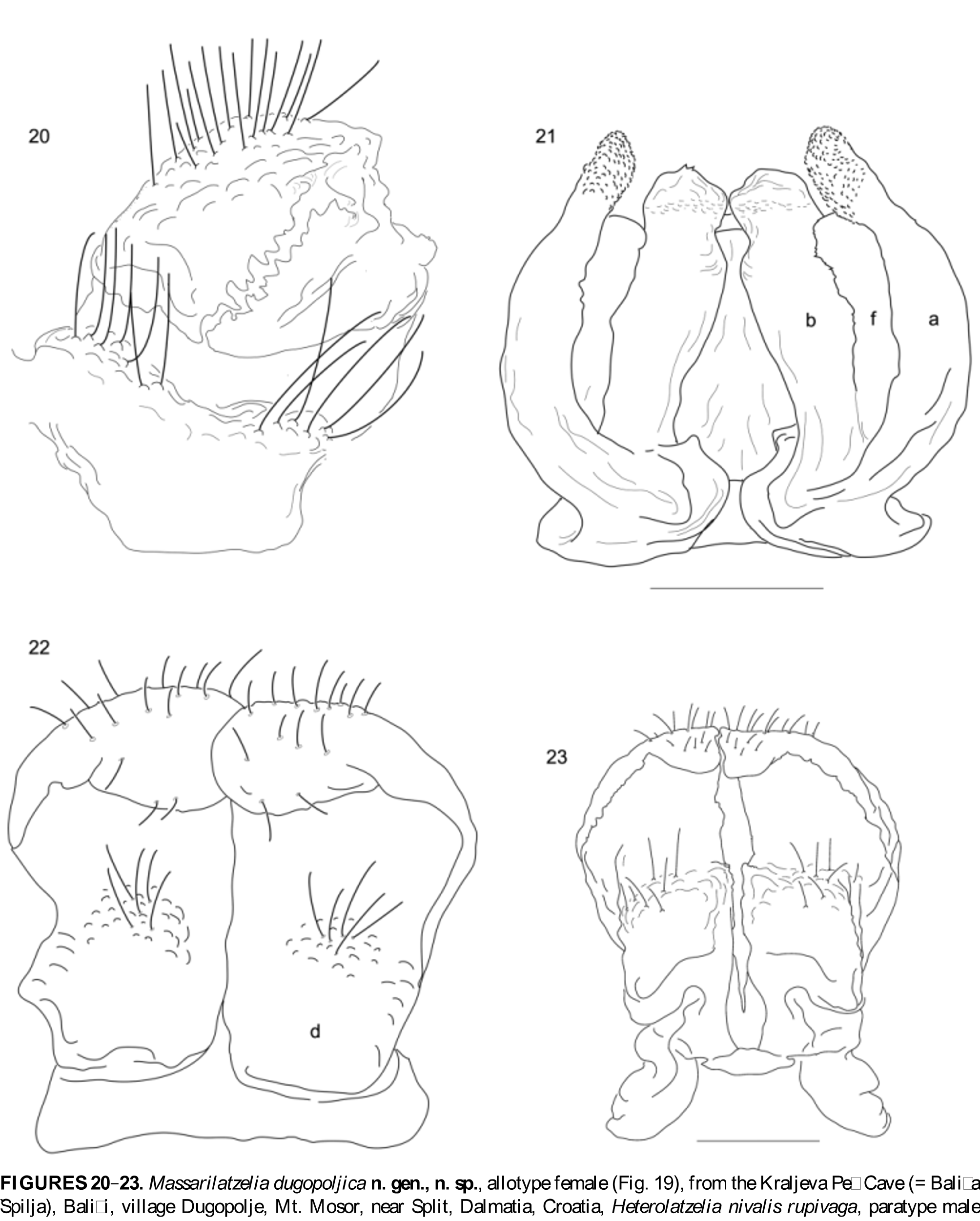
Remarks. After reexamination of the type material we interpret the structure of the anterior gonopods in the following way. The outer anterior coxal finger-shaped process (sensu Mršiċ 1992) or ‘Aussenfinger’ (sensu Verhoeff 1897, 1899) we assigned as angiocoxite impaling its development from the wall of the coxal podomere ( Fig. 21 View FIGURES 20 – 23 , a). We assume that ‘Innenarm’ (sensu Verhoeff 1897, 1899) represents a colpocoxite, derived from the coxal glands ( Fig. 21 View FIGURES 20 – 23 , b). We found that the medial process, inner anterior coxal process (sensu Mršiċ 1992) or even ‘Grundtheil eines Aufsatzgebildes mit der Sperma…’ (sensu Verhoeff, 1897, plate XVIII, Fig. 10 View FIGURES 2 – 12 , sp) represents an independent structure from both the colpocoxites and the angiocoxites, and even may be the result of the spreading of the lateral side of the sternite ( Fig. 21 View FIGURES 20 – 23 , f). Comb-like process (‘Femoroide’ sensu Verhoeff 1897, 1899) is fused with the base of the angiocoxites on the caudal side (possibly derived from coxa). The posterior gonopods consist of a unique ladle-shaped coxite ( Fig. 22 View FIGURES 20 – 23 , d), devoid of the apical process characteristic for the new genus. Such interpretation of the gonopodal elements is useful in the interpretation of phylogeny within heterolatzeliids, as well as within related genera belonging to different families.
In the original description of H. n. rupivaga , as one of the main characters for distinguishing the new subspecies from its nominal species, Verhoeff (1899) noted that both coxites on the posterior gonopods touch each other on the mesal sides. The posterior gonopods in heterolatzeliids definitely have only a protective role for the anterior apparatus. The position of the coxites or their contact on the mesal side or not ( Figs. 22–23 View FIGURES 20 – 23 ) is insufficient for designation as a character of a specific level. Moreover, we indicate in the description of the new genus that the anterior gonopods, especially the angiocoxites and colpocoxites, show a certain degree of intraspecific or individual variability. Between the three subspecies, H. nivalis nivalis , H. nivalis rupivaga , and H. nivalis absoloni there exists only a very modest variation in the shape of the angiocoxites and colpocoxites, as well as the position of the posterior coxites. This variation seems to be nothing more than interpopulational or even individual, as revealed by our comparative analyses of the type material and comparison with the original description. Also, in all analyzed specimens the exterior macrochaetae is positioned posteriorly in relation to the anterior and median ones. Values for the macrochaetal index CIX (15) are = 0.48–0.71 ( H. nivalis nivalis ) and 0.4–0.5 ( H. nivalis rupivaga ), median index MIX (15) = 0.50–0.57 ( H. nivalis nivalis ) and 0.40–0.49 ( H. nivalis rupivaga ), and values for the macrochaetal angle are MA (15) ≈ 155 ( H. nivalis nivalis ) and 159 ( H. nivalis absoloni ). These facts lead us to synonymize all the subspecies ( H. n. rupivaga , H. n. absoloni ) under the nominal Heterolatzelia nivalis species.
Heterolatzelia durmitorensis Guli č ka, 1968
Syn .: Heterolatzelia cornutum Gulička, 1968 View in CoL
Material examined. Six juveniles from Stoška Peċina Cave, Stozi, Mt. Golija, Montenegro, 10. viii 2010 by Iva Njunjiċ (IZB 1224–1229); one juvenile from Crnogorska Jama Cave, Višnjiċa Do, Mt. Golija, Montenegro, 7. viii 2010 by Iva Njunjiċ (IZB 1230); two juveniles from Crno Jezero Lake, Mt. Durmitor, Montenegro, 7. vi 1995 by Vladimir Pešiċ (IZB 1231–1232).
Remarks. Mršiċ (1992) examined numerous samples of H. durmitorensis from different localities on Mt. Durmitor, and found that Gulička’s (1968) description of H. cornutum is probably the result of damaged gonopods. Moreover, he explained that Attems’ (1951) determination of a species from Mt. Durmitor (referred to as H. nivalis nivalis ) was erroneous because it actually represents a H. durmitorensis species.
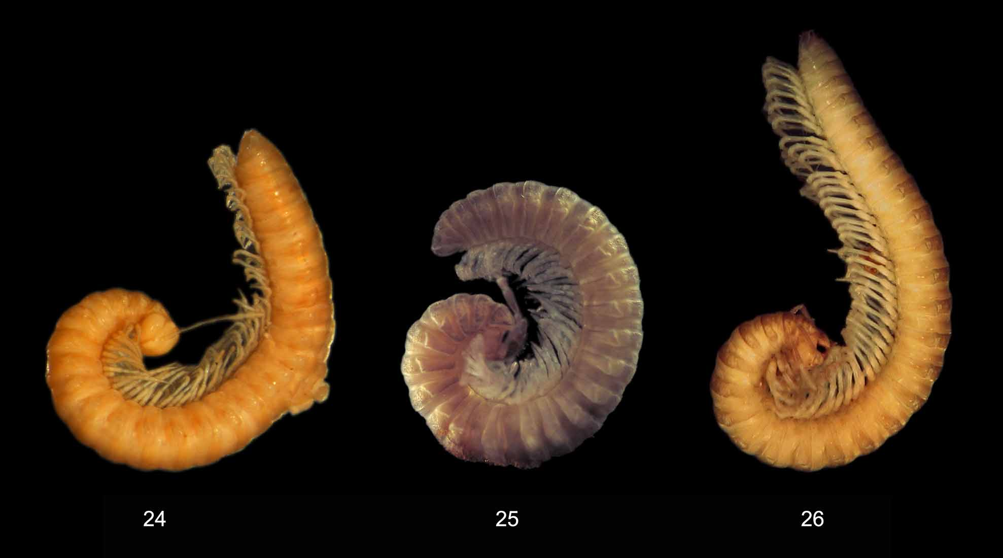
H. durmitorensis is small heterolatzeliid, 9–12 mm long, with small lateral keels ( Fig. 25 View FIGURES 24 – 26 ). The exterior macrochaetae is positioned considerably posteriorly in relation to the anterior and median ones. Macrochaetal index CIX (15) = 1.05. Median index MIX (15) = 1. Macrochaetal angle MA (15) ≈ 140 (all values are for juveniles in stadium VI). Angiocoxites (= outer anterior coxal finger-shaped process) are elongated, almost straight, and considerably longer than other gonopodial elements (in H. nivalis angiocoxites are C-shaped and almost the same height as colpocoxites). Colpocoxites are more or less triangular, supplied with medial coxal shred ( Mršiċ, 1992; Fig. 13 View FIGURES 13 – 19 , C and D) (in H. nivalis colpocoxites are subquadrangular and without coxal shred). Posterior gonopods are similar to the nominal species. According to the characteristics of the body architecture and structure of anterior gonopods, we retained species status for this heterolatzeliid.


No known copyright restrictions apply. See Agosti, D., Egloff, W., 2009. Taxonomic information exchange and copyright: the Plazi approach. BMC Research Notes 2009, 2:53 for further explanation.
|
Kingdom |
|
|
Phylum |
|
|
Class |
|
|
Order |
|
|
Family |
|
|
Genus |
Heterolatzelia nivalis Verhoeff, 1897
| Makarov, Slobodan E., Ćurčić, Božidar P. M., Tomić, Vladimir T., Rađa, Tonći, Rađa, Biljana, Ćurčić, Srećko B., Mitić, Bojan M. & Lučić, Luka R. 2011 |
Heterolatzelia cornutum Gulička, 1968
| Gulicka 1968 |
Heterolatzelia nivalis absoloni
| Attems 1951 |
Heterolatzelia nivalis rupivaga
| Verhoeff 1899 |
Heterolatzelia nivalis nivalis
| Verhoeff 1897 |