
Leuclathrina translucida, Voigt & Ruthensteiner & Leiva & Fradusco & Wörheide, 2018
|
publication ID |
https://doi.org/ 10.11646/zootaxa.4382.1.5 |
|
publication LSID |
lsid:zoobank.org:pub:B222C2D8-82FB-414C-A88F-44A12A837A21 |
|
DOI |
https://doi.org/10.5281/zenodo.5950835 |
|
persistent identifier |
https://treatment.plazi.org/id/1075FC2E-0BC4-46A7-94FE-9DE49A769442 |
|
taxon LSID |
lsid:zoobank.org:act:1075FC2E-0BC4-46A7-94FE-9DE49A769442 |
|
treatment provided by |
Plazi |
|
scientific name |
Leuclathrina translucida |
| status |
sp. nov. |
Leuclathrina translucida View in CoL sp. nov.
( Figs. 2 A–I View FIGURE2 , 3 A–F View FIGURE 3 ; Table 2)
Diagnosis. Leuclathrina with a yellow cormus composed of two distinct parts: an inhalant body part with macroscopically visible inhalant openings, and an exhalant body part, consisting of several transparent tubes, which emerge from the inhalant body part and occasionally reunite. The skeleton consists of equiradiate triactines lying tangentially in the sponge wall and forming a thick-layered skeleton in the inhalant body parts, and a much thinner layer in the wall of the exhalant tubes. The choanosome is free of spicules and restricted to the inhalant body part.
Type material. Holotype: SNSB-BSPG GW3934 (1 specimen), outer reef wall close to Vavvaru, Lhaviyani Atoll, Maldives (5.4203° N; 73.3508° E), coll. O. Voigt at 23 m on 9 Sept 2015; Paratypes: SNSB-BSPG GW4029 (1 specimen), outer reef wall close to Magoodhoo, Fafuu Atoll, Maldives (dive site 3.0748° N; 72.9663° E), coll. G. Wörheide at 20 m on 12 Oct 2016; SNSB-BSPG GW30279 (4 specimens) & SNSB-BSPG GW30280 (1 specimen), reef wall close to Magoodhoo, Fafuu Atoll, Maldives (dive site Wallino, 3.0870° N, 72.9558° E) coll. O. Voigt & B. Fradusco at 15 –17 m on 29 Sep 2017; SNSB-BSPG GW3 0 292 (1 specimen), inner reef close to Magoodhoo, Fafuu Atoll, Maldives (dive site Coral Garden, 3.09170° N, 72.96787° E), coll. O. Voigt & B.
Fradusco at 20 m on 29 Sep 2017 ; SNSB-BSPG GW30328 (1 specimen), outer reef, Fafuu Atoll , Maldives (dive site Beyrufushi, 3.11282° N, 73.02108° E), coll. O. Voigt & B. Fradusco at 17 m on 1 Oct 2017 GoogleMaps ; SNSB-BSPG GW30350 (5 specimens), reef at Bileiydhoo , Fafuu Atoll, Maldives (dive site Wall Street, 3.12061° N, 72.97958° E), coll. O. Voigt & B. Fradusco at 17 m on 2 Oct 2017 GoogleMaps ; SNSB-BSPG GW30361 (1 specimen), reef between Magoodhoo and Bileiydhoo , Fafuu Atoll, Maldives (dive site M& Ms, 3.12429° N, 72.99201° E), coll. O. Voigt & B. Fradusco at 15 m on 2 Oct 2017 GoogleMaps ; SNSB-BSPG GW30377 (3 specimens), reef wall close to Magoodhoo , Fafuu Atoll, Maldives (dive site Wallino, 3.0870° N, 72.9558° E) coll. O. Voigt, B. Fradusco & G. Wörheide at 20 m on 4 Oct 2017 GoogleMaps ; SNSB-BSPG GW30419 (2 specimens), outer reef, E of Magoodhoo , Fafuu Atoll, Maldives (dive site Route 66, 3.07902° N, 72.97797° E), coll. O. Voigt & B. Fradusco at 15 m on 2 Oct 2017 GoogleMaps .
Type locality. Vavvaru , Lhaviyani Atoll, Maldives
Etymology. Named for its translucent exhalant tubes.
Colour. Yellow in life ( Figs. 2 A, B View FIGURE2 ) and in 80 % ethanol.
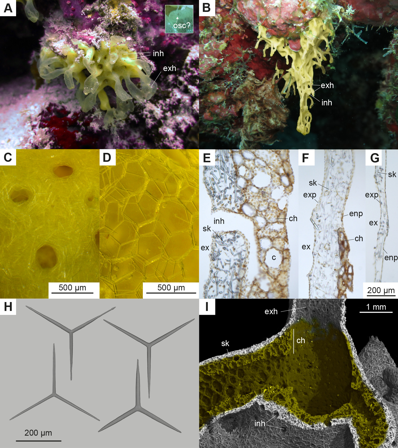
Growth form. Two different growth forms were observed. The majority of sponges have a cushion-shaped cormus, which is attached to the substrate with its base ( Fig. 2 A View FIGURE2 ). The holotype measures 3.5 cm x 2.3 cm (fixed as two fragments). Several translucent exhalant tubes (about 2 -2.7 mm wide in the holotype) emerge from the base and sometimes reconnect at their upper parts ( Fig. 2 A View FIGURE2 ). Only on few occasions, small openings were visible in the exhalant tubes of living specimens, which are most likely oscula ( Fig. 2 A View FIGURE2 , inset). In fixed material, the oscula are not visible, either because of contraction or due to damage of the delicate exhalant tubes. Two specimens showed a modified growth form and were hanging from ceilings of small or larger overhangs ( Fig. 2 B View FIGURE2 ). In the larger specimen of this growth form the inhalant body parts were forming a mesh-like structure in some parts of the cormus ( Fig. 2 B View FIGURE2 ). Exhalant tubes were less developed or contracted in comparison to the more typical growth form ( Fig. 2 B View FIGURE2 ). In both growth forms, the inhalant body parts have inhalant openings of about 150 to 270 µm in diameter ( Figs. 2 A, C View FIGURE2 ), which are lacking in the exhalant tubes ( Fig. 2 D View FIGURE2 ).
Skeleton. The body wall of the inhalant part of the sponge and at the transition between the inhalant and exhalant body parts is supported by a thick layer of tangential triactines ( Figs. 2 E, F View FIGURE2 ). Towards the more distal parts of the exhalant tubes, the skeleton is getting much thinner ( Fig. 2 G View FIGURE2 ), consisting of only a thin layer of triactines ( Figs. 2 C, G View FIGURE2 ). In this region the triactines leave approximately hexagonal shaped areas that are free of spicules, resulting in the transparent appearance of the exhalant tubes ( Fig. 2 D View FIGURE2 ).
Spicules. Equiangular, equiradiate triactines ( Fig. 2 H View FIGURE2 ). The actines are straight and conical, with sharp or blunt tips. In some triactines, the actines are locally slightly thickened near the tip ( Fig. 2 H View FIGURE2 ). Spicule sizes of the holotype are: inhalant region: actine length: 147–194.4–247 µm, actine width: 12–14.4–18 µm; exhalant region: actine length: 143–212.1–256 µm, actine width: 9–14.2–19 µm. Spicules measurements of three paratypes are provided in Table 2.
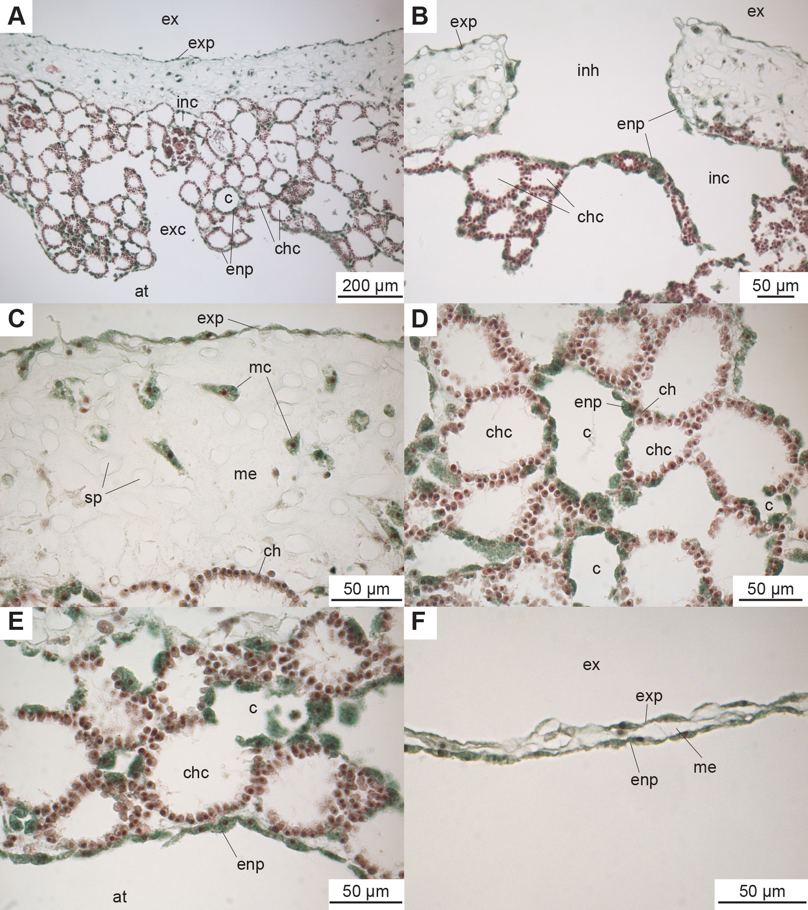
Histology and organisation of the aquiferous system. The sponge wall of the inhalant body part consists of a thick skeletal layer and the spicule-free choanosome ( Figs. 2 E, I View FIGURE2 , 3 A View FIGURE 3 ). The skeletal layer is delimited by an exopinacoderm on the outer side and is perforated by inhalant openings, which are lined by endopinacocytes and lead to narrower inhalant canals ( Figs. 2 E View FIGURE2 , 3 B View FIGURE 3 ). The skeletal layer contains only few mesohyl cells ( Fig. 3 C View FIGURE 3 ). The choanosome layer comprises spherical choanocyte chambers, between which inhalant and exhalant canals occur, which are lined by endopinacocytes ( Fig. 3 D View FIGURE 3 ). The exhalant canals unite and increase their diameter until they open into the central atrium ( Fig. 3 A View FIGURE 3 ). The 3D reconstruction reveals that the diameter of the openings to the atrium increases with the thickness of the choanosome ( Fig. 2 I View FIGURE2 ). A layer of endopinacocytes delimits the wall of the central atrial cavity ( Fig. 3 E View FIGURE 3 ). Towards the transition of the inhalant to the exhalant tubes the choanoderm decreases its width until it disappears, while the thickness of the skeletal layer remains the same in this region ( Figs. 2 F View FIGURE2 , 3 I View FIGURE 3 ). The latter however decreases in the more distal parts of the exhalant tubes, until it only contains a thin layer of spicules within a thin mesohyl ( Fig. 2 G View FIGURE2 , Fig. 3 F View FIGURE 3 ). Oscula were not visible in the fixed material.
Ecology and distribution. The specimens were growing on hard substrate, in most cases on vertical parts of reef walls, on living or dead corals, or hanging from ceilings of small overhangs, in depths between 15 and 23 m. The samples in this study originated from the Maldives; a wider distribution in the Indo-Pacific is mentioned by Coleman (2001), but not further specified.
Remarks. Photographs of Leuclathrina translucida sp. nov. have been published previously, e.g. as ‘ Clathrina sp.’ in the field guide ‘Marine Life of the Maldives’ ( Coleman 2001, page 36).
Leuclathrina translucida View in CoL sp. nov. differs from the type species of the genus, L. asconoides View in CoL in several aspects and can easily be distinguished. Leuclathrina asconoides View in CoL is smaller (3.5 mm) and has a globular shape; its exhalant tubes (‘chimneys’) are shorter (a few millimetres, Borojević & Boury-Esnault 1987). Triactines of L. asconoides View in CoL can grow to a larger size compared to L. translucida View in CoL sp. nov.: The length of the actines varies between 156–598 µm, and their width between 13-36 µm ( Borojević & Boury-Esnault 1987).
No known copyright restrictions apply. See Agosti, D., Egloff, W., 2009. Taxonomic information exchange and copyright: the Plazi approach. BMC Research Notes 2009, 2:53 for further explanation.
|
Kingdom |
|
|
Phylum |
|
|
Class |
|
|
Order |
|
|
Family |
|
|
Genus |
Leuclathrina translucida
| Voigt, Oliver, Ruthensteiner, Bernhard, Leiva, Laura, Fradusco, Benedetta & Wörheide, Gert 2018 |
Leuclathrina translucida
| Voigt & Ruthensteiner & Leiva & Fradusco & Wörheide 2018 |
L. translucida
| Voigt & Ruthensteiner & Leiva & Fradusco & Wörheide 2018 |
L. asconoides
| Borojevic & Boury-Esnault 1987 |
Leuclathrina asconoides
| Borojevic & Boury-Esnault 1987 |
L. asconoides
| Borojevic & Boury-Esnault 1987 |