
Amitermes beaumonti Banks, 1918
|
publication ID |
https://doi.org/ 10.11646/zootaxa.4751.1.4 |
|
publication LSID |
lsid:zoobank.org:pub:778B13DA-851C-4CE8-B283-08B8F07E997A |
|
DOI |
https://doi.org/10.5281/zenodo.3718064 |
|
persistent identifier |
https://treatment.plazi.org/id/987B995A-FFE0-FFE4-FF51-FA20FBB4F928 |
|
treatment provided by |
Plazi |
|
scientific name |
Amitermes beaumonti Banks, 1918 |
| status |
|
Amitermes beaumonti Banks, 1918
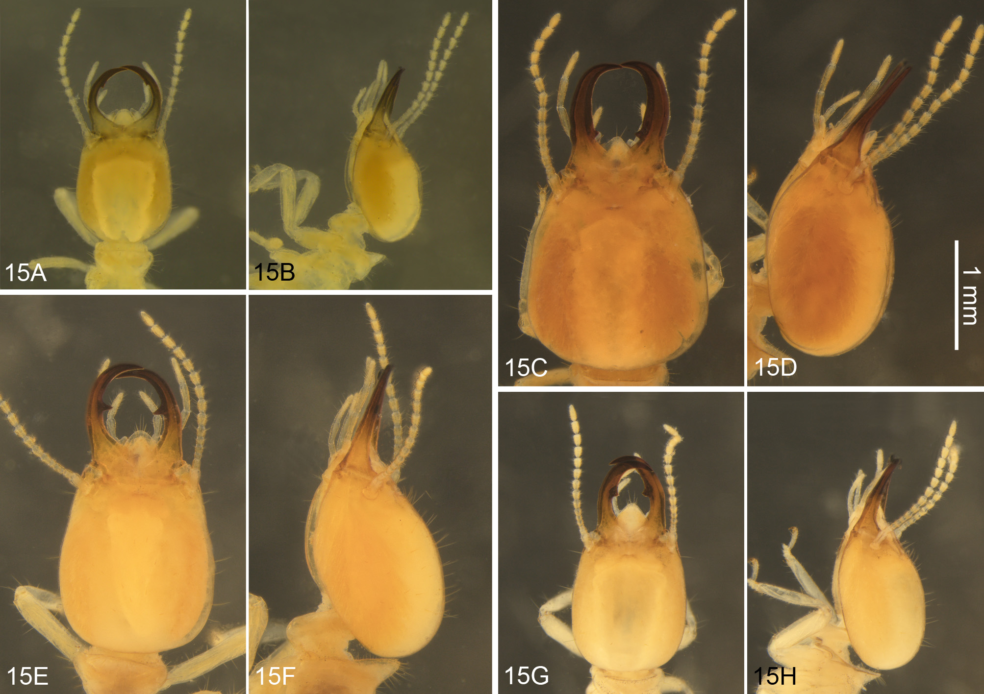
Soldier ( Figs. 15A, 15B View FIGURE 15 ): Described by Banks (1918), with a redescription by Light (1932). Additional illustrations are provided here.
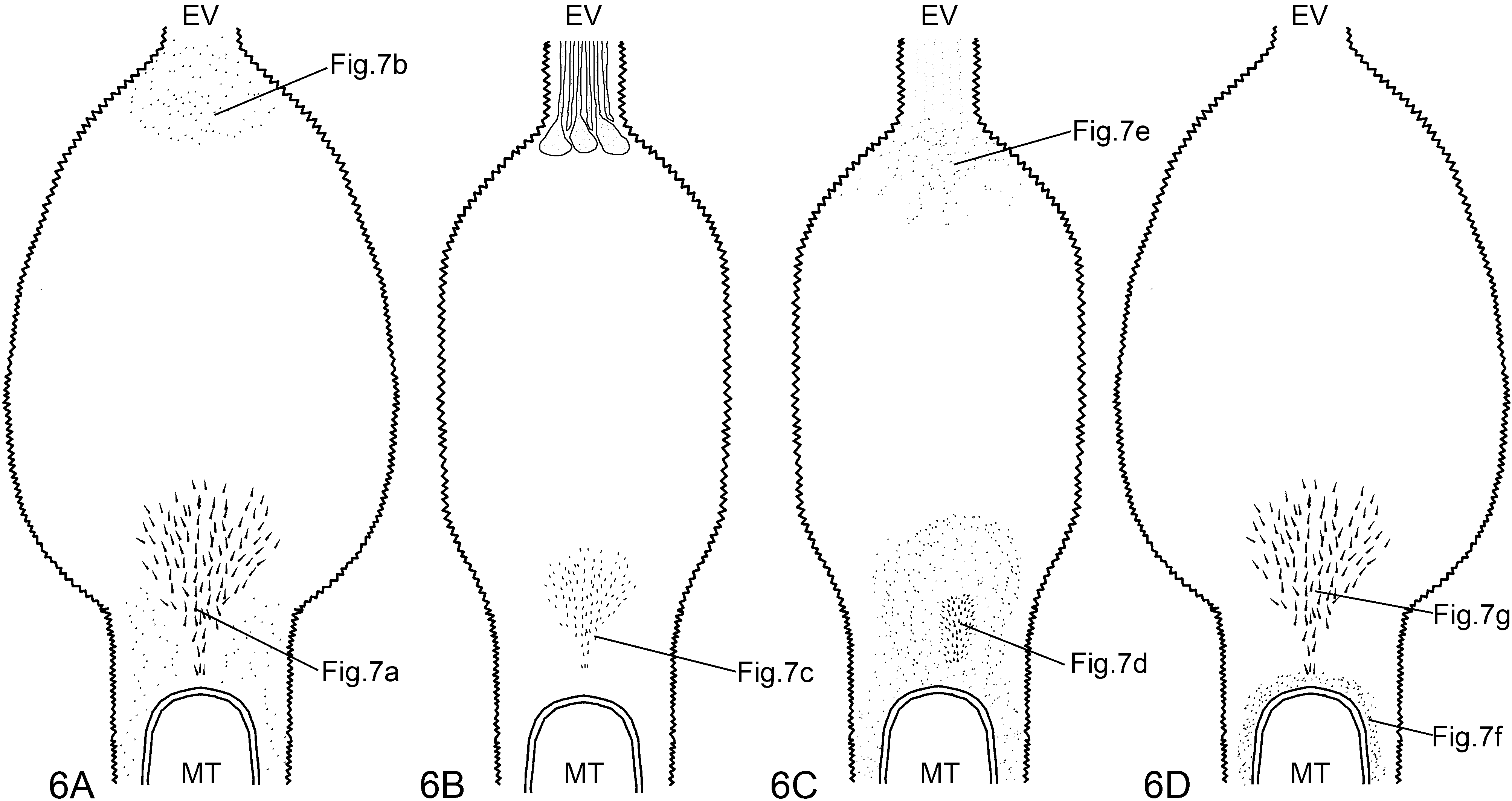
Gut anatomy ( Figs. 6C View FIGURE 6 , 7D, 7E View FIGURE 7 , 16A View FIGURE 16 , 17 View FIGURE 17 , 18A View FIGURE 18 ): Crop slightly asymmetrical, covered internally with pectinate scales before gizzard. Gizzard of the generalized type, with columnar and pulvillar belts subequal in length; firstorder pulvilli conspicuous, with tiny pectinate scales, second-order pulvilli reduced ( Fig. 16A View FIGURE 16 ). Stomodeal valve inserted in apex of mesenteron. Mesenteric tongue internal to mesenteric arch, filiform proximally and gradually enlarging to tip, ending above dilated portion of first proctodeal segment (P1) and enlarged distally ( Figs. 17 View FIGURE 17 A–17C). P1 fusiform, crossing abdomen diagonally in dorsal view, over paunch (P3), inserted in P3 dorsally at right side of abdomen, but partially occluded by P4 segment ( Fig. 17E View FIGURE 17 , arrow). P1 cuticular ornamentation composed of sparse small spines around mesenteric tongue at mixed segment, followed by elongated area of spines ( Figs. 6C View FIGURE 6 , 7D View FIGURE 7 ), and sparse spines in distal portion, just before enteric valve ( Figs. 6C View FIGURE 6 , 7E View FIGURE 7 ). Enteric-valve armature (P2) composed of six digitiform cushions ornamented with small spines distally, and with broadly coverage of small spines proximally ( Fig. 18A View FIGURE 18 ). P3 well developed and globose, with a marked pouch after P2 ( Figs. 17A, 17B View FIGURE 17 , arrows). Isthmus easily recognizable. Fourth proctodeal segment (P4) with short U-turn and located dorsally ( Fig. 17A View FIGURE 17 ).
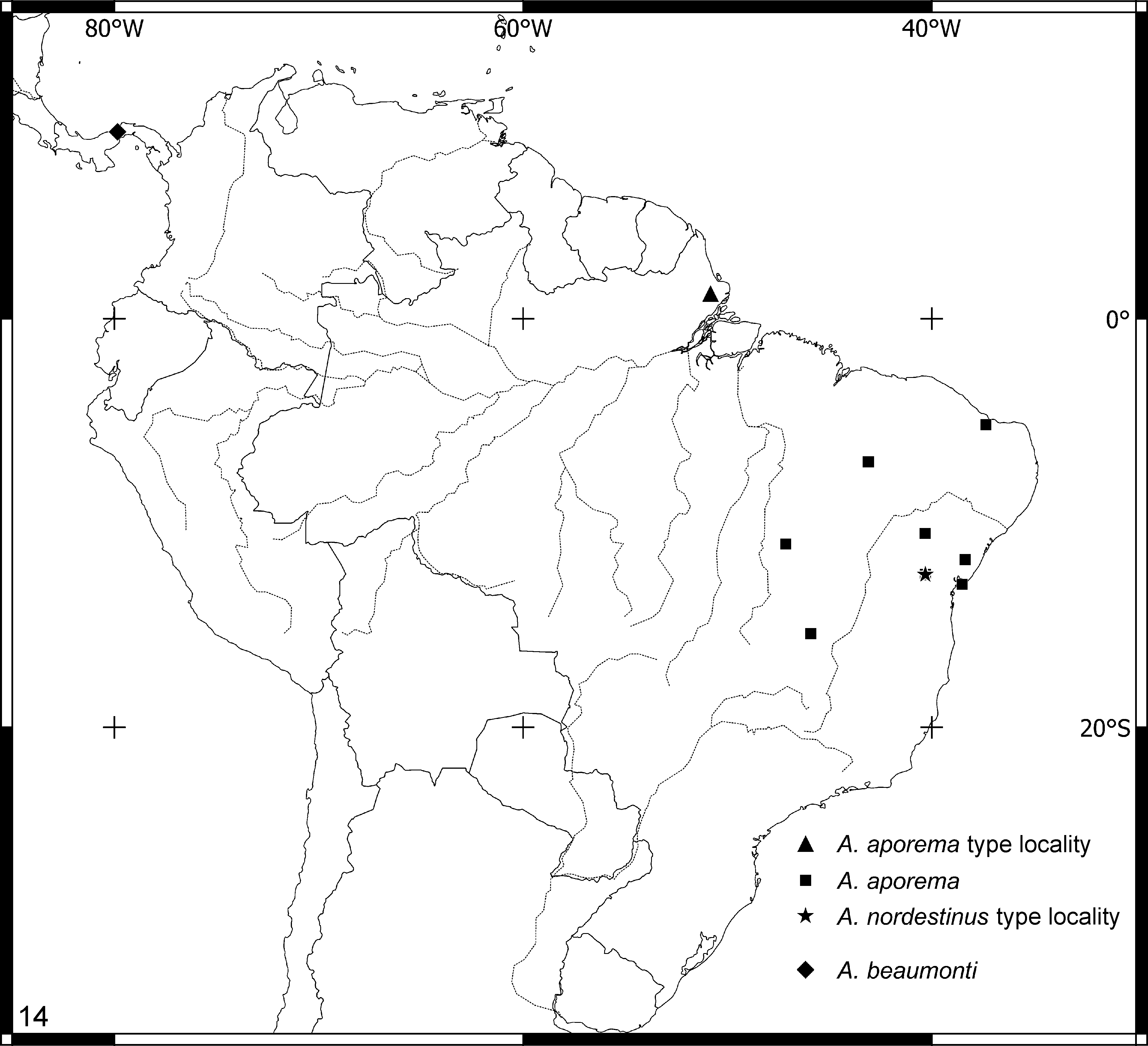
Comments. Although the available specimens for this study are from Panamá ( Fig. 14 View FIGURE 14 ), we agree with one of the reviews suggestions and include in the paper, since this species distribution overlaps with A. foreli , and maybe reachs the west of Colombia.
Material examined. PANAMA. Barro Colorado Island , 26.v.1966 , E.K.P. Silveira (1113), 21.vi.1966 (1114) , A.E. Emerson , 01.v.1935 (3446*).
Amitermes excellens ( Silvestri, 1923)
Soldier ( Figs. 15C, 15D View FIGURE 15 ): Described by Silvestri (1923); a brief redescription and illustrations were provided by Emerson (1925). Additional illustrations are provided here.
Gut anatomy ( Figs. 6D View FIGURE 6 , 7F, 7G View FIGURE 7 , 16B View FIGURE 16 , 18B View FIGURE 18 , 19 View FIGURE 19 ): Crop asymmetrical, with oesophagous insertion displaced to left side, covered internally with pectinate scales before gizzard. Gizzard of the generalized type, with columnar and pulvillar belts subequal in length; first-order pulvilli conspicuous, with tiny pectinate scales, second-order pulvilli reduced ( Fig. 16B View FIGURE 16 ). Stomodeal valve inserted in apex of mesenteron. Mesenteric tongue external to mesenteric arch, of uniform width along its length, ending before dilated portion of first proctodeal segment (P1) ( Figs. 19C, 19D View FIGURE 19 ). P1 globose, crossing abdomen diagonally in dorsal view, over paunch (P3), inserted in P3 dorsally at right side of abdomen, but occluded by P4 segment ( Fig. 19E View FIGURE 19 , arrow). P1 cuticular ornamentation composed of sparse small spines around mesenteric tongue at mixed segment ( Figs. 6D View FIGURE 6 , 7F View FIGURE 7 ), followed by elongated area of spines with sclerotized bases, and located proximally ( Figs. 6D View FIGURE 6 , 7G View FIGURE 7 ). Enteric-valve armature (P2) composed of six digitiform cushions ornamented with small spines ( Fig. 18B View FIGURE 18 ). P3 well developed and globose. Isthmus not recognizable. Fourth proctodeal segment (P4) with short U-turn and located dorsally ( Figs. 19A, 19B View FIGURE 19 ).
Material examined. BRAZIL. Amazonas. São João near to Taperucuara, AM ( SA 20 , 0-65d), 08.xi.1972, P.E. Vanzolini (5181); Beruri: Purus river, 06.iv.1967, Departamento de Zoologia (1259); Itacoatiara : AM-010, km 232 (between Manaus and Itacoatiara), 21–28.v.1977, A.G. Bandeira (8307, 8311, 8312, 8314, 10449), 29.ix.1977, A.G. Bandeira (7463), 04.xi.1977, A.G. Bandeira (7464*, 8313); Manaus : 20.viii.1976, A.G. Bandeira (7543), Sítio Conceição de Manaus , Rio Amazonas , 10.xi.1953, C. R. Gonçalves (4583), EEST-Alojamento, 05.iv.1991, A.G. Bandeira (9477); Presidente Figueiredo: Cachoeira Iracema, 12.iii.2016, R. G. Santos (24344); Pará. U.H.E São Luís do Tapajós , 17.i.1979, J.M.F. Camargo & P. Mazucato (26960), Between Santa Maria and Itaituba, 18.i.1979, J.M.F. Camargo & P. Mazucato (26973); Belterra: ii.1949, C. R. Gonçalves (3160*), 31.i.1949, C. R. Gonçalves (3165*); Óbidos: ii.1949, C. R. Gonçalves (3854); Santarém: 12.xi.1945, C. R. Gonçalves (4584, 4587); Rondônia. UHE, Santo Antônio, Módulo de Jaci Paraná, 06.iii.2012, R. G. Santos, J. Cabral & T. F. Carrijo (23727); Roraima. Manaus-Boa Vista Km 810 Macujaí, 14.v.1977, A.G. Bandeira (8306), Ilha de Maracá , ix.1987, E.M. Cancello & O.F.F. Souza (9074), Arumim, 13.iii.2016, J.P. Constantini (24325), R. G. Santos (25239); Amajari: 13.iii.2016, T. F. Carrijo (24326), R. G. Santos (24920); Boa Vista : 08.xi.1953, C. R. Gonçalves (4585, 4586), Passarão, 12.iii.1972, K. Kitayama (5313,5316); Bonfim: 15.iii.2016, T. F. Carrijo (24328, 24334, 24335, 24926*), R. G. Santos (24338, 24343), J.P. Constantini (24342, 25764); Caracaraí: 15.iii.2016, J.P. Constantini (24324), T. F. Carrijo (24340), RR-km 639, Manaus-Boa Vista, 14.v.1977, A.G. Bandeira (8309); Mucajai: 15.iii.2016, R. G. Santos (24345), J.P. Constantini (24919); Uraricoera: E. Ecol. de Maracá , 20.v.2015, G. Biffi & R. Falashi (23400) ; GUIANA. Cuyuni- Mazaruni. Kamakusa (Mountain?), 03.xii.1922, H. Lang (3448), Kartabo : 10.viii.1919, A.E. Emerson (3449*, type colony) .
Amitermes foreli Wasmann, 1902
Soldier ( Figs. 15E, 15F View FIGURE 15 ,): originally described by Wasmann (1902), with a brief description of the external morphology of the soldier, worker and nymph. A good redescription was provided by Light (1932), based on the type specimens. Additional illustrations are provided here.
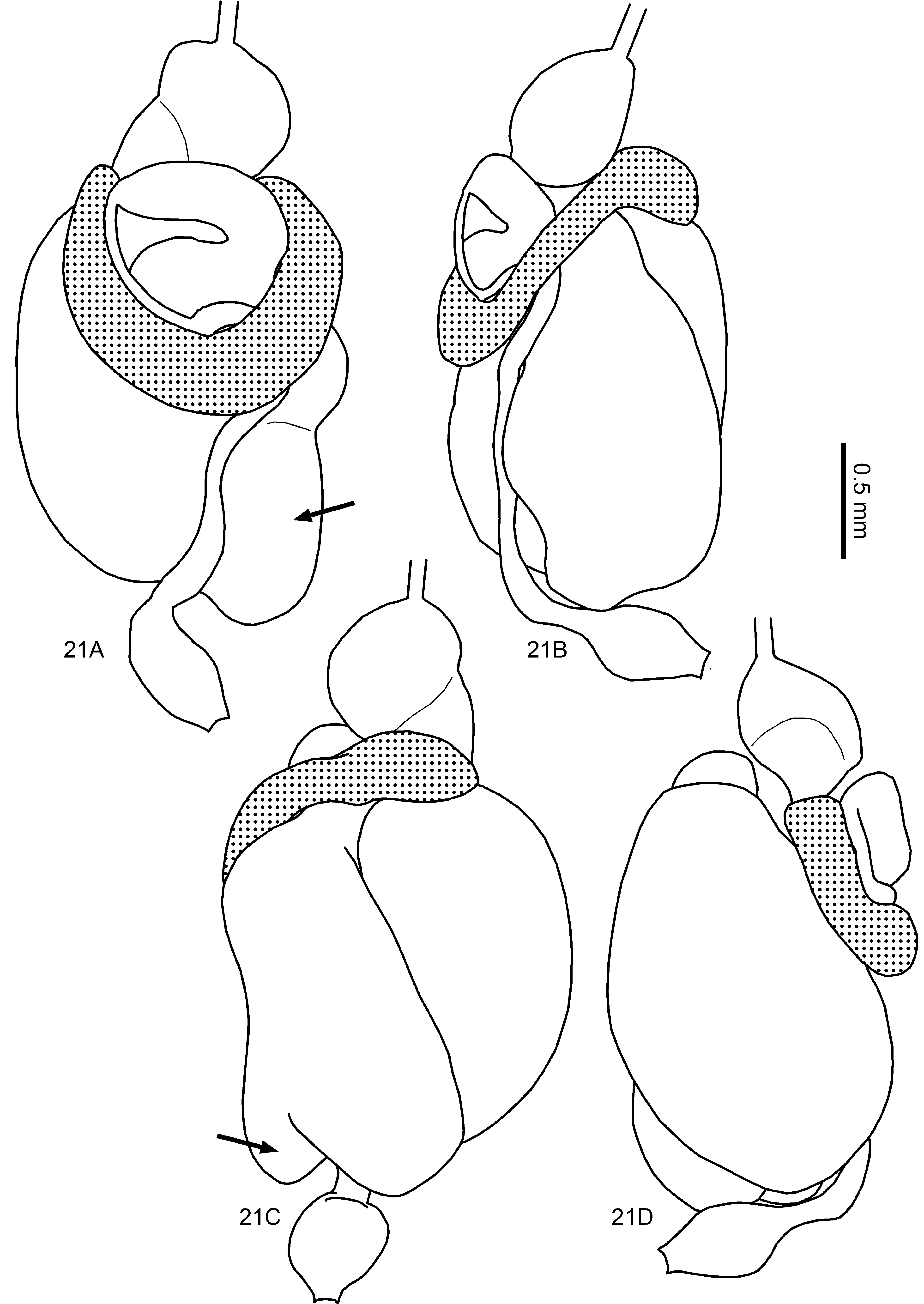
Gut anatomy ( Figs. 20A View FIGURE 20 , 21 View FIGURE 21 , 22A View FIGURE 22 , 23A View FIGURE 23 , 24A View FIGURE 24 ): Crop asymmetrical, with oesophagous insertion displaced to left side, covered internally with pectinate scales before gizzard. Gizzard of the generalized type, with columnar and pulvillar belts subequal in length; first-order pulvilli conspicuous, with tiny pectinate scales, second-order pulvilli reduced ( Fig. 20A View FIGURE 20 ). Stomodeal valve inserted in apex of mesenteron. Mesenteric tongue external to mesenteric arch, of uniform width along its length, ending above dilated portion of first proctodeal segment (P1) and enlarged distally ( Fig. 21C View FIGURE 21 ). P1 strongly globose, crossing abdomen diagonally in dorsal view, over paunch (P3), inserted in P3 dorsally, at right side of abdomen, but occluded by P4 segment. P1 cuticular ornamentation composed of small spines restricted to border of mesenteric tongue, remaining surfaces glabrous ( Figs. 22A View FIGURE 22 , 23A View FIGURE 23 ). Enteric-valve armature (P2) composed of six elongated cushions ornamented with small spines ( Fig. 24A View FIGURE 24 ). P3 well developed and
globose, with a partial pouch after P2 ( Figs. 21A, 21C View FIGURE 21 , arrows). Isthmus easily recognizable. Fourth proctodeal segment (P4) with short U-turn, located dorsally ( Fig. 21A View FIGURE 21 ).
Material examined. COLOMBIA. Atlántico. Barranquilla : 29.viii.1938, C. Seevers (3450) .
Amitermes lunae Scheffrahn & Huchet, 2010
Soldier ( Figs. 15G, 15H View FIGURE 15 ): Originally described by Scheffrahn & Huchet (2010), with photographs of the soldier and worker´s enteric valve, but without a description of the gut coiling.
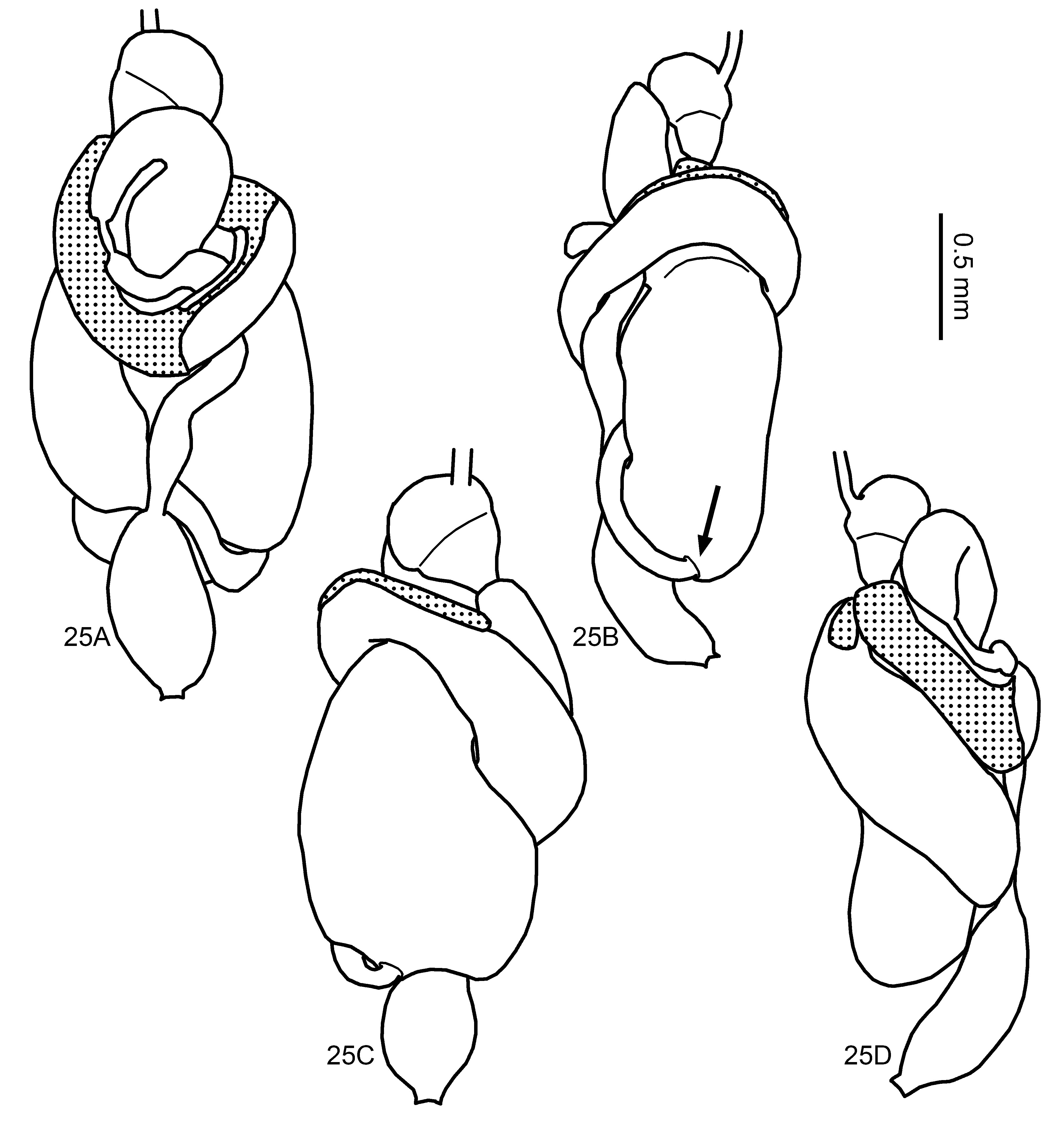
Gut anatomy ( Figs. 20B View FIGURE 20 , 22B View FIGURE 22 , 23B View FIGURE 23 , 24B, 24C View FIGURE 24 , 25 View FIGURE 25 ): Crop asymmetrical, with oesophagous insertion displaced to left side, covered internally with pectinate scales before gizzard. Gizzard of the generalized type, with columnar and pulvillar belts subequal in length; first-order pulvilli conspicuous, with tiny pectinate scales, second-order pulvilli reduced ( Fig. 20B View FIGURE 20 ). Stomodeal valve inserted in apex of mesenteron. Mesenteric tongue internal to mesenteric arch, narrower proximally and gradually enlarging to tip, ending before dilated portion of first proctodeal segment (P1) ( Fig. 25C View FIGURE 25 ). P1 fusiform, crossing abdomen diagonally in dorsal view, over paunch (P3), with long narrow neck, inserted ventrally at P3 ( Fig. 25B View FIGURE 25 , arrow). P1 cuticular ornamentation composed only of elongated area of spines with sclerotized base, located proximally ( Figs. 22B View FIGURE 22 , 23B View FIGURE 23 ). Enteric-valve armature (P2) weakly sclerotized, with six elongated cushions ornamented with small scattered spines ( Figs. 24B, 24C View FIGURE 24 ). P3 well developed. Isthmus easily recognizable. Fourth proctodeal segment (P4) with short U-turn and located dorsally ( Fig. 25A View FIGURE 25 ).
Comment. Only a few specimens were available to prepare the enteric valve slides and we did not succeed in obtaining a perfect preparation; however, the original description includes pictures.
Material examined. PERU. Huaca de la Luna , 05.vi.2009, J.B. Huchet (27469) .
Fig. 26 shows the MZUSP collection records of A. excellens , A. foreli and A. lunae .


| AM |
Australian Museum |
| R |
Departamento de Geologia, Universidad de Chile |
| T |
Tavera, Department of Geology and Geophysics |
| MZUSP |
Museu de Zoologia da Universidade de Sao Paulo |
No known copyright restrictions apply. See Agosti, D., Egloff, W., 2009. Taxonomic information exchange and copyright: the Plazi approach. BMC Research Notes 2009, 2:53 for further explanation.
|
Kingdom |
|
|
Phylum |
|
|
Class |
|
|
Order |
|
|
Family |
|
|
Genus |