
Clubiona kai Jäger & Dankittipakul, 2010
|
publication ID |
https://doi.org/ 10.11646/zootaxa.4679.2.1 |
|
publication LSID |
lsid:zoobank.org:pub:FF25178E-7343-4544-9C70-BEA506A4CD99 |
|
DOI |
https://doi.org/10.5281/zenodo.3797839 |
|
persistent identifier |
https://treatment.plazi.org/id/995687C0-FF89-CA03-CDC8-2329FD73CDD2 |
|
treatment provided by |
Plazi |
|
scientific name |
Clubiona kai Jäger & Dankittipakul, 2010 |
| status |
|
Clubiona kai Jäger & Dankittipakul, 2010 View in CoL
Figs 7–8 View FIGURE 7 View FIGURE 8
Clubiona kai Jäger & Dankittipakul, 2010: 25 View in CoL View Cited Treatment , figs 4–12 Material examined. CHINA: Yunnan: Xishuangbanna, Mengla County, Menglun Town, Menglun Nature Reserve, 1♂, Lvshilin Forest Park, Limestone tropical seasonal rain forest (N21º54.714′, E101º16.935′, 660 m), 16 November 2009, leg. G. Tang and Z.Y. Yao (Tang-Yao_No.11); 3♂, 7♀, Lvshilin Forest Park, Limestone tropical seasonal rain forest (N21º54.555′, E101º16.860′, 610 m), 29 November 2009, leg. G. Tang and Z.Y. Yao (Tang-Yao_No.33).
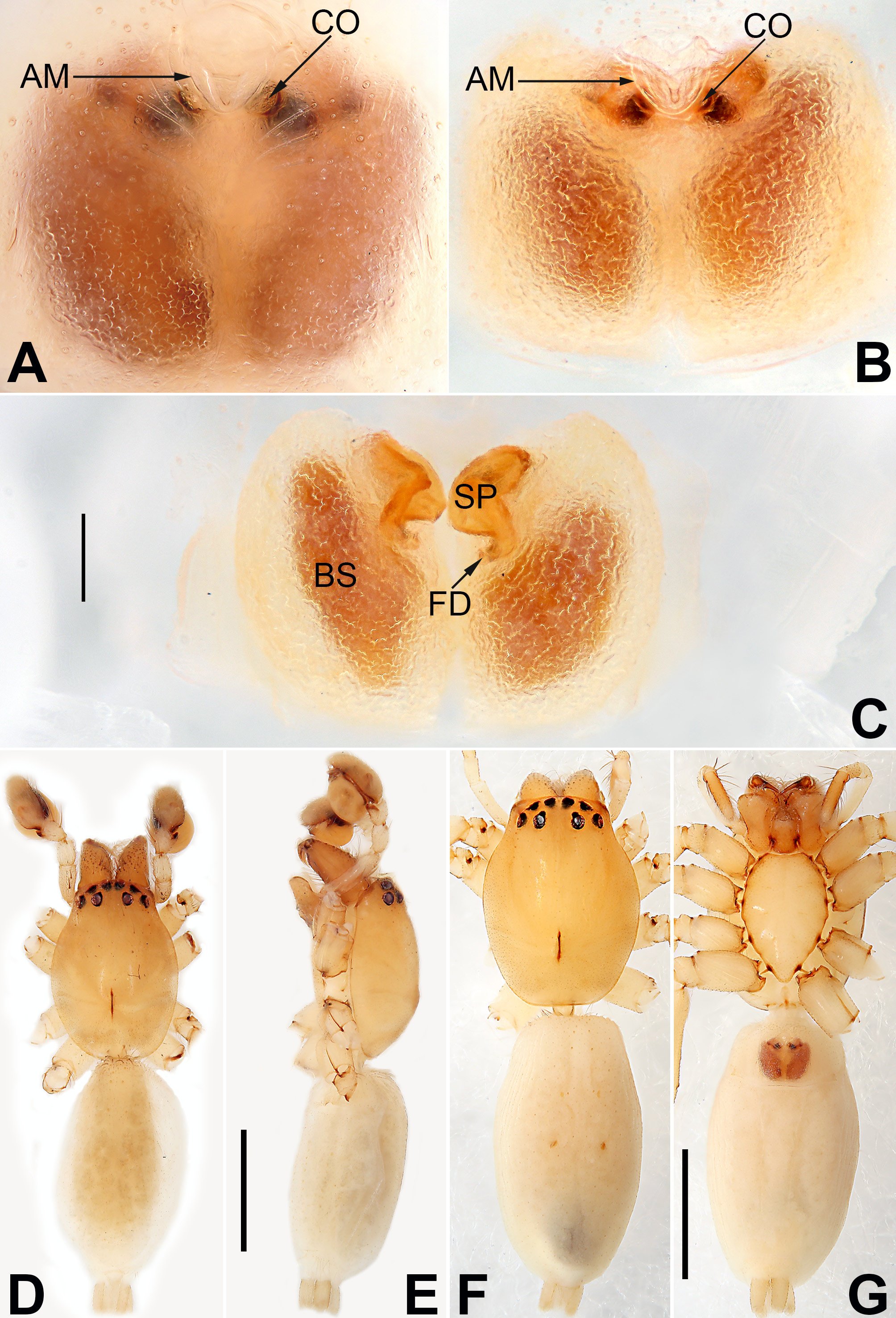
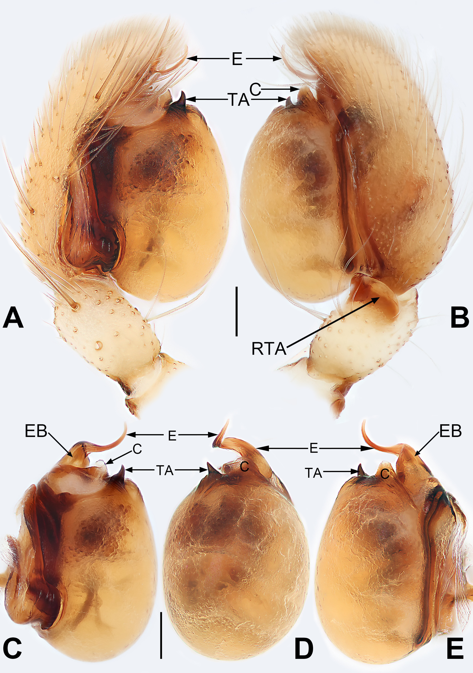
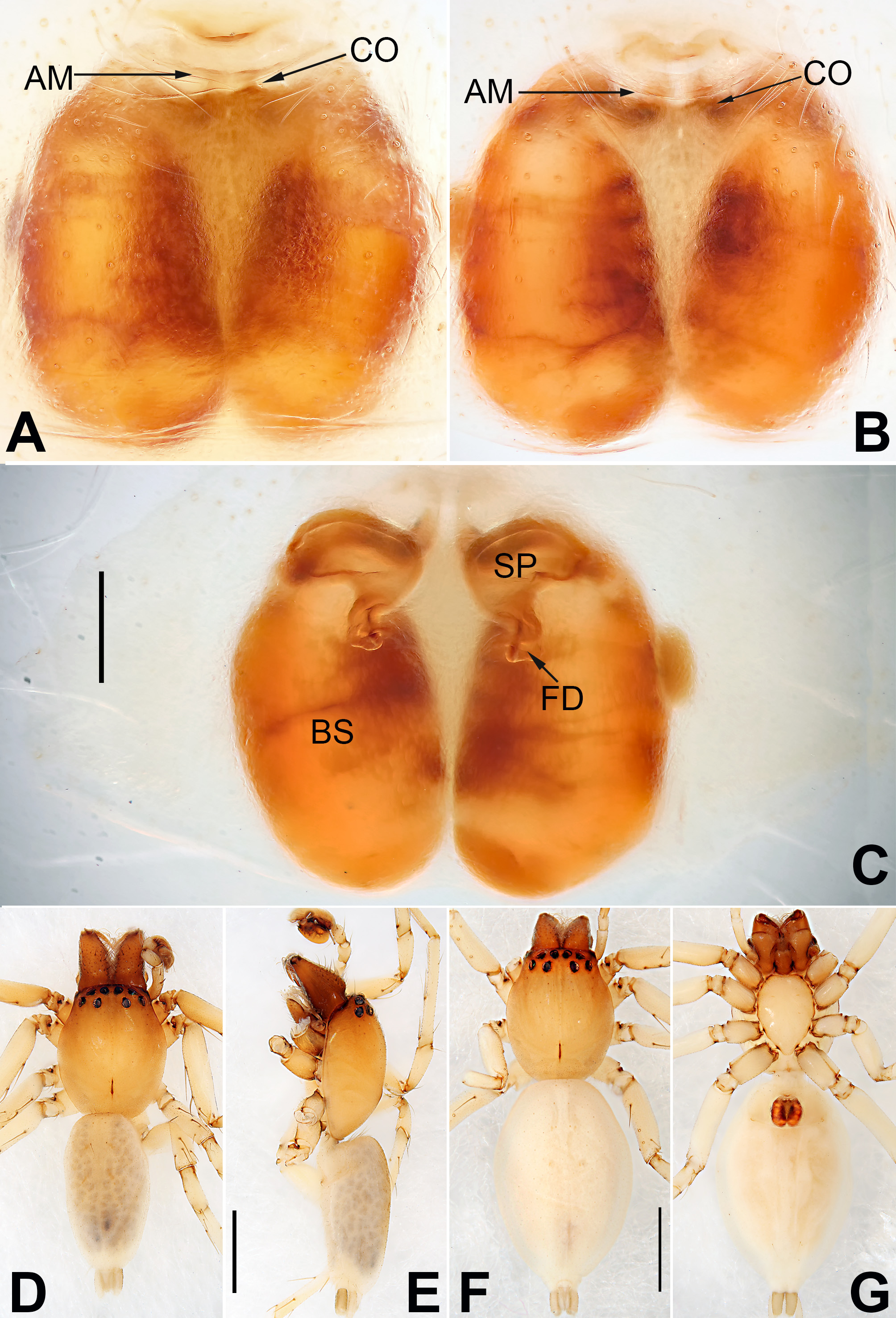
Diagnosis. This species can be easily distinguished from those of the other species belonging to the corticalis - group, with the exception of C. didentata ( Figs 5–6 View FIGURE 5 View FIGURE 6 ), by their bulged bulb with tegular apophysis and the epigynal plate with atrial membrane (tegular apophysis and atrial membrane are usually absent in other corticalis -group species). It can be separated from C. didentata by the larger, twisted embolus, smaller and partly membranous conductor, smaller and teeth-shaped tegular apophysis ( Figs 7 View FIGURE 7 A–E vs. Figs 5 View FIGURE 5 A–E), and by the disc-shaped atrial membrane and the smooth bursae ( Figs 8 View FIGURE 8 A–C vs. Figs 6 View FIGURE 6 A–C).
Description. Male: Total length 3.60; prosoma 1.69 long, 1.32 wide; opisthosoma 1.30 long, 1.94 wide. Prosoma ( Figs 8 View FIGURE 8 D–E), ovoid in dorsal view, ocular region slightly narrowed, widest between coxae II and III; in profile, slightly higher between ocular area and longitudinal fovea, gradually sloping towards pars cephalica; integument smooth, clothed with short fine hairs. Carapace brown, darker anteriorly, without distinct color pattern; fovea reddish. Chelicerae protruding and wine-coloured, with five promarginal and two retromarginal teeth. Labium and endites light brown, longer than wide. Sternum yellowish white. Eyes: AER slightly recurved, PER wider than AER and slightly procurved in dorsal view. AME dark, other eyes light; with black rings. Eye sizes and interdistances: AME 0.12, ALE 0.13, PME 0.14, PLE 0.13, AME–AME 0.05, AME–ALE 0.07, PME–PME 0.21, PME–PLE 0.12, MOQL 0.16, MOQA 0.15, MOQP 0.33. Legs yellowish-white, without distinct color markings. Leg formula: IV, II, III, I; leg measurements: I 3.60 (0.96, 1.68, 0.80, 0.38), II 4.26 (1.30, 1.80, 0.79, 0.48), III 3.68 (1.11, 1.16, 1.02, 0.37), IV 4.73 (1.37, 1.73, 1.45, 0.52). Opisthosoma ( Figs 8 View FIGURE 8 D–E) elongate-oval, marked with numerous brown spots. Palp ( Figs 7 View FIGURE 7 A–E): tibia short, only with retrolateral apophysis; RTA broad, flat and with a semicircular flange; bulb oval and strongly bulged, extending prolaterally and retrolaterally beyond cymbium, with several fanshaped markings; sperm duct inconspicuous; embolus with basal torsion and distal sickle-shaped bend; conductor small and papilliform, with membranous tip; tegular apophysis small and tooth-shaped, pointing prolatero-distally.
Female: Total length 4.24; prosoma 1.60 long, 1.26 wide; opisthosoma 2.61 long, 1.63 wide. General color lighter than in male ( Figs 8 View FIGURE 8 F–G). Eye sizes and interdistances: AME 0.10, ALE 0.12, PME 0.13, PLE 0.11, AME– AME 0.02, AME–ALE 0.03, PME–PME 0.22, PME–PLE 0.10, MOQL 0.16, MOQA 0.15, MOQP 0.32. Legs yellowish-white, without distinct color markings. Leg formula: IV, I, III, II; leg measurements: I 3.56 (0.98, 1.46, 0.74, 0.38), II 3.03 (1.09, 0.98, 0.59, 0.38), III 3.34 (1.05, 1.07, 0.90, 0.32), IV 4.86 (1.29, 1.62, 1.45, 0.52). Epigyne ( Figs 8 View FIGURE 8 A–C): Epigynal plate distinctly longer than wide, margin not rebordered; atrium small, covered by atrial membrane; atrial membrane disc-shaped, located on the anterior margin of atrium; bursae prominently visible through epigynal plate in ventral view; copulatory openings small, located at basolateral atrial borders, leading to short copulatory ducts which descend obliquely to connect with spermathecae; spermathecae with bean-shaped proximal part and tubular distal part; fertilization ducts short and curved, acicular; bursae oblong, translucent, surface smooth except several transversal depressions.
Distribution. Laos, China (Xishuangbanna, Yunnan Province). The new record presented here extends the known range of this species to the northwest.

No known copyright restrictions apply. See Agosti, D., Egloff, W., 2009. Taxonomic information exchange and copyright: the Plazi approach. BMC Research Notes 2009, 2:53 for further explanation.
|
Kingdom |
|
|
Phylum |
|
|
Class |
|
|
Order |
|
|
Family |
|
|
Genus |
Clubiona kai Jäger & Dankittipakul, 2010
| Yu, Hao & Li, Shuqiang 2019 |