
Tyrannosaurus rex Osborn, 1905
|
publication ID |
https://doi.org/ 10.2307/3889334 |
|
DOI |
https://doi.org/10.5281/zenodo.5224644 |
|
persistent identifier |
https://treatment.plazi.org/id/9A3A87D0-0B58-0DE5-FA0D-A2503C52F408 |
|
treatment provided by |
Plazi |
|
scientific name |
Tyrannosaurus rex Osborn, 1905 |
| status |
|
Tyrannosaurus rex Osborn, 1905
DESCRIPTION: SKULL AND MANDIBLE
Skull—General Form and Preservation
The skull of FMNH PR2081 is nearly complete ( Figs. 2—7 View FIGURE 2 View FIGURE 3 View FIGURE 4 View FIGURE 5 View FIGURE 6 View FIGURE 7 ). The left temporal region is damaged, and much of the left postorbital had broken off and was preserved in isolation. The snout is compressed ventrally immediately in front of the orbits, and the nasals have been pressed into the external naris. As a result, vertical structures in the preoccipital region—such as the descending lacrymal process and postorbital bar—are compressed.
As originally collected, the skull appeared to be severely crushed toward the left side. The left mandibular ramus had been broken and pulled away from the skull prior to burial, but the right ramus was still in place, albeit compressed into the palate. As a result, the skull listed to the left, where the quadrate and toothrow were no longer being supported by the jaw. The left maxilla’s palatal shelf is severely compressed, and in ventral view the vomer appears to abut the toothrow. This also caused the ventral skull elements on the right side to be pushed dorsally into the adductor chamber, and in ventral view the left pterygoid, ectopterygoid, and palatine appear to “stick out” more than their right counterparts.
Some of the damage on the left side has been interpreted in the popular media as evidence for a bite wound from another tyrannosaur ( Glut, 2000; Hanna, 2000; Larson, 2002). There is no conclusive evidence in support of this idea, and although there are shallow circular depressions on the left squamosal, they are not regularly spaced and do not immediately suggest bite marks.
Major Cranial Openings
The external naris is an anteroposteriorly long oval with its long axis sloping anteriorly. It is bound anterodorsally by the premaxilla and dorsally by the nasal. The nasal forms the posterior margin of the naris, but the extent to which it formed the ventral margin cannot be determined at present.
The antorbital and maxillary fenestrae sit within a broad antorbital fossa. The antorbital fenestra is bound anteriorly, ventrally, and dorsally by the maxilla, posterodorsally by the lacrymal, and posteroventrally by the jugal. It opens dorsolaterally, and when the dorsal aspect of the skull is viewed, the internal choanae are visible. The ascending processes of the palatines and, on the right side, the palatine recess, are clearly visible through them laterally. The fenestra’s anterior corner is acute on the left side as a result of compression.
The promaxillary fenestra (“first antorbital fenestra” of Osborn, 1912 and Carpenter, 1992) is a small, anteriorly facing circular opening at the anterior corner of the antorbital fossa ( Fig. 8 View FIGURE 8 ). It opens into the promaxillary recess and is not visible externally in lateral view, as it is in Albertosaurus .
The maxillary fenestra (“second antorbital fenestra” of Osborn, 1912) is a roughly circular opening entirely within the maxilla ( Fig. 8 View FIGURE 8 ). Its natural shape is partly preserved only on the right side, though even here it has been dorsoventrally compressed. Its anterior border is inset from the cheek.
The orbit is a dorsoventrally tall opening, wider dorsally than ventrally, and is bound anteriorly by the lacrymal, posteriorly and dorsally by the postorbital, and ventrally by the jugal. Exclusion of the frontal from the orbital margin has been regarded as a tyrannosaurid synapomorphy ( Gauthier, 1986; Holtz, 1994), although there may be a narrow gap between the postorbital and lacrymal at the dorsal margin of the orbit exposing the frontal in some forms ( Russell, 1970), and the extent of lacrymal-postorbital contact may vary systematically and ontogenetically (Holtz, 2001a). There is a broad flange of the postorbital, a feature typical of derived tyrannosaurids (e.g., Osborn, 1912; Maleev, 1974; Molnar, 1991; Holtz, 2001a), covering the ventral half of the orbit. The postorbital flanges of this specimen are far more extensive than any yet described and, on the right side, virtually fill the ventral half of the orbit (Fig. 2).
The infratemporal fenestra is a keyhole-shaped opening bound dorsally by a long, slender process of the squamosal, posteriorly by the squamosal and quadratojugal, and anteriorly and ventrally by the jugal. The infratemporal flange comprised of the squamosal and quadratojugal—a tyrannosaurid synapomorphy ( Gauthier, 1986; Holtz, 1994, 2001a)—virtually bisects the fenestra, although on the right side its anterior extent has been exaggerated by postmortem compression. Its relative length would have been similar to that of other large T. rex skulls (Osborn, 1912).
The supratemporal fenestrae are rectangular in dorsal view ( Fig. 3 View FIGURE 3 ). The postorbital horns do not obscure the anterolateral corner, as they do in AMNH 5027 View Materials (Osborn, 1912)—in this sense, FMNH PR2081 is more like published material of Tarbosaurus (Maleev, 1974). The frontal and parietal form the medial wall separating the fenestrae, which are bound laterally by the squamosal and postorbital; anteriorly by the postorbital and, to a limited extent, the frontal; and posteriorly by the parietal.
The internal choanae, in ventral view, are bound anteriorly by the maxillae, laterally and posteriorly by the palatines, and medially by the fused vomer ( Fig. 4 View FIGURE 4 ). Previous reconstructions (Osborn, 1912; Molnar, 1991) suggested small choanae that would not extend anteriorly beyond the anterior limit of the antorbital fenestrae, leading Holtz (1998) to describe tyrannosaurids as having a secondary palate. The choanae of FMNH PR2081 are relatively larger than those described by previous authors, and are more than half as large as the antorbital fenestrae.
The posttemporal fenestrae are long, thin grooves on the posterior surface of the skull. The paroccipital process forms the ventromedial margin, and the dorsolateral margins are formed by the posteroventral process of the parietal and, ventrally, the squamosal.
The foramen magnum is rectangular in posterior view and bound ventrally by the basioccipital, dorsolaterally by the exoccipital s, and dorsally by a thin slip of the supraoccipital. The exoccipital-supraoccipital suture is most visible on the left side, and the exoccipital-basioccipital sutures are not visible at all in this region.
The right stapes was preserved in place ( Fig. 9 View FIGURE 9 ), entering the external otic recess, which is a crescentic slit on the lateral braincase wall bound anterolaterally by the prootic and posteromedially by the opisthotic. The opening identified by Osborn (1912) as the fenestra ovalis is actually the caudal tympanic recess; the actual fenestra ovalis is not visible externally.
Premaxilla
Because of crushing, the subnarial region of each premaxilla is flattened and oriented dorsally. When reconstructed, the subnarial regions would have projected anterolaterally. A series of small mental foramina runs parallel to the toothrow along the ventral margin of each premaxilla, and although neither element is as rugose laterally as the maxillae, the bone surface is not smooth.
The premaxillae each bore four conical, almost incisiform teeth. All four teeth are present in the right element, each displaced slightly to the right. Only a single crown—the first in the series—remains in the left premaxilla, and it has been shifted against the right premaxillary teeth, giving the illusion that the right premaxilla bore five teeth ( Fig. 6 View FIGURE 6 ). Although the crowns for the remaining left premaxillary teeth are gone, their roots are still preserved within their alveoli; these can be seen in the CT animations, especially in coronal and sagittal sections. As with other tyrannosaurids, the premaxillary teeth are “Dshaped” in cross section, with a flattened posterior surface (Leidy, 1868; Bakker et al., 1988; Molnar, 1989, 1991). Fine serrations occur along the lateral and medial margins of the posterior surface. The alveoli are bordered medially by square interdental plates, similar to those bordering the maxillary and dentary toothrows.
Each premaxilla bears a cylindrical ascending process that forms the anterior rim of the naris. This projects posteriorly at its dorsal tip, tapering posteriorly to a point and passing medially along an anterior projection of the nasal for approximately 8 cm. About half of the posterior portion is broken and displaced ventrally on both premaxillae.
The premaxilla forms most of the naris’ ventral border ( Fig. 10 View FIGURE 10 ). It is unclear if it meets the nasal ventrally, as it does in most other theropods—the nasals are damaged in this region. But if it did, the processes involved were thin and would have been primarily visible in dorsal view, as with AMNH 5027 View Materials . Premaxilla-nasal contact ventral to the naris had been viewed previously as a feature diagnosing Albertosaurus (e.g., Russell, 1970; Carpenter, 1992), but in fact such contact occurs in all sufficiently-preserved tyrannosaurids (including other T. rex specimens).
The palatal surface is deeply vaulted, and the alveoli project ventral to the palate itself. Each premaxilla bears a slender, rodlike process on the medial edge of the palatal ramus, projecting from the posteromedial corner anteriorly and covering the region that would have been perforated by an incisive foramen. These processes meet at the midline and do not reach the interdental plates ( Fig. 4 View FIGURE 4 ). Sutural separation between the premaxillae and vomer is indistinct, but the anterior tip of the vomer lies below these processes. Osborn (1912) and Molnar (1991) suggested less overlap of the vomer under the processes. Similar processes are present on the premaxillae of Daspletosaurus (NMC 8506), but they appear not to have been midline structures, and Russell (1970) did not reconstruct this portion of the Daspletosaurus skull.
We cannot tell if palatal subnarial foramina were present between the premaxillae and maxillae, as reconstructed by Carr (1999). A subnarial foramen is found on the lateral surface of the cheek.
Nasal
The nasals are restricted to the dorsal surface of the snout. The surface of the joined nasals is generally flat, although they are gabled toward the naris. They are fused to each other for much of their length, but a clear separation between right and left elements can be seen anteriorly over the naris and posteriorly between the lacrymals and prefrontals. They are expanded laterally posterior to the external naris, and each ends anteriorly in an acute process that would have contacted an ascending process of the premaxilla, although as preserved they were depressed into the narial chamber. The ascending premaxillary process would have passed medially, and the two nasals in articulation form a V-shaped suture with the premaxillae.
At first, there appears to be no nasal-premaxillary contact in lateral view, and the maxillae seem to form the ventral borders of the narial apertures. Based on published figures, this would appear to be true for other T. rex specimens ( Osborn, 1912:pl. 1 View Osborn, 1912: pl. 1 ) and Daspletosaurus torosus ( Russell, 1970) . In Tarbosaurus and Albertosaurus , a slender process of the nasal passes around the naris, contacting a corresponding posterior process of the premaxilla and prevents the maxilla from participating in the naris. A ventral process is laterally visible in an immature T. rex skull (Gilmore, 1940; Bakker et al., 1988).
Closer inspection of the skull figured by Osborn ( AMNH 5027 View Materials ) shows that the nasal did bear a slender ventral process contacting the premaxilla and excluding the maxilla from the narial border, but that this process lay along the very lip of the naris and is only visible dorsally ( Osborn, 1912:pl. 2 View Osborn, 1912: pl. 2 and fig. 2 View Osborn 1912: Fig. 2 ). Whether this was also true for FMNH PR 2081 is not known ( Fig. 10 View FIGURE 10 ). Maximum extent of the lateral flaring occurs immediately posterior to the naris, and the anterior edge of this flaring is concave at the posterior angle of the narial aperture, but if there was a discrete process, it has broken away.
The nasals taper posteriorly, and their sutural relationship with the lacrymals is complex. Each nasal bears two posterior processes—a short, ventrolateral process that projects into the lacrymal, and a longer dorsomedial process extending back to the frontal and prefrontals. They are constricted by the lacrymals and are narrow when they make contact with the frontal. Because of maxillary-lacrymal contact, the nasals are excluded from the antorbital fenestral border.
Carr (1999) showed that the dorsal surface of the nasal is rugose throughout most of posthatching ontogeny in Albertosaurus , but that an ontogenetic increase in rugosity may occur in Tyrannosaurus . Given the probable maturity of this specimen, the bosses on the nasals are surprisingly small. The surface of the joined nasals is wrinkled, especially anteriorly, but the prominent nasal protuberances seen in most other tyrannosaurids are restricted to a patch directly over the antorbital fenestra. The most prominent “boss” is a V-shaped crest over the fenestra. These protuberances appear to extend further anteriorly in most known Tarbosaurus and Albertosaurus specimens, though the type of Daspletosaurus torosus is comparable to that of FMNH PR 2081 . Other T. rex skulls preserve protuberances nearly approaching the naris.
Based on CT images, the ventral surface of each nasal is smooth. This is congruent with isolated tyrannosaurid nasals (e.g., LACM 23845, MOR 555).
Maxilla
The maxilla is laterally broad, and the toothrow is ventrally convex. There are twelve alveoli in the series, as with some other adult T. rex maxillae (Osborn, 1912; Molnar, 1991). The alveoli extend dorsally either to the dorsal rim of the bone or, posteriorly, to the antorbital fossa. As a consequence, anterior maxillary teeth have extremely long roots—one isolated tooth ( Fig. 11A View FIGURE 11 ) bears an 8 cm long crown, but a 20 cm long root. Maleev (1974) indicated twelve alveoli in Tarbosaurus , but observation of PIN 551-1 clearly shows thirteen. Thirteen alveoli are also found in some immature T. rex (Bakker et al., 1988 and pers. obs.). Albertosaurus typically has fourteen or fifteen; Lambe (1917) reconstructed the Albertosaurus maxilla with thirteen, and Russell (1970) suggests as many as seventeen. Mature Daspletosaurus maxillae have as many as sixteen alveoli.
The maxillary teeth are laterally compressed and recurved, and the first is much larger than any of the premaxillary teeth. All are double-serrated. The length of the crowns decreases posterior to the sixth.
A single row of 2 to 5 mm wide mental foramina parallels the toothrow ( Fig. 12A View FIGURE 12 ). This can be referred to as the alveolar row. Another row—the circumfenestral row—can be traced from the subfenestral ramus anterodorsally, curving around the ventral margin of the antorbital fossa. Shorter rows of three to five foramina are arranged between the alveolar and circumfenestral rows. Natural molds for the rami of the maxillary nerve are visible ventral to some of these foramina, including one preserving a ramification in the exiting nerve ( Fig. 12 B View FIGURE 12 ).
Internally, CT images show only a single consistent channel probably associated with the maxillary nerve, located immediately dorsal to the alveoli. One is tempted to associate the alveolar and circumfenestral rows to the palatine and nasal rami of the maxillary nerve, respectively; but smaller channels can be followed from the main maxillary channel dorsal to the alveoli to mental foramina along the maxillary surface. All mental foramina relate to the same ramus. This same channel communicates with small openings on the dorsomedial comer of the maxilla. Another set of openings penetrates the maxilla medially, dorsal to the palatal ramus and ventral to the antorbital fossa. All of these are labeled “V2” in Figure 13 View FIGURE 13 . Presumably, maxillary nerve branches enter the maxilla through these medial openings, pass to the common channels, and then pass laterally through the mental foramina. There is no single opening for the maxillary nerve posteriorly, as there would be in a crocodylian.
A subnarial foramen (“third antorbital fenestra” in Osborn, 1912) lies on the maxillary-premaxillary suture ventral to the naris ( Figs. 2, 10 View FIGURE 2 View FIGURE 10 ). This is distinct from the subnarial foramen identified on the palatal surface by Madsen (1976) and reconstructed on the T. rex palate by Carr (1999). This opening has a broad distribution among archosauriforms ( Juul, 1994) and was apparently not related to the craniofacial air sac system (Witmer, 1997). The subnarial foramen is located closer to the toothrow in Albertosaurus (Lambe, 1917; Russell, 1970) and Tarbosaurus (Maleev, 1974 and pers. obs.), but the placement between the toothrow and naris seen in FMNH PR2081 is also true for Daspletosaurus ( Russell, 1970) . None of the immature T. rex specimens currently published preserve this feature.
The maxilla is reduced to a slender posterior process behind the twelfth alveolus. This process passes ventral to the jugal and is separated from the tooth-bearing part of the maxilla by a concavity on the ventral outline of the cheek. This process is slender, as in most tyrannosaurids but in contrast to the rather robust process indicated for the holotype by Osborn ( 1912:fig. 1 View Osborn 1912: Fig. 1 ). The process on AMNH 5027 View Materials was much longer and more slender, but the posterior tip may be broken off, as indicated in the photograph published by Osborn ( 1912:pl. 1 View Osborn, 1912: pl 1 ), giving the appearance of a shorter structure.
The surface of the maxilla dorsal to the toothrow is heavily sculpted. All mature tyrannosaurid skulls have sculpted lateral maxillary surfaces, but the degree of rugosity in FMNH PR 2081 is greater than that of any other observed, and is best developed anterior to the antorbital fossa. Surficial rugosity continues along the subfenestral ramus and the interfenestral bar between the antorbital and maxillary fenestrae.
The lateral surface of the maxilla within the antorbital fossa is very smooth, and the demarcation between sculpted and unsculpted bone is very sharp, especially dorsally and anteriorly, where the margin of the maxillary fenestra is deeply inset from the cheek. The hourglass-shaped column separating the maxillary and antorbital fenestrae (the interfenestral pillar; Witmer, 1997) is broken in the middle, giving it the appearance of a compound structure comprised of two bones. This would normally have a V-shaped anterior outline and broadly concave posterior edge bordering the antorbital fenestra (Osborn, 1912; Maleev, 1974).
Several internal structures are well preserved and are observable in CT imagery. The promaxillary recess fills the anteriormost fifth of the bone, and is connected to the maxillary antrum by a triangular promaxillary fenestra. Witmer (1997) described a vestibular bulla in the anterodorsal corner of the promaxillary recess in Albertosaurus , and an isolated T. rex maxilla (UCMP 118742) preserves this feature; the bulla is not obvious in the FMNH PR 2081 slices, but the nasals may have been pushed in to obscure them. The promaxillary fenestra, which opens posteriorly, lies close to the anterior border of the maxillary fenestra ( Figs. 8 View FIGURE 8 , 14 View FIGURE 14 ); indeed, the tyrannosaurid promaxillary fenestra is usually visible through the maxillary fenestra when the antorbital space is prepared or the skull is disarticulated.
There is also a large caudal antromaxillary fenestra, parallel with the promaxillary fenestra, bordered laterally by the interfenestral pillar and medially by an anteroposteriorly slender postantral pillar. As with Albertosaurus (Witmer, 1997) and other T. rex maxillae (e.g., UCMP 118742, CM 9380), the caudal antromaxillary fenestra lies at the anterior comer of the antorbital fenestra ( Figs. 13D View FIGURE 13 , 14 View FIGURE 14 ); in Tarbosaurus (Maleev, 1974, pers. obs.), it is closer to the anteroventral margin.
The palatine ramus of the maxilla is a thin sheet flooring the antorbital cavity ( Fig. 13 View FIGURE 13 ). It forms the anterior and lateral margins of the internal choanae, which are relatively larger than reconstructed by Osborn (1912) or Molnar (1991) and more like those figured by Russell (1970) for Daspletosaurus .
Most disarticulated tyrannosaurid maxillae have a thin ridge on the medial surface, running anteroposteriorly dorsal to the maxillary fenestra ( Fig. 14A View FIGURE 14 ). As this region houses the maxillary antrum, which is bound ventrally by the palatal ramus and posteriorly by the postantral pillar, Witmer (1997) suggested that a cartilaginous medial wall was attached to this ridge. CT images reveal a thin bony wall rising from where this ridge should be on each maxilla in FMNH PR2081 ( Figs. 13 View FIGURE 13 , 14 View FIGURE 14 ; labeled “maw”). Each is approximately two millimeters thick and is attached to the maxilla in two places—dorsally, above the maxillary fenestra, and ventrally on the palatal ramus. There are indications of such a structure anteriorly, toward and perhaps within the promaxillary recess.
A few other tyrannosaurid maxillae preserve this structure. One is MOR 590, a specimen referred to Daspletosaurus from the Two Medicine Formation, which includes the left maxilla. In this specimen, a sheet of bone covered the maxillary antrum medially prior to preparation, but it was drilled through by preparators who evidently did not expect bone there. It terminates at the preantral pillar. Another is the left maxilla of a mature Albertosaurus at the RTMP (unnumbered at the time of my visit), which has a similar wall covering both the maxillary antrum and promaxillary recess. In this case, long oval gaps perforate the wall ventrally near its base at the palatal ramus. Similar structures are also seen in Troodon (e.g., MOR 5535) and may account for the curved walls of the “accessory sinuses” noted by Ruben et al. (1996).
Early reports of turbinates in tyrannosaurids ( Bakker, 1992) may have been based on these structures. They lie within the region of the snout where turbinates may be expected, and resemble them in many respects. The space between these structures and the cheek is enclosed anteriorly and posteriorly, isolating them from the nasal passages that they would have filled had they been turbinates.
Sutural relationships between the maxilla, lacrymal, and nasal are rather complex ( Fig. 15 View FIGURE 15 ). The maxilla forms nearly all of the antorbital fenestra’s dorsal margin. Prior to preparation, the nasal seemed to participate in the fenestra’s rim, but the nasal is excluded from the fenestra in all other tyrannosaurids, and in this case the nasal disrupted the maxillary-lacrymal border of the fenestra because of dorsoventral crushing. The posterodorsal ramus of the maxilla is deeply cleft, with a notch accepting a slender anterior process of the lacrymal.
Vomer
The vomer is a unified rodlike midline structure with a broad, diamond-shaped anterior expansion. As with the T. rex vomer described by Molnar (1991), it is split posteriorly, the only remaining evidence that the vomer is a compound structure.
It is rodlike for most of its length and separates the choanae, and the anterior expansion is present ventral to the promaxillary recesses. Palatal reconstructions by Osborn (1912) and Molnar (1991) indicate an anterior processes on the medial margin of each premaxilla, with the anterior tip of the vomer lying posterior to this; these structures can be seen in FMNH PR 2081 , but the vomer passes ventral to these and lies against the premaxillae ( Fig. 4 View FIGURE 4 ).
The anterior expansion is thin, but concave dorsally. It is large compared with those figured by previous workers (Osborn, 1912; Molnar, 1991; Carr, 1999), and whereas the anterior expansion is a simple diamond in other T. rex specimens, the anterior tip is elongate, approximating a spear point in the present specimen. Posteriorly, ventral to the maxillary fenestrae, the vomer bears a thin ventral keel. This keel becomes thicker and less prominent between the choanae, and all but disappears ventral to the antromaxillary fenestrae. But a deep dorsal furrow appears where the keel disappears, running along the length of the vomer to the posterior margin of the choanae. As the vomer approaches the ascending process of the palatine, it expands dorsally and, to a lesser extent, laterally. There is a broad surface for contact with the palatine on each side, demarcated ventrally and laterally by a low ridge. The facet for the palatine can be seen on the left side, where the palatine has shifted posteriorly.
A circular 2-cm-wide opening perforates the vomer’s anterior expansion. This was formed when a tooth from the right dentary was pushed up into the palate after death, and a tooth was found in place during preparation. A similar perforation is found on the right maxilla behind the vomer’s anterior expansion. Entry of dentary teeth into these openings is clearly visible in the CT sections ( Fig. 16 View FIGURE 16 ), as these were generated before the jaw was removed. These are the only demonstrable bite marks on the skull, and were self-inflicted.
The vomer splits and becomes dorsoventrally thin immediately behind the palatine ascending process, with thin posterior rods extending toward their respective anterior pterygoid flanges ( Fig. 17 View FIGURE 17 ). A similar arrangement was reconstructed by Molnar (1991), with a narrow separation between rami. Although Osborn (1912) reconstructed the T. rex vomer as splitting posteriorly, he reconstructed the rami as remaining adjacent and in contact with each other until meeting the anterior pterygoid flanges. The isolated vomer Molnar studied is incomplete, and the posterior rami he figured were not available in the LACM collections at the time of my visit. However, CT imagery of FMNH PR2081 corroborates part of Molnar’s reconstructionthe rami diverge slightly and project posteroventrally, merging with a slight medial thickening in the anterior pterygoid flanges.
However, the split vomer extends posteriorly nearly to the posteriormost extent of the pterygoids, forming a rodlike medial border to each pterygoid ( Fig. 18 View FIGURE 18 ).
Palatine
Anteriorly, each palatine takes the form of a thin rod medial to the palatine ramus of the maxilla, forming the lateral border of the choana. Each expands in diameter posteriorly, and a narrow embayment is present laterally for the maxilla, so that close to the ascending process, the palatine appears to grip the palatine ramus of the maxilla.
The ascending process rises from the maxilla along a thick, hollow pillar (see below) at the posterior corner of the antorbital fenestra. It expands dorsally and anteriorly, forming a large egg-shaped palatine process. There is a shallow fossa—identified by Witmer (1997) as a lateral pneumatic recess in more derived theropods—anterior to the foramen.
Coronal CT images confirm the presence of a large palatine recess. Tyrannosaurid palatines, when found in isolation, are usually crushed; the recesses in FMNH PR 2081 fill the ascending process and lie next to the expanded vomer ( Fig. 19 View FIGURE 19 ). They terminate adjacent to the lacrymal descending process. A single large foramen lies on each recess’ dorsal surface, near its lateral edge. This can be seen on the right palatine (left-hand side of figure) in Figure 19B View FIGURE 19 as a discontinuity in the palatine’s outline.
The recess roof is very thin—perhaps no more than two millimeters thick—but the floor is much thicker, approaching a centimeter in thickness. The foramen in FMNH PR 2081 , and in other T. rex specimens, is large relative to the foramen in Albertosaurus ; Daspletosaurus and Tarbosaurus palatines have foramina comparable in relative size to those of T. rex , at least when mature specimens are considered.
Posteriorly, each palatine bears a dorsoventrally thin pterygoid process that broadly contacts the pterygoid nearly from the midline for approximately 10 cm laterally. The posterior margin of the palatine is concave lateral to the pterygoid contact, where it is reflected posteriorly at the cheek to form the jugal lamina and lie medially against the maxilla and jugal. The left palatine bears a deep concavity in the outline of the lamina contacting the maxilla; this may be pathological, and the right palatine is too damaged to allow determination of this feature’s presence.
Both palatines bear large circular tuberosities on the ventral surface immediately anterior to the pterygoids ( Fig. 4 View FIGURE 4 ). The only muscle attaching directly to the palatine in crocodylians is the dorsal (anterior) pterygoideus ( Schumacher, 1973), but its origination is usually broad and fleshy—one does not see a discrete tubercle on one bone. However, thin slips of both the dorsal and ventral pterygoideus attach to the palatine in birds, and the origination for both can be tendonous ( George and Berger, 1966). Topographically, the tuberosity in FMNH PR 2081 corresponds more closely with a posterior palatine origination for the ventral pterygoideus in birds.
These tuberosities were not observed on other tyrannosaurid palatines. Indeed, I am unaware of any published theropod in which this has been described. However, the pterygoid ramus of the palatine is delicate and usually imperfectly preserved in disarticulated tyrannosaurid skulls.
The functional basis for the antorbital fenestra is debated. Historically, expanded insertion surface for the dorsal pterygoideus musculature has been the explanation, but more recently Witmer (1995, 1997) argued that rostral air sacs were important. The lateral surface of the palatine process in FMNH PR 2081 is rugose and covered in Sharpey’s fibers, which would suggest the attachment of a muscle mass, and the hyolingual musculature would have attached posterior and ventral to this structure.
Lacrymal and Prefrontal
The lacrymal consists of a descending process separating the antorbital fenestra and orbit, an anterior process bordering the nasal and forming part of the dorsal roof of the antorbital cavity, and a massive posterior process forming the dorsal border of the orbit.
In dorsal view ( Fig. 20 View FIGURE 20 ), the lacrymals are semilunate structures constricting the nasals and partially fused frontal. A small D-shaped prefrontal ossification can be discerned between each lacrymal and frontal, with indistinct borders. Unlike some tyrannosaurids (e.g., Daspletosaurus , Albertosaurus ) and some nontyrannosaurid theropods (e.g., Madsen, 1976; Currie and Zhao, 1993a; Madsen and Welles, 2000; Currie and Carpenter, 2000), there is no prominent lacrymal “horn” or cornual process ( Carr, 1999).
The presence of a prefrontal in tyrannosaurids has been debated (Osborn, 1912; Russell, 1970; Bakker et al., 1988). It is reasonable to conclude that the prefrontals are ontogenetically ephemeral structures that gradually merge with the lacrymals, and they are represented by a set of depressions on the lacrymal in FMNH PR 2081 ( Fig. 19 View FIGURE 19 ). Whereas Osborn (1912) placed the postorbitals completely between the lacrymals and frontal, those of FMNH PR2081 clearly make broad contact with the nasal.
The lacrymal descending process is bowed anteriorly, though on the right side the convexity is exaggerated by dorsoventral crushing. The anterior edge is normally convex in tyrannosaurids, but not to this degree. It meets the jugal at the posteroventral comer of the antorbital fenestra and the anteroventral comer of the orbit. It is D-shaped in cross-sectional area, acute anteriorly and broad posteriorly adjacent to the orbit.
The dorsal surface of the lacrymal is rugose, especially along the posterior process. The lacrymal rugosity joins seamlessly with its postorbital counterpart. Normally, even in T. rex , the lacrymal and postorbital horns are separated by a distinct notch dorsal to the orbit, but this notch is absent from FMNH PR2081 .
The lacrymal recess opens anteriorly through a 2-cm wide circular opening at the posterodorsal corner of the antorbital fossa. It is very small for a skull of this size, as with other T. rex and Daspletosaurus skulls (Osborn, 1912; Russell, 1970; Molnar, 1991), but in contrast with the comparatively larger openings in some Tarbosaurus (Maleev, 1974) and Albertosaurus (Lambe, 1917; Russell, 1970). This may be an ontogenetically variable feature; the lacrymal of “ Nanotyrannus ” is imperfectly preserved, but it appears to have a large opening, as do those of skulls referred to Maleevosaurus lancinator (Maleev, 1974; Carpenter, 1992), which is likely an immature version of Tarbosaurus bataar ( Carr, 1999) .
The recess itself can be seen in CT images (e.g., Figs. 18 View FIGURE 18 , 19 View FIGURE 19 ) and is large, virtually filling the dorsal portion of the bone. There is also a large vacuity filling the descending process, though there is no obvious connection between the two vacuities ( Fig. 18B View FIGURE 18 ). A small tyrannosaurid lacrymal collected with FMNH PR2081 shows a large dorsal opening, as in other theropods, but also a set of small openings on the anterior and posterior surfaces of the descending process. Two of these form narrow tunnels and were probably vascular structures, but one of them—the dorsalmost on the posterior surface—has no anterior counterpart. Each lacrymal of FMNH PR2081 has a pair of openings in this position.
The lacrymal’s anterior process forks anteriorly, with a smaller ventral process penetrating the maxilla and a larger dorsal process wedged between the nasal and maxilla. The ventral process meets a posterior process of the maxilla and excludes the nasal from the antorbital fenestra.
Jugal
The jugal can be divided into a horizontal body forming much of the ventral margin of the cheek and a pair of ascending processes anterior and posterior to the orbit. The posterior ascending process forms part of the postorbital bar and the anterior margin of the infratemporal fenestra.
The lateral surface of the jugal ventral to the orbit is inflated, giving the skull the appearance of “cheeks.” The forward-facing orbits characteristic of Tyrannosaurus result, in part, from jugal curvature. Osborn (1912) reconstructed the jugals with much more curvature than Molnar (1991), and subsequent discoveries—including this one—support Molnar’s reconstruction.
The body of the jugal overlaps the maxilla ventral to the antorbital fenestra. At the posteroventral comer of the fenestra, the large, circular jugal foramen opens into a large embayment that extends ventrally and posteriorly, as shown in CT imagery ( Fig. 17B View FIGURE 17 ). The left jugal bears a small depression anterior to the jugal foramen that may be pathological. Its ventral margin is convex, with a prominent, rugose ventral tuberosity immediately behind the orbit. One of the vascular foramina on the jugal ventral to the orbit is surrounded by exostotic bone, possibly indicating infection.
The anterior ascending process formed the ventral one-third of the antorbital pillar, but the lacrymal descending process has been compressed, so the details of the lacrymal-jugal contact in this region are destroyed.
The concavity between the ascending processes is deep and narrow, largely filled medially by a thin lamina projecting from the postorbital. As with AMNH 5027 View Materials , the jugals do not contribute to the orbital laminae, as they do in MOR 555 and some Albertosaurus skulls (e.g., RTMP 86.144.1).
The posterior ascending process is acute at its dorsal tip. It is flattened posteriorly and bears a broad facet for the descending postorbital process anteriorly. There is a low rugosity on the posterolateral surface that probably contacted the anterior squamosal/quadratojugal laminae. Jugal-postorbital contact is thus a lap joint, with the postorbital overlapping the jugal anteriorly. Externally, the lineation between the postorbital and jugal cuts the postorbital bar diagonally from anteroventral to posterodorsal. The jugal’s ascending process has been displaced posteriorly, but it would have terminated at the dorsal rim of the infratemporal fenestra, meeting the squamosal and excluding the postorbital from the infratemporal fenestra anteriorly. This is an unusual arrangement for a theropod, though the reconstructed jugal ascending process is tall in tyrannosaurids generally (Osborn, 1912; Russell, 1970; Bakker et al., 1988).
Posterior to the ascending process, the jugal forms the ventral floor of the infratemporal fenestra, bearing a short, thin lamina projecting dorsally into the infratemporal space at the fenestra’s posteroventral corner. The quadratojugal passes laterally over a long, broad facet on the jugal, which deepens and ultimately divides the posterior jugal process into a pair of thin cylindrical rods. The dorsal rod is short, terminating just behind the infratemporal fenestra, but its ventral counterpart nearly reaches the quadrate.
Frontal
The frontals are partially fused in FMNH PR2081 , with indistinct grooves indicating the former sutural zone anteriorly. The faint trace of the former suture zone can be seen in coronal CT images ( Fig. 21 View FIGURE 21 ).
The anterior margin, in contact with the nasals, is complex ( Fig. 20 View FIGURE 20 ). The anterior margin of each frontal ossification is concave along its contact with the lacrymal and prefrontal in dorsal view, and the two ossifications meet to form an acute anterior process. The frontonasal suture suggests a fleur-de-lys pattern, with the nasals flaring laterally along the frontal, but also passing between the frontals with narrow and short posterior processes. The anteriormost tip of the frontal can be seen as a pair of thin wedges arising between the crescentic nasals in coronal CT images ( Fig. 17B View FIGURE 17 ).
Russell (1970) placed great systematic weight on the nature of this suture in different tyrannosaurid taxa— Tarbosaurus has a broad medial nasal process (pers. obs.), but it is said to be absent from some putative species of Albertosaurus and slender in others and in Daspletosaurus ( Russell, 1970) . Given the subtlety of the medial nasal processes in FMNH PR2081 , much of the difference between specimens could easily be taphonomic rather than phylogenetic.
The dorsal surface of the united frontal is generally smooth and concave, and it slopes ventrally into the supratemporal space from the anterior and medial supratemporal borders. Its contact with the postorbital is V-shaped, with the concavity on the frontal. Its sutural relationship with the parietal is difficult to trace, but both frontal and parietal are reflected dorsally at their medial contact, forming the distinct peak that can be seen when the skull is viewed laterally in all tyrannosaurids, although the peak is restricted to the parietal in Albertosaurus . There is a pair of distinct linear ridges radiating from the medial angle of the frontal laterally, either of which may be related to the frontoparietal suture.
Ventrally, the frontal bears a pair of deep fossae for the enlarged olfactory bulbs, which in FMNH PR 2081 have a larger diameter than the foramen magnum. The fossae continue laterally to the ventral surface of the lacrymal and are separated, at least posteriorly, by the sphenethmoid ( Fig. 21 View FIGURE 21 ); they narrow posteriorly as they approach the ventral surface of the parietal.
Parietal
The parietals are fused at the midline, and no trace of the interparietal suture can be seen, even in CT images. They meet the fused frontals anteriorly; the frontoparietal suture is not distinct, but there is a tall crest where they meet.
The parietal bears an acute midsagittal ridge separating the supratemporal fenestrae. The parietal slopes broadly posterolaterally within the supratemporal space, forming the medial walls of the fenestrae. This ridge intersects a tall posterior wall projecting over the skull roof and separated from the squamosals by distinct notches. Most large nonavian theropods have a structure like this, though it is more gracile in Allosaurus (Madsen, 1976) . This is responsible for the second peak, behind that between the frontal and parietal, visible in the lateral profile of all tyrannosaurids.
Sutural relationships within the supratemporal fenestrae cannot be seen, except for the slender posteromedial process of the squamosal that passes over the parietal on the fenestra’s posterior wall. In disarticulated tyrannosaurid parietals (e.g., LACM 23845, RTMP 94.143.1), the parietal is deeper posteriorly than anteriorly, where the laterosphenoids comprise much of the medial wall of the supratemporal fenestra.
In posterior view, the posterior wall forms a pair of broad winglike tabs with concave surfaces. Each bears an acute ventrolateral process separating the paroccipital process from the squamosal. There are discrete circular depressions medially, including one on the midline and two broader sulci medial to the dorsal tabs of the supraoccipital.
Ventrally, the parietal forms the bony roof for the midbrain and is pierced by foramina interpreted here as serving the dorsal cephalic venous system. This is apparent in sagittal sections of the skull, where the dorsal expansion of the endocranial cavity indicates the approximate location of the cerebellum ( Fig. 22 View FIGURE ). The parietal is apneumatic.
As currently prepared, the relationship between the parietal and left squamosal is unnatural. The left squamosal was removed during preparation to permit reattachment of the left postorbital. It proved impossible to reattach the squamosal internal to the parietal, which is why the squamosal is now broadly exposed posteriorly ( Fig. 5 View FIGURE 5 ).
Postorbital
The right postorbital is preserved intact on the skull, but most of the left postorbital was disarticulated ( Fig. 23 View FIGURE 23 ). The left descending process was found lying within the orbit, and was later reattached to the dorsal portion of the bone.
Attention has been directed to alleged evidence for bite trauma from another tyrannosaurid on the left cranial bones of FMNH PR2081 . Some accounts ( Glut, 2000; Larson, 2002) claim that the left postorbital was wrenched out of place during intraspecific combat. There are no bite marks, or even depressions that could be interpreted as such, on either postorbital; disassociation of the left element is not demonstrably a perimortem effect.
The postorbital forms the anterior and lateral walls of the supratemporal fenestrae. The surface of the fenestra is smooth, with shallow mediolaterally long striations along the anterior wall. The squamosal overlaps the postorbital medially, and a narrow fragment of the squamosal prior to preparation was preserved posteriorly, immediately dorsal to the rim of the infratemporal fenestra, on the left postorbital. Four bones contact the postorbital anteriorly and medially—the frontal, lacrymal, parietal, and laterosphenoid.
The anteromedial sutural surface is complex. Articulation with the lacrymal occurs anteriorly within a thin sulcus that splits the peak of the horn. The frontal contacts the postorbital along a broad sutural surface adjacent to the anterior wall of the supratemporal fenestra. There is a long, deep sulcus on the anteromedial surface of the postorbital bar, immediately ventral to the sutural surface, for articulation with the capitate process of the laterosphenoid. Contact with the parietal is modest and, on the postorbital, is represented by a small process dorsal to the sulcus for the laterosphenoid.
The most prominent feature of the postorbital is the large protuberance, or “horn,” at the dorsal end of the postorbital bar. The horn covers the dorsal rim of the anterolateral comer of the bone, and peaks dorsally where the lacrymal and postorbital meet. The horn bears an anterior process ventral to this peak that would have projected a short distance into the orbital space. Several 5 mm wide circular openings perforate the postorbital ventral to the horn, but none of them is the clear homologue of the temporal artery foramen seen on the postorbital of crocodylians.
The postorbital posterior process is acute and slender, as in other tyrannosaurids, despite Osborn’s reconstruction (1912) of a more robust process. It externally divides the squamosal dorsal to the infratemporal fenestra, and bears a broad sutural surface for the squamosal dorsally that brings the squamosal into direct contact with the postorbital horn.
The descending process of the postorbital forms the anterodorsal half of the postorbital bar. It is broadly crescentic, tapering to a ventral point and bearing an anterior flange with a rugose anterior margin and distinct anteroposterior grooves ventral to the horn. A pair of small (presumably vascular) foramina pierces the anterior surface just dorsal to the anterior flange. The medial surface of the flange is striated dorsoventrally.
The postorbitals of both Tarbosaurus and Tyrannosaurus have anterior flanges penetrating the orbital space. More or less linear bars, lacking the anterior flange, have been described for Daspletosaurus , Albertosaurus ( Russell, 1970) and “ Nanotyrannus ” (Bakker et al., 1988, although that of Nanotyrannus appears much stouter than in its larger relatives and expression of the postorbital flange is thought to vary ontogenetically— Chure, 2000). Close examination of specimens confirms its absence in Albertosaurus , but the anteroventral margin of the bar in Daspletosaurus is rugose and suggests an incipient flange; indeed, the surface of the jugal along the orbit’s ventral margin is also rugose, suggesting that the modest flange included a jugal contribution, as in some T. rex (including the present specimen).
The anterior flanges on FMNH PR2081 are not bilaterally symmetrical. On the left, the flange has a complex anteroventral margin not unlike the disrupted anterior margin of the left surangular. The right postorbital has been compressed dorsoventrally on the right, and as a result the flange appears to close off the orbit ventrally. The flange has been displaced anteriorly, and the ascending process of the right jugal is displaced posteriorly; shifting these into life position, the orbit is keyholeshaped, as in other derived tyrannosaurids.
Keyhole-shaped orbits with postorbital flanges characterize several large theropods ( Chure, 2000), but the morphological details differ between groups. In derived tyrannosaurids, the ventral portion of the postorbital descending process is splayed anteriorly to form a flange. A similar flange is seen in abelisaurids ( Bonaparte and Novas, 1985; Bonaparte et al., 1990; Sampson et al., 1998), but in these cases the flange is a discrete process and does not extend to the ventralmost tip of the postorbital’s descending process, as it does in tyrannosaurids. Osborn (1912) figured a similar morphology, but in AMNH 5027 View Materials (as in other tyrannosaurids) the corresponding ascending jugal process is very long. The orbits of some allosauroids are also keyhole-shaped, but this results in part from a posterior flange on the descending process of the lacrymal ( Chure, 2000); this is best developed in Acrocanthosaurus (Currie and Carpenter, 2000) , but also evident in some other forms (e.g., Sinraptor, Currie and Zhao, 1993a ).
Quadratojugal
In lateral view, the quadratojugal is an hourglass-shaped bone with a broadly concave posterior margin and a deeper sulcus dividing the anterior border into two distinct processes—a dorsal process that, along with the squamosal, forms the flange bisecting the infratemporal fenestra, and a longer, narrower ventral process overlapping the jugal. The lateral surface of the dorsal process is distinctly concave, and the ventral process is cylindrical in cross-section.
The dorsal process in most other tyrannosaurids is longer anteroposteriorly than its ventral counterpart, and the sulcus separating them is deepest adjacent to the ventral process; as a result, the quadratojugal appears to flare dorsally from a ventral base. The degree of flare is greatest in Daspletosaurus ( Russell, 1970) and Tyrannosaurus (Osborn, 1912 and the present specimen), and appears least in Tarbosaurus (Maleev, 1974) , although it is unclear if these differences are not reflecting preservational differences between specimens. The anterior sulcus appears more U-shaped and the bone as a whole more symmetrical in FMNH PR 2081 than in AMNH 5027 View Materials as described by Osborn (1912).
An infratemporal flange comprised of the quadratojugal dorsal process and a corresponding ventral process of the squamosal may be a tyrannosaurid synapomorphy ( Holtz, 1994, 2001). In FMNH PR2081 it evidently bisected the infratemporal fenestra by contacting the postorbital bar. A rugosity on the posterior ascending process of the jugal appears to have contacted the flange, although the bones in this region have shifted from life position. Those of other tyrannosaurids constricted the infratemporal fenestra but did not contact the postorbital bar. The quadratojugal component passed laterally over its squamosal counterpart.
There are two rugosities on the lateral surface of the quadratojugal. Both are two centimeters in diameter and resemble muscle attachment scars. The first is located near the junction of the two processes, and the second is on the ventral process, immediately ventral to the infratemporal fenestra’s rim.
The ventral process extends anterior to the infratemporal fenestra, as in Daspletosaurus ( Russell, 1970) and “ Nanotyrannus ” (Bakker et al., 1988), but unlike the ventral processes of Albertosaurus and Tarbosaurus that terminate ventral to the infratemporal fenestra.
The quadratojugal forms the lateral margin of the quadrate foramen.
Squamosal
In articulation, most of the squamosal is not visible. Only the dorsal surface, which is broadly convex and slopes posteroventrally, and a portion of the anterolateral process are seen. The left squamosal was removed during preparation ( Fig. 24 View FIGURE 24 ), allowing description of the ventral surface.
The anterolateral process is forked, with dorsal and ventral projections. Only the dorsal projection is visible in an articulated skull, lapping laterally over the postorbital and terminating at the postorbital horn. The ventral projection extends as far anteriorly as its dorsal counterpart, but laps over the postorbital medially, within the supratemporal fenestra. The fragment of squamosal found attached to the disarticulated left postorbital was the anteriormost tip of this projection. Other tyrannosaurid squamosals (e.g., MOR 555, RTMP 94.143.1) appear to lack this projection, but it may have been broken off.
Dorsally, the squamosal meets the parietal near the posterolateral corner of the supratemporal fenestra. There is a distinct notch along the supratemporal rim where the two bones meet. The squamosal forms part of the lateral margin of the supratemporal fenestra, and bears a slender anteromedial process that passes along the anterolateral surface of the parietal. It would thus be hidden by the parietal in an articulated skull; it is exposed on the present specimen on the left side as a result of restoration in that area.
The ventrolateral margin of the squamosal, which defines the dorsal rim of the infratemporal fenestra, is a simple arc in most tyrannosaurids. In the left squamosal of FMNH PR 2081 ( Fig. 24 View FIGURE 24 ), there is a short ventral tuberosity immediately posterior to the ventral projection of the anterior process, and the ventral margin behind it is comprised of two concavities—a small anterior concavity and a larger posterior concavity. The process and anterior concavity would be covered laterally by the postorbital. The right squamosal does not have a pair of concavities, but bears a small ventral tuberosity. The tuberosity may contact the dorsalmost tip of the large ascending process of the jugal, which forms the posteroventral half of the postorbital bar.
The posterior process of the postorbital fits within a deep, acute groove on the lateral surface of the squamosal. This groove is confluent with the margin of the infratemporal fenestra anteriorly with the postorbital removed. The lateral surface of the squamosal is rugose dorsal to the postorbital facet, perhaps for attachment of the depressor mandibulae musculature.
The squamosal bears a prominent descending process that, along with a corresponding process of the quadratojugal, forms a flange within the infratemporal fenestra. The squamosal component of the flange is dorsoventrally narrower, but anteriorly longer, than the quadratojugal component, and the squamosal component is rugose at its anterior tip. It also bears a shallow lateral depression at the posterodorsal corner of the infratemporal fenestra.
There is a deep fossa immediately posterior to the descending process that articulates with the head of the quadrate ( Fig. 24C View FIGURE 24 ). On the left squamosal, there is a deep, oval perforation within the fossa that is absent on all other tyrannosaurid squamosals observed for this study.
The squamosal extends posteriorly with a process that is square in lateral view and teardrop-shaped in posterior view. The acute dorsal tip extends anteriorly as a thin crest. The expanded ventral portion of this process forms part of the roof for the quadrate fossa. Its medial surface articulates broadly with the opisthotic.
The disarticulated left squamosal reveals the large circular recess described for the Daspletosaurus squamosal by Witmer (1997:fig. 24D). It is deepest posteromedially, adjacent to the anteromedial process. This is also present on the squamosal of MOR 555. A shallow depression lies on the ventral surface of the squamosal lateral to the recess.
Quadrate
The quadrate forms most of the posterior quadrate foramen’s border—only the lateral margin is formed by the quadratojugal.
The mandibular condyle projects ventrally from the quadrate body on a columnar descending condylar process. The condyle is transversely expanded and divided by a deep, anterolaterallyoriented trochlea, imparting a saddle-shape to the condylar surface. The hemicondyles are spherical in shape. The lateral hemicondyle is much larger than its medial counterpart.
The pterygoidal flange is a thin wall projecting anteriomedially toward the pterygoid and epipterygoid. It is flat laterally and bears a broad sulcus medially. It seems to lap over the epipterygoid on the left side, but this probably results from postmortem compression.
The hollow (and presumably pneumatic) nature of the tyrannosaurid quadrate has long been appreciated (Molnar, 1985, 1991), as has the degree of variation within species, and even within individuals, for pneumatic features (Britt, 1993). As with other tyrannosaurids, a long, deep sulcus extends from the posteroventral surface of the pterygoidal flange to the anterior surface of the condylar process in the present specimen. Externally, one can see the sulcus leading to two foramina, one on the condylar process and the other restricted to the pterygoidal flange ( Fig. 25 View FIGURE 25 ). The foramen on the condylar process is circular and can be seen in CT imagery to open into a broad chamber (herein termed the condylar recess) extending ventrally into the medial hemicondyle (and possibly the lateral hemicondyle as well, though this is not clear from the images) and dorsally into the quadrate body. The foramen on the pterygoidal flange opens into a chamber (herein termed the quadrate recess) filling the posterior third of the pterygoidal flange. On the right side, the quadrate and condylar recesses do not communicate with each other within the quadrate itself; but on the left side, the foramen on the pterygoidal flange is compound, and within a centimeter of the foramen’s rim it divides into an anterior branch leading to the quadrate recess and a posterior branch merging with the dorsalmost extension of the condylar recess. Neither of the recesses within the quadrate communicates with any other cranial recess.
This description seems to differ somewhat from that of Molnar (1991), but this is largely because we are orienting the quadrates differently. Molnar (1991) described the right quadrate of LACM 23845 as bearing a large foramen on the anteromedial surface. This corresponds to the sulcus on FMNH PR2081 on the posteromedial surface of the pterygoidal flange; I describe it as posteromedial because it covers the posteriormost third or so of the flange, but it faces anteriorly rather than posteriorly. Molnar (1985, 1991) described a pair of recesses within the quadrate, one medial to the other. It is unclear whether this is a reference to the distinct condylar and quadrate recesses (which actually have an anteroposterior relationship) or whether he was observing a different phenomenon; again, this may relate to differences in anatomical orientation. Molnar (1991:151) also figured a small opening on the anterior face of the quadrate just below the head, which he indicated as leading to some sort of chamber. This opening was not observed on other tyrannosaurid quadrates. The CT images for FMNH PR2081 are ambiguous about the presence of such an opening—there is considerable damage in this region on both sides. If such a foramen exists, it might allow the quadrate recess to communicate with the ventral squamosal recess.
The single quadrate head is spherical and projects dorsally from the pterygoidal flange. It is visible on the left side, where the squamosal was displaced after death. The quadrate and squamosal are not sutured to each other, as suggested by Osborn (1912; see Molnar, 1991).
Ectopterygoid
The jugal ramus of the right ectopterygoid is crescentic in anterior view and circular in cross-section. It meets the jugal laterally and projects dorsally before curving ventrally to meet the pterygoid along what Molnar (1991) described as the “pterygoid limb.” It would have been visible through the orbit in the absence of the jugal-postorbital flanges.
Most of the pterygoid limb projects ventrolaterally into the palate and, along with a corresponding process of the pterygoid, constitutes the “pterygoid flange” or “wing.” The flange is wedge-shaped in anterior view and rectangular when viewed ventrally. The anterior and lateral margin is rugose, and there is a shallow pit on the ventral surface of the flange, 18 mm long, 40 mm from the lateral edge.
The most prominent feature of the ectopterygoid is the siphonial opening. On the right side, the opening is a single 10 cm long, 2.5 cm wide oval slit on its ventral surface near the junction between the flange and the pterygoidal and jugal rami. On the left side, there are two smaller openings. These lead into deep recesses that largely fill the entire bone, extending into the jugal ramus ( Fig. 26 View FIGURE 26 ). Gauthier (1986) listed this as a coelurosaurian synapomorphy, although he regarded tyrannosaurids as outside Coelurosauria. The opening corresponds to a visible expansion in the flange’s outline when viewed anteriorly. It opens into a large cavity that can be traced in CT imagery—it extends for a short distance into the pterygoidal ramus and all but fills the ascending portion of the jugal ramus. It does not appear to connect with chambers in other bones.
Although Osborn (1912) only indicated a fossa on the ectopterygoid flange of AMNH 5027 View Materials , Molnar (1991) correctly placed a deep opening in his reconstruction of the T. rex ectopterygoid. The siphonial opening on LACM 23844 is more extensive on the flange, nearly approaching its lateral edge.
Pterygoid
The dorsoventrally flat palatine process contacts the palatine anteriorly and the vomer medially. There is a slender anterior process—the vomerine process of Molnar (1991)—that lies ventral to the vomer lateral to the midline ( Fig. 17 View FIGURE 17 ). The vomer is in contact with the palatine process’ dorsomedial edge along its entire length. The medial margins diverge posteriorly to form the interpterygoidal vacuity.
The quadrate process is anteroposteriorly flat and projects dorsally along the anteromedial surface of the quadrate. The morphology of the dorsal margin is imperfectly preserved, and was covered dorsomedially by the epipterygoids. The short, flat caudal processes have been reflected dorsally, lying against the posterior surface of the basisphenoid; in life, they would have projected posteriorly, as in other tyrannosaurids.
The ectopterygoid process is rarely preserved intact in disarticulated pterygoids, and Molnar (1991) was only able to describe its base. In FMNH PR 2081 , the ectopterygoid process is a slender blade lying against the posteromedial surface of the ectopterygoid. This is homologous with the pterygoid flange of crocodylians, though in tyrannosaurids the ectopterygoid forms the bulk of the flange. It appears to terminate in an acute point in ventral view, but there is a thin blade dorsal to this visible within the adductor chamber. There is a shallow sulcus running along its ventral surface from the palatine process to within 3 cm of the posterolateral tip.
Epipterygoid
The epipterygoid is a teardrop-shaped bone lying on the lateral surface of the braincase, adjacent to the laterosphenoid and, posteriorly, the prootic ( Fig. 27 View FIGURE 27 ). The dorsal tip is rounded and displaced posteriorly ( Fig. 27B View FIGURE 27 ), and it appears to contact the laterosphenoid, though whether this is natural or the result of compression is uncertain. Other tyrannosaurid epipterygoids (e.g., FMNH PR 308) appear not to extend as far dorsally.
The epipterygoids expand ventrally as they lap over the quadrate processes of the pterygoids. There is a rugosity on the anterolateral surface just dorsal to where the bone expands. The expanded portion is concave laterally, with a very thin anterior margin to the concavity.
The epipterygoids of BHI 3033 (“Stan;” casts observed at RTMP and ANSP) are very different from those of FMNH PR 2081 —rather than a teardrop, each is truncated ventrally and has a sharply concave ventral border. Indeed, at first glance each appears to be a compound element, with ventral and dorsal ossifications, but the “ventral ossification” is actually the ascending process of the pterygoid, which is exposed on each side in this specimen. The morphology of the epipterygoids in Stan appears to be unique for a tyrannosaurid; the longer teardrop-shape seen in FMNH PR2081 is also seen in FMNH PR308 and RTMP 94.143.1. The only other published occurrence of a truncated tyrannosaurid epipterygoid was that figured by Kurzanov (1976:fig. 7) for Alioramus, though it is not clear from that figure whether the concavity is natural or the result of damage.
Braincase—General Comments on Description and Terminology
Parts of the braincase of FMNH PR2081 were prepared, but many surface details remain obscured either by matrix or by surrounding bones, such as the quadrate or epipterygoid. Many details are revealed by CT imagery. Unfortunately, some details are simply not discernable on the scans—in particular, the sutural relationships between braincase ossifications. For this reason, I have added details from other tyrannosaurid braincases to the following discussion. In particular, I relied heavily on two well-preserved T. rex braincases analyzed by Osborn (1912)—AMNH 5029 and, with particular attention, AMNH 5117 ( Fig. 28 View FIGURE 28 ). The braincases of MOR 555 and MOR 008 were also examined, but these were not as informative as the two AMNH specimens. I also include information from other tyrannosaurids, especially Daspletosaurus', one immature specimen, RTMP 94.143.1, includes an especially well-preserved braincase ( Fig. 29 View FIGURE 29 ).
As discussed by Molnar (1991), the terminology used by Osborn (1912) varies from that applied by contemporary theropod morphologists. What Osborn termed the orbitosphenoid is more commonly called the laterosphenoid by archosaur workers. This is an ossification within the embryonic pila antotica in crocodylians and birds. Similar ossifications in the pila antotica in snakes may be called the “pleurosphenoid” or “laterosphenoid,” but this element is not homologous with that found in archosauriforms ( Clark et al., 1993). Osborn’s "?presphenoid” and “ethmoid” encompass what we would now call the “orbitosphenoid.” The “ethmoid” of Osborn appears to include the anterior half of the orbitosphenoid and the sphenethmoid, which is a midline element.
Figure 30 View FIGURE 30 presents a schematic reconstruction of a T. rex braincase. This is based in large part on AMNH 5117, but with details from other specimens (primarily AMNH 5027 View Materials , MOR 555, and FMNH PR2081 ) added. Detailed CT studies of the endocranial cavity with particular attention to the cranial nerves are presented in Figures 31 View FIGURE 31 and 32. View FIGURE 32
A description of the endocast has been presented elsewhere (Brochu, 2000). This endocast is shown in Figure 33 View FIGURE 33 . This is largely consistent with previously-described tyrannosaurid endocasts (Osborn, 1912; Maleev, 1965; Molnar, 1978).
Laterosphenoid
Externally, only the anterolateral surface of the laterosphenoid in front of the epipterygoid is visible on FMNH PR2081 . One can see a small foramen—interpreted as the exit foramen for the ophthalmic nerve (Brochu, 2000)—anterior to the narrow dorsal portion of the epipterygoid. The laterosphenoids underlie the frontal and parietal, although the sutures are not clearly visible. The laterosphenoid’s suture with the orbitosphenoid is linear and oriented mediolaterally.
Separate exit foramina for the trochlear and oculomotor nerves are not visible externally, but a matrix-filled anterolateral groove can be seen toward the laterosphenoid’s anteromedial margin, in the region where these nerves exit in other tyrannosaurids. CT images ( Fig. 32 View FIGURE 32 ) show that the oculomotor foramen is small and immediately adjacent to the trochlear foramen. The oculomotor tract can also be seen as a slender object dorsal to the much more robust trochlear tract on the endocast ( Fig. 33 View FIGURE 33 ). This suggests an arrangement much like that figured by Russell (1970) for Daspletosaurus . Based on CT images, the trochlear foramen is dorsal to the oculomotor foramen and would be very small, but it would not be set within a deep sulcus, as in the type of Daspletosaurus . The foramina are visible externally on the braincase of AMNH 5117 (Molnar, 1991) and the two MOR braincases (008 and 555), although Osborn (1912) only figured a single opening in this part of the laterosphenoid, which he interpreted as the exit foramen for the oculomotor. If Osborn did not see the second foramen (one genuinely has to look closely to see it), he may have concluded that the trochlear exited through a foramen further back on the braincase—an opening here interpreted as one of the exit foramina for the trigeminal system (Brochu, 2000). Molnar (1991) described the trochlear foramen for AMNH 5117 as ventral to the oculumotor, but the present specimen suggests the opposite relationship, as suggested by Maleev (1965) for Tarbosaurus .
In at least one immature Daspletosaurus braincase (RTMP 94.143.1), the foramina are externally distinct and do not lie within a common groove. The more mature Daspletosaurus holotype braincase (NMC 8506) confirms Russell’s observation. The foramina for the trochlear and oculomotor do not lie within a common groove in AMNH 5117; the braincase of MOR 555 is similar to that of FMNH PR 2081 in that they lie within a shallow groove. The presence or absence of a common groove may thus be an ontogenetically variable feature in at least some tyrannosaurids, or it may simply represent individual variation.
FMNH PR 2081 confirms that a single anteroventrally-projecting midline opening for the optic nerve perforates the braincase, formed by deep semicircular concavities on the midlines of each laterosphenoid. This can be seen in the CT images in Figure 31 View FIGURE 31 . The laterosphenoid thus participates in the border for this opening (contra Molnar, 1991). The braincase of AMNH 5117 ( Fig. 28 View FIGURE 28 ; see also Osborn, 1912: fig. 8 View Osborn 1912: Fig. 8 ) suggests the presence of a slender posterior process of bone extending dorsally and passing in front of the optic foramen, secondarily dividing it. The braincases associated with MOR 008 and MOR 555 do not suggest such a structure, nor is it unambiguously present in any other tyrannosaurid braincase observed for this study. It is thus unclear whether the process seen in AMNH 5117 is normal (a structure herein termed the optic bar, and broken away in other T. rex braincases) or a displaced piece of the parasphenoid. The braincase of FMNH PR 2081 is ambiguous in this regard; it does not appear in coronal sections, but there is an ephemeral bony strut visibly bifurcating the optic tract in horizontal sections. This is why the optic nerve appears to split in the digital endocast ( Fig. 33 View FIGURE 33 ; Brochu, 2000). Visual inspection of the braincase shows a circular opening dorsolateral to the parasphenoid on both sides, with what looks like a bony separation ( Fig. 34 View FIGURE 34 ); this could be an optic bar, or it could be a combination of preservational artifact and postmortem asymmetry.
There is a broad sulcus on the lateral surface between the epipterygoid and parasphenoid. Matrix covers much of this, obscuring the sella turcica. From CT images, the sella turcica of FMNH PR 2081 is much like that of other tyrannosaurids (see below), and the laterosphenoids form part of the dorsolateral roof for the structure.
The ophthalmic branch of the trigeminal nerve passes through the laterosphenoid and exits on its anterodorsal surface, behind the epipterygoid ( Fig. 31 View FIGURE 31 ), as in other tyrannosaurid braincases ( Russell, 1970; Kurzanov, 1976; Molnar, 1991) and at least some other coelurosaurian theropods (Colbert and Russell, 1969; Currie and Zhao, 1993b; Clark et al., 1994), including birds ( Bubien-Waluszewska, 1981). As discussed by Brochu (2000), this is at odds with the arrangement indicated by Osborn (1912) for T. rex , in which all branches of the trigeminal system left the braincase through a set of two adjacent foramina on the laterosphenoid-prootic suture. I concur that the maxillary and mandibular branches are related to these paired openings, but the tract for the ophthalmic is entirely within the laterosphenoid.
Supraoccipital
Most of the sutural margins for this bone are not visible, presumably because the braincase elements have begun coossifying. However, two parallel sutures can be seen emerging from the dorsal margin of the foramen magnum, where the supraoccipital forms a small portion of the foramen’s roof. They begin to diverge dorsolaterally about 1 cm dorsal to the foramen magnum, and are lost within the occipital plate.
Most of the supraoccipital’s surface in this region is convex, forming the broad transition between the posteroventrally-facing occipital plate around the occipital condyle and the posterodorsally-facing occipital surface dorsal to the paroccipital processes.
The dorsal margin of the supraoccipital, where it contacts the parietal, can also be seen. The supraoccipital forms a broad ascending process that diverges dorsally, forming a pair of flat tabs lapping externally over the parietal. Two small foramina lie in the sulcus forming the lateral supraoccipital-parietal contact, which connect in CT imagery with slender channels best interpreted as vascular features, probably for the dorsal cerebral vein.
The supraoccipital is hollow and bears a large, tripartite sinus that arches over the endocranial cavity. This was immediately apparent when the skull arrived at the Field Museum, as a large hole in the supraoccipital showed its posterior wall to be no thicker than two or three millimeters. The supraoccipital recess lies above the medullary region of the endocranial cavity and behind the cerebellar region ( Figs. 22, 35 View FIGURE 22 View FIGURE 35 ). It communicates with recesses in the exocippitals and extends beyond the endocranial cavity roof, where it branches dorsally into a complex set of passages. At its dorsalmost extent, the recess is reduced to two slender dorsal channels on either side of the midline. Unlike a large recess over the endocranial cavity recently described for an oviraptorid (Clark et al., 2002), the supraoccipital recess does not extend forward to penetrate the parietal or frontal.
No such recess was noticed by Osborn (1912) in T. rex or by Maleev (1974) in Tarbosaurus . The braincase figured by Osborn is damaged in this region, and so he may simply have missed something that would have been present in better-preserved specimens, but it was most likely absent. Close examination of the Tarbosaurus braincase suggests that it really is absent from that specimen. It is also evidently absent from the supraoccipital of Itemirus ( Kurzanov, 1976a) , which is sometimes regarded as a possible tyrannosaurid relative (Holtz, 2000). However, it is present in some other tyrannosaurid braincases ( Russell, 1970 and personal observation); as with other skeletal pneumatic features (Britt, 1993), this may simply reflect interspecific variation.
Exoccipital-Opisthotic
Sutural separation of the exoccipitals and basioccipital is only visible ventrallv, lateral to the basal tubera. These sutures pass dorsomedially toward the occipital condyle, but cannot be traced dorsal to the ventralmost subcondylar recess of the basioccipital. The exoccipitals themselves do not bear subcondylar recesses.
The exoccipitals and opisthotics are fused completely. The opisthotics form the broad paroccipital processes, each of which bears a rounded knob distoventrally, ventral to the squamosal. A broad ridge separates the dorsal and posteriormost margins, which face posteromediodorsally, from the occipital plate itself, which faces posteromedioventrally.
A large pneumatopore perforates the dorsal surface on the right side. The paroccipital process is hollow, and the recess appears not to be confluent with any of the circumcranial recesses filling the prootic, basisphenoid, or basioccipital ( Fig. 36 View FIGURE 36 ). The only other cranial recess communicating with these are in the supraoccipital ( Fig. 35 View FIGURE ). If these recesses were pneumatic, they probably did not communicate with the respiratory system directly through the pharynx, as other cranial recesses seem to have done. They may instead have been connected with the pneumatic recesses in the presacral vertebrae; the axis bears large anterolaterally-facing pneumatopores on the neural spine, within the lateral axial pneumatic chamber, that could have been involved in exoccipital pneumaticity.
A large caudal tympanic recess also pierces the opisthotic. This opens into the prootic sinus anteriorly, passing lateral to the recesses in the basioccipital. The caudal tympanic recess in T. rex is much larger than in any other tyrannosaurid, where it usually takes the form of an anteroposteriorly long slit (e.g., Daspletosaurus , Fig. 29 View FIGURE 29 ). This was thought to be the fenestra ovalis by Osborn (1912), but in fact the fenestra ovalis in tyrannosaurids is internal, and the stapes passes through a narrow, crescentic external otic recess between the opisthotic and prootic ( Figs. 9 View FIGURE 9 , 28 View FIGURE 28 , 29 View FIGURE 29 ) in all tyrannosaurids in which the braincase is known.
Prootic
Little can be said of the present specimen’s prootic from external observation. Sutural separation of the prootic from surrounding bones (basisphenoid, laterosphenoid, and opisthotic) is indistinct, as is true for most theropods—the braincase usually fuses in mature individuals.
From CT images, the prootic of FMNH PR 2081 closely resembles those of other tyrannosaurids. There is a depression along the ventral portion of the prootic-laterosphenoid suture including a trigeminal exit foramen and a probable pneumatic recess. There was a thin posterolaterally-projecting wing ventral to the exit foramen for the facial nerve. Within the sulcus bound by the wing, a dorsoventrally long prootic recess opens into a wide cavity that fills most of the prootic and may penetrate the basisphenoid. The prootic recess extends dorsally to communicate with the tympanic cavity.
In hemisected tyrannosaurid braincases (Osborn, 1912; Maleev, 1974) the facial (VII) and both branches of the vestibulocochlear (VIII) nerves exit through three small foramina in the prootic. These are visible in Osborn’s endocast as tiny nubs. Exits for the facial and the vestibular branch of the vestibulocochlear nerves are typically located immediately posteroventral to the trigeminal ganglion and directly ventral to the floccular recess. Externally, the facial foramen is typically a circular hole within the depression posteroventral to the laterosphenoid. Identifying channels for these nerves in the CT data has proven difficult (Brochu, 2000), and at present I am unwilling to specify any particular structure in the slices as the external facial foramen.
The anteriormost of the paired foramina on the laterosphenoid-prootic suture is for the maxillary-mandibular nerves, and the posteriormost opens into a recess. This may be pneumatic in nature, and it is confluent anteroventrally with the tympanic cavity. Carr and Williamson (1999) described the prootic of a new species of Daspletosaurus as “inflated”; this may relate to the sinus seen in the prootic of FMNH PR 2081 .
The semicircular canals are visible within the prootic internally, along with the utricular sinus ( Figs. 31 View FIGURE 31 , 33 View FIGURE 33 ). The external otic recess opens into a long, slender channel that directs the stapes to the otic capsule. There is no unambiguous separation between the fenestra ovalis and fenestra pseudorotunda; a slender bar of bone may represent the interfenestral crista ( Fig. 37A View FIGURE 37 ).
Stapes
The right stapes ( Fig. 9 View FIGURE 9 ) is approximately 2 mm in diameter. Little can be said of its morphology; it evidently passes through the external otic recess, but the foot plate cannot be seen in the CT images. The stapes is only barely visible in coronal CT sections ( Fig. 37 B, C View FIGURE 37 ) and does not appear at all in the horizontal slices. Indeed, upon receipt of the CT data, this author assumed the stapes was not preserved on either side; he was proved wrong when the preparators asked him to identify a slender rod of bone projecting from the braincase. He then returned to the data set and was able to pinpoint the stapes—an example of reciprocal illumination in comparative anatomy. The stapes is approximately as thick as the original slice thickness and nearly in the same plane; it was very easy to miss. This demonstrates that however powerful modem CT technology may be, it still cannot replace a well-trained preparator.
Sphenethmoid
There is a slender sphenethmoid ossification separating the olfactory nerves. This is visible only in CT images (e.g., Fig. 21 View FIGURE 21 ). It takes the form of a mediolaterally thin midline wall connected to the ventral surface of the frontal. Clear sutural separation can be seen between the sphenethmoid and orbitosphenoids.
Parasphenoid
The parasphenoid rostrum is a rectangular bladelike process extending anteriorly from the braincase ventral to the pituitary fossa ( Figs. 27 View FIGURE 27 , 34 View FIGURE 34 , 38 View FIGURE 38 , 39 View FIGURE 39 ). It is distorted and, in horizontal sections, takes on a sigmoidal outline as a result of breakage, with the anterior tip of the rostrum accordioned back on itself. CT images also suggest that the rostrum is hollow.
The rostrum passes posteroventrally along the surface of the basisphenoid, forking to form a deep, triangular subsellar recess ( Fig. 34 View FIGURE 34 ).
Basioccipital /Basisphenoid
The basioccipital forms most of the occipital condyle in tyrannosaurids, and in the present specimen the exoccipital-basioccipital sutures on the condyle simply cannot be seen. The condyle is spherical, and CT imagery shows that, internally, the bone is not dense ( Fig. 22 View FIGURE ).
Lateral to the occipital condyle, the basioccipital should bear a pair of foramina—the exit foramen for the hypoglossal nerve and the jugular foramen, through which pass the vagus and accessory nerves and the jugular vein. Based on other tyrannosaurid braincases, the jugular foramen is lateral to the hypoglossal foramen; but in FMNH PR 2081 , there is too much matrix filling the sulci lateral to the occipital condyle and the foramina cannot be distinguished externally. They can be seen in CT images diverging as in other tyrannosaurid braincases. Hypoglossal foramina are single on both sides.
The basioccipital expands laterally ventral to the occipital condyle. It bears at least one long subcondylar recess ventral to each hypoglossal foramen. The basal tubera are short and separated by a broad wall of bone (the “basituberal web” of Bakker et al., 1988; Fig. 3).
The subcondylar recesses are elongate slits ventrolateral to the occipital condyle. They lead to a complex set of three dorsoventrally elongate chambers—one medial, two lateral—within the body of the basioccipital ( Fig. 39 View FIGURE 39 ). They ramify internally, leading to short diverticulae within the occipital condyle posteroventrally and merging with the prootic recess anteriorly. This set of basioccipital recesses resembles the Eustachian passages of eusuchian crocodyliforms ( Owen, 1850; Colbert, 1946; Rowe et al., 1999), but the connections to the pharynx would have been different. Rather than a set of three foramina between the basioccipital and basisphenoid, as in crocodylians, these open primarily through the paired subcondylar recesses piercing the basioccipital.
The basisphenoid recess (“basicranial boxwork” of Bakker et al., 1988) is collapsed in FMNH PR 2081 —the basisphenoid has been shifted posteriorly, and so the basipterygoid processes are in contact with the basal tubera. Even in CT imagery, it is not clear if the basisphenoid recess was a blind sac or a connection between the pharynx and the prootic sinus. The basal tubera are not hollow, as they are in some other theropods (e.g., Chure and Madsen, 1998).
Most details on the basisphenoid itself are hidden from view, and separating the basisphenoid from the prootic on the CT images is not easy. The basisphenoid is pierced by a slender channel interpreted here as the pathway for the abducens nerve (Brochu, 2000), though this is not a certain identification. The basipterygoid processes are subspherical ( Fig. 4 View FIGURE 4 ).
Mandible—General Form and Preservation
The left mandibular ramus ( Fig. 40A, B View FIGURE 40 ) is preserved, but was not collected intact. It was rotated laterally and counterclockwise along its axis such that it lay on its lateral side. The dentary was attached to the skull upon collection, although only the anterior third was still in place upon arrival at the Field Museum. The postdentary bones were collected as a separate block.
The tooth-bearing portion of the jaw is narrow, with an acute anterior tip. The mandibular outline expands dorsally behind the last alveolus, in approximately the region where the intramandibular joint would be located in other theropods—unfortunately, this cannot be seen in this specimen at present.
The right ramus ( Fig. 40C, D View FIGURE 40 ) was compressed into the palate, and the anteriormost teeth were pushed against—and, in three cases, into—the premaxillae. As a result, the palate is bowed dorsally in sagittal CT sections. This also resulted in compression to the right postdentary bones; the Meckelian fossa is nearly ten centimeters shorter on the right than on the left, and the right posterior surangular foramen appears much smaller.
Dentary
The dentary is a narrow, laterally compressed element. It is most slender at its midpoint, flares posteriorly toward the postdentary bones, and flares anteriorly, albeit less conspicuously, toward the symphysis. It is linear in dorsal view to the fourth alveolus, anterior to which it curves inwardly. At present, the ventral half of the dentary is missing from the symphysis to the sixth alveolus, and posterior to the thirteenth alveolus the dentary is still attached to the postdentary block.
The dentary is D-shaped in cross-section with a convex lateral surface. Laterally, the surface is perforated by numerous 2 to 5 mm wide mental foramina. These are most abundant anteriorly, and are arranged in dorsal and ventral rows posterior to the 8th alveolus.
The symphysis takes the form of an oval, rugose, beveled surface on the medial surface of the dentary, which extends posteriorly only to the level of the second alveolus. The symphysis is said to be very weak in other tyrannosaurids, and a mobile joint between the mandibular rami may have remained (Molnar, 1991), though this has been disputed ( Hurum and Currie, 2000). There is a small foramen intramandibularis oralis near the ventral tip of the symphysis.
The dentary bears thirteen alveoli, as in other T. rex mandibles (Osborn, 1912; Molnar, 1991), but in contrast to the fourteen to sixteen seen in other tyrannosaurids ( Lambe, 1903, 1917; Russell, 1970; Maleev, 1974; Carpenter, 1992; Carr, 1999; pers. obs.). The third through fifth alveoli are the largest, and the posteriormost are much smaller. The teeth are curved and double-serrated, in most cases with the serrations along the anterior and posterior margins of the crown to within a centimeter of the root anteriorly and more nearly to the root posteriorly. None shows the double carinae seen in some other T. rex teeth ( Erickson, 1995).
Although most of the teeth are laterally compressed, they are not nearly as flattened as in most other nonavian theropods. In smaller theropods, the teeth are three or four times broader than long, but the fifth dentary tooth of FMNH PR 2081 is only twice as long. The degree of flattening increases toward the back of the toothrow, and the first dentary tooth is nearly cylindrical in shape.
The first dentary tooth is incompletely preserved, but the serrations appear to have been on the medial and lateral edges. The lines of serration on the second tooth are anteroposterior at the tip and curve mediolaterally toward the root. The first tooth on the left is broken, but some of this damage may have occurred while the animal was still alive, as there appear to be occlusal facets radiating from the crown. Facets of this nature are occasionally seen in crocodylian teeth—they do not indicate precise occlusion in the mammalian sense, but merely suggest continued use of a broken tooth. Given the overbite seen in FMNH PR2081 and other theropods, the beveling was likely caused by impact with hard food items rather than a corresponding premaxillary tooth.
Although the alveoli are discrete and bear clear interdental plates, the left plates cannot be observed medially because of the supradentary ossification. On the right, a series of inverted triangles can be seen between each alveolus as the interdental plates flare medially and are truncated by a groove running ventral to the toothrow ( Fig. 41 View FIGURE 41 ). The supradentary lies against the interdental plates and completes the alveoli medially. Posterior to the toothrow, the dentary bears a beveled surface for the supradentary.
The medial surface dorsal to the Meckelian fossa is flattened. The fossa continues anteriorly within the dentary, and ventral to the fossa’s anterior margin, there is a deep, rugose concavity for the ventral process of the splenial. The Meckelian groove passes anteriorly along the medial surface of the dentary from the Meckelian fossa to the symphyseal region.
Both dorsal and ventral surfaces of the dentary diverge posterior to the last alveolus, and the dentary as a whole takes on a flared outline in lateral view. The beveled surface for the supradentary tapers and terminates within this area. Dorsal flare is much greater than ventral.
Both jaws preserve interesting pathological features that may represent healed trauma ( Fig. 40 View FIGURE 40 ). These are most extensive on the left and, on both rami, on the postdentary bones. On the right, the only pathology on the dentary is a small pit near its posterior end. But on the left, four or five of the unusual healed trauma features are found on the dentary toward its posterior end. The largest of these is a complete perforation located toward the middle of the dentary 10 mm anterior to the intramandibular joint. It is 35 mm in diameter laterally and medially, circular in lateral outline, ovoid medially, and forming a channel directed ventrolaterally. The lateral surface of the bone is expanded around the perforation; expansion medially is minimal, but one can see distinct Sharpey’s fibers surrounding the opening. Two smaller openings are located 30 mm from the dorsal surface, one adjacent to the intramandibular joint; another is located adjacent to the ventral rim immediately ventral to the mandibular fenestra, exposing the angular in lateral view; and yet another perforates the process of the dentary forming the dorsal roof of the mandibular fenestra. All are characterized by exostotic expansion laterally and, to a lesser extent, medially, with distinct changes in bone texture surrounding them medially. One of those toward the dorsal edge is contiguous with the intramandibular joint and may correspond with a small opening figured by Maleev (1974) in Tarbosaurus .
Contact between the dentary and surangular occurs along an anterodorsal-posteroventral line tracing the intramandibular joint. On the left, it is disrupted by pathological embayments; the right intramandibular joint is difficult to describe because the posterior half of the ramus is crushed. Dorsally, contact between the bones is robust, though not interdigitating—the contact surface of the dentary is rugose, but movement between the dentary and surangular would not be constrained. There is an 85 mm long oval perforation on the left dentary, oriented dorsolaterally, 47 mm from the dorsal edge of the jaw interrupting the dentary-surangular contact, corresponding with similar openings figured for other tyrannosaurids (Osborn, 1912; Russell, 1970; Maleev, 1974). Robust contact between the dentary and surangular continues for another 50 mm, ventral to which is one of the possibly pathological holes.
Ventral to the perforation, the posterior surface of the dentary is concave and smooth. Smooth surfaces of this nature have been figured for tyrannosaurid jaws before ( Russell, 1970; Maleev, 1974), and Osborn (1912) implied a gap between the bones in this area contiguous with the mandibular fenestra. There is too much damage in this region to tell if the gap was actually connected to the fenestra in FMNH PR2081 , but there was definitely a constriction caused by an anterior process of the surangular between the gap and fenestra. This region will here be informally called the hinge gap, as it occurs within the intramandibular joint.
The degree to which the dentary borders the mandibular fenestra dorsally is difficult to trace on FMNH PR2081 , either because the relevant parts of the jaw were found separately (left) or the mandible has been compressed (right). The “normal” sutural relationships may have been compromised not only by postmortem distortion, but also by the abovementioned perforations. In “ Nanotyrannus , ” the dentary borders the fenestra anteriorly only—the dorsal roof is formed entirely by the surangular. A similar relationship is suggested for other, presumably mature, theropod jaws, although a short posterior process of the dentary appears to form a small part of the dorsal margin of the fenestra in Tarbosaurus (Maleev, 1974) and in the AMNH T. rex skull (Osborn, 1912). The left dentary of FMNH PR 2081 bears a posterior process, the dorsal margin of which is contiguous with the dentary margin of the hinge gap. When the relevant fragments are put together, the mandibular fenestra resembles a wide concave-dorsal crescent, with the dentary forming the anterior quarter of the mandibular fenestra.
Ventral to the mandibular fenestra, the dentary overlaps the angular and takes the form of an acute posterior process. The angular forms the actual ventral border of the mandibular fenestra.
Splenial
Contact between the dentary and splenial was ligamentous; contact surfaces were smooth, as in most archosaurs. The splenial made up much of the Meckelian fossa’s medial wall.
The anterior mylohyal foramen in theropods is usually a small notch along the ventral margin of the splenial. In FMNH PR 2081 the splenial completely encircles the foramen, which is large for a mandible of this size and is located beneath the twelfth and thirteenth dentary alveoli ( Figs. 40, 42 View FIGURE 40 View FIGURE 42 ). A ventral process of the splenial passes under and then dorsally around the anterior comer of this opening, meeting the remainder of the splenial with a discrete separation at the junction. The ventral process appears not to meet the rest of the splenial anteriorly in at least some Tarbosaurus (Maleev, 1974) , and the main splenial body instead passes along the anterior foramen margin toward the ventral process. Osborn (1912) figured a similar arrangement for T. rex , although he did explicitly indicate a junction between process and main splenial body. Extent of foramen enclosure by the splenial appears to vary within tyrannosaurid species, perhaps ontogenetically (Currie and Dong, 2001a).
The splenial continues anteriorly and tapers to a point. Although the anteriormost point is not preserved, it would be ventral to the eighth or ninth alveolus. On the right, there is an unusual semicircular notch along its ventral margin that may have been pathological.
The splenial is in direct contact with the dentary only adjacent and anterior to the mylohyal foramen. The ventral process is mediolaterally expanded at its anterior tip, and there is a sulcus between it and the main splenial body. In articulation, the sulcus fits over the medial wall of a pit ventral to the Meckelian fossa on the dentary, locking the splenial in place. The lateral surface of the ventral process is beveled along its ventral margin.
There is a notch along the dorsal margin of the splenial ventral to the eleventh alveolus. It corresponds with a ventral process on the supradentary. This probably corresponds with a deep swale in the dorsal outline of the splenial of several other tetanurines, such as Allosaurus (Madsen, 1976) .
Posteriorly, the splenial forks and forms the anteroventral margin of the Meckelian fossa. It passes ventrally along the fossa for approximately 20 cm and tapers to a rounded point. Most of the lateral surface of the splenial, forming the medial wall of the Meckelian fossa, is flat ( Fig. 42C View FIGURE 42 ); but ventral to the Meckelian fossa, there is a prominent ridge on the lateral side, extending from the posterior tip for 10 cm.
The posterior margin of the splenial anterior and dorsal to the Meckelian fossa is blunt and flares dorsally where the mandible as a whole expands behind the toothrow. There is a large vascular foramen ventral to the top of the bone near the region of this expansion. On the right side, the posterior margin bears a slender dorsally-projecting spur adjacent to the foramen, and the lateral surface between the vascular and mylohyal foramina is rugose. The posterodorsal portion of the splenial is beveled and rugose posterior to this foramen where it overlaps the supradentary medially. It contacts the anterior ascending process of the prearticular posteriorly.
Coronoid and “Supradentary”
In most archosaurs, the coronoid is a small, roughly triangular membranous ossification on the medial surface of the jaw. Indeed, because it attaches to the jaw ligamentously and falls away easily, the coronoid is rarely figured for any theropod. Another thin ossification, termed the “supradentary” (Osborn, 1912; Gilmore, 1920; Hurum and Currie, 2000) or “intercoronoid” (Brown and Schlakjer, 1940) has been described in the jaw of some dinosaurian lineages. However, the supradentary is not universally present within Dinosauria, and the morphology of FMNH PR2081 casts doubt on the separateness of this bone. For this reason, the coronoid and supradentary are described together.
Osborn (1912) figured a boomerang-shaped coronoid on the jaw of AMNH 5027 View Materials at the anterodorsal comer of the Meckelian fossa, separated from the supradentary by the prearticular. Similar morphology has been described for other tyrannosaurids ( Hurum and Currie, 2000).
The corresponding ossification in FMNH PR2081 is concave dorsally and convex ventrally, with an uneven medial surface ( Fig. 40 View FIGURE 40 ). The posterior and anterior tips of the coronoid—assuming that the coronoid terminates at the tip of the prearticular—are upturned, giving the bone a boomerang shape. These upturned tips will be termed the cornua of the coronoid. The surface of the bone is generally smooth, with a few rugosities. It is thickest dorsally and tapers to a thin sheet ventrally.
The U-shaped ventral margin is uneven. A pair of shallow centimeter-wide pits is found along the posteroventral margin of the bone, one near the ventral comer and another 45 mm from the posterior tip. There is a 6 mm long prominent ridge on the anteroventral margin, 18 mm from the ventral comer, that continues as a 5 mm long process projecting from the margin of the bone. A ridge like this is suggested in Osborn’s photograph of the jaw of AMNH 5027 View Materials ( Osborn, 1912:fig. 18 View Osborn 1912: Fig. 18 ). The anteroventral margin would have made a broad contact with the prearticular.
The posterior cornu has a flat dorsal surface for 38 mm, along which it contacts the surangular. The remainder bears a concave dorsal surface. A 2.1 cm long groove extends from the margin, starting at the anterior end of the flat surface diagonally toward the center. A shorter groove occurs closer to the midline. The cause of these grooves is not known, but in crocodylians various branches of the mandibular ramus of cranial nerve V extend from foramina at the anterior margin of the coronoid (or within the coronoid in caimans) along the medial surface of the jaw, both anteriorly and posteriorly ( Poglayen-Neuwall, 1953). These grooves could indicate the pathway of one of these branches.
The anterior cornu continues the concave dorsal surface anteriorly. As it approaches the tip of the prearticular, it bears a broad sulcus along its dorsal rim. The morphology of the sulcus posteriorly is obscured by matrix, but anteriorly it bears laterally such that it passes along the lateral surface of the coronoid. The anterior cornu is rugose medial to this sulcus. In cross section, the anterior cornu is heart-shaped and narrow ventrally. The dorsal margin of the cornu curves anteriorly as it grades into the supradentary.
The supradentary is a thin plate of bone bordering the toothrow medially from the thirteenth alveolus anteriorly through the third ( Figs. 40, 42A, B View FIGURE 40 View FIGURE 42 ). It is broadly U-shaped in medial view along the toothrow, with its maximum thickness ventral to the sixth and seventh alveoli. It bears a ventral process ventral to the eleventh alveolus on the left, where the surface of the bone is rugose. This portion of the supradentary is less rugose on the right, but a rugosity is nonetheless present, and it corresponds with a notch along the dorsal margin of the splenial. The supradentary is narrow posterior to the toothrow. It is overlain medially by the splenial. The bone curves ventrally and grades into the coronoid behind the splenial-prearticular contact. It is perforated by a 2 cm wide foramen near its anterior tip on the right. The lateral surface bears broad dorsoventral grooves, representing the lateral margins of the dentary alveoli.
Osborn (1912) suggested that the supradentary in Tyrannosaurus is an “outgrowth” of the splenial. He was not clear on the meaning of “outgrowth:” this could have meant an anterior extension of the splenial, or the result of some sort of division in the ossification center for that bone. The splenial is not an endochondral or perichondral bone, but segmentation is a possibility. In any case, an absolutely clear separation between the supradentary and splenial can be seen in FMNH PR 2081 —if the supradentary is part of a more general mandibular element, it is not the splenial.
Brown and Schlakjer (1940), however, suggested fusion in “later ceratopsians” between the supradentary (intercoronoid in their terminology) and the dentary and coronoid. However, it is not clear that the intercoronoid described by Brown and Schlakjer (1940) is homologous with the supradentary described for tyrannosaurids. A purported supradentary has been described in Plateosaurus , but no non-ceratopsian omithischian is known to possess this structure. In ceratopsians, the intercoronoid extends from an anteroventral projection of the coronoid. Other ornithischians are not known to have a supradentary or intercoronoid per se, but the coronoids of Stegosaurus (Berman and Macintosh, 1986) and at least some omithopods ( Galton, 1974) possess a distinct anterior process that could represent a shorter version of what we see in ceratopsians, outwardly resembling the coronoids of sauropterygian reptiles. However, in these taxa the process is on the anterodorsal comer of the coronoid, not the anteroventral corner.
The relationships between the supradentary and postdentary bones cannot be determined in Ceratosaurus ( Gilmore, 1920), but continuity between the supradentary and coronoid is evident in Monolophosaurus (Zhao and Currie, 1993), some dromaeosaurids (Currie, 1995), and Ornitholestes (pers. obs.). In addition, it has been described in other tyrannosaurid mandibles ( Hurum and Currie, 2000). Supradentary-coronoid continuity may not be a general feature for Dinosauria, but it may have a broad distribution at some level within Theropoda.
There is no clear separation between the coronoid and supradentary in FMNH PR2081 ( Fig. 43 View FIGURE 43 ). There are several cracks that might represent the separation near the anterior tip of the prearticular on the left, but none resemble a suture under magnification. On the right, the coronoid and supradentary are clearly unified. It appears as though the supradentary expands ventrally after passing through the bottleneck formed by the prearticular and surangular. Similar morphology is seen in other tyrannosaurids where relevant elements are preserved.
This raises several possibilities. The most obvious is that the supradentary is an anterior extension of the coronoid, and that no separate ossification ever existed. This is Galton’s interpretation (1990) of the Plateosaurus supradentary—he simply labels it the coronoid. Alternatively, they might be separate bones that have fused together. This was Hurum and Currie’s interpretation (2000). If they fused, they did so early in ontogeny, as not even the trace of a suture can be seen, and in at least one immature T. rex skull (“ Nanotyrannus ”), clear continuity between these elements can be seen in CT images. Moreover, Hurum and Currie (2000) did not cite any immature specimens in which these elements are unambiguously separate, and Carr (1999) makes no mention of such a transition during tyrannosaurid ontogeny. Still, the great maturity of this specimen, as suggested by several other skeletal features, makes this possibility viable. Alternatively, the coronoid and supradentary might represent an ontogenetic segmentation of an originally continuous precursor, and segmentation failed to occur in this individual. A fourth, and least likely, possibility is that the separation between the two bones has been obscured by postmortem damage. Unfortunately, neither of the living archosaur groups has a structure of this nature.
Prearticular
The prearticular is a broadly U-shaped element that suturally contacts the articular posteriorly and lies against the medial face of the angular ventrally ( Fig. 40 View FIGURE 40 ). Lap contacts with the splenial, supradentary, and (probably) the coronoid are made anteriorly ( Fig. 43 View FIGURE 43 ). It forms the ventral margin of the Meckelian fossa. It will here be separated into posterior and anterior ascending rami separated by the central bend of the bone, described informally by Molnar (1991) as the “central segment.”
The anterior ramus, which passes dorsally along the anterior margin of the Meckelian fossa to meet the supradentary and coronoid, is flat and bladelike. The anterior and posterior edges appear to be parallel, but the anterodorsal margin of the bone is damaged, and based on the prearticulars figured by Osborn (1912) and Molnar (1991), the blade would normally have flared somewhat dorsally. However, based on these and the jaw figured by Maleev (1974:151), little of the left prearticular is missing in FMNH PR2081 . The medial surface is beveled slightly where the anterior margin is preserved.
Anteriorly, the prearticular and splenial maintain a complicated relationship. The ascending ramus contacts the medial surface of the supradentary and then meets the splenial. There is a short segment (23 mm) along which this contact is an interdigitating suture, and both the prearticular and splenial flare medially. Dorsal to this suture, the prearticular seems to slip lateral to the splenial; ventral to it, the prearticular and splenial are separated by a narrow circular gap. Ventral to this, and anterior to the Meckelian fossa, the splenial and prearticular are separated by a broad posteroventral-to-anterodorsal gap marking the medial expression of the intramandibular joint.
The central segment is narrower dorsoventrally than the anterior ramus, but mediolaterally thicker. The ventral margin is rugose, and distinct Sharpey’s fibers can be seen that lap over to the medial surface posteriorly, toward the posterior ramus.
The rugosities along the ventral margin of the central segment grade into a shallow fossa at the ventral base of the posterior ramus. Molnar (1991) described a similar structure in LACM 23844 and suggested that it might be related to branchiomandibularis musculature. Its lateral edge is irregular, with a 20 mm wide, 8 mm long lateral process near the middle.
The posterior ascending ramus flares dorsally as it approaches the articular. There is a long sulcus within the Meckelian fossa along the prearticular-surangular contact. The prearticular’s posterior surface is flattened immediately ventral to its suture with the articular. The suture itself is concave dorsally, with long processes of the prearticular extending posteriorly toward the articular and anteriorly toward a medial process of the surangular.
Surangular
The anterior margin of the surangular is broadly convex and forms the posterior hinge of the intramandibular joint. The oval perforation near the dorsal margin, on the dentary-surangular contact, lies ventral to a centimeter-wide anterior process of the surangular. The margin of the surangular ventral to this opening matches that of the dentary—robust and rugose for approximately 10 cm, followed by a smooth border for the hinge gap, continuous with the dorsal rim of the mandibular fenestra. The surangular border of the external mandibular fenestra is convex, but on the left is interrupted by a 13 mm wide perforation, similar to those seen on the dentary and elsewhere on the surangular.
Two primary landmarks on the surangular occur toward the posterior end of the bone. One is the posterior (or caudal) surangular foramen, which is large (47 mm in diameter on the left) and circular in outline. The lateral surface ventral and anterior to the foramen is rugose. All tyrannosaurids have large posterior surangular foramina (Maleev, 1974; Kurzanov, 1976b; Molnar, 1990; Holtz, 2001a), in contrast to the much smaller channels seen in nearly all other theropods (Molnar, 1990). Stovall and Langston (1950) reconstructed the allosauroid Acrocanthosaurus with a large, tyrannosaur-like foramen; a recent review of Acrocanthosaurus suggested the presence of a large foramen on a fragmentary surangular ( Harris, 1998), but figures of this specimen are not clear, and the foramen might have been much smaller. A more complete specimen described by Currie and Carpenter (2000) has a small foramen.
There is a small perforation on the caudal surface of the surangular channel. This is a consistent feature in other tyrannosaur surangulars, and CT imagery through the right element on FMNH PR2081 shows it to be the opening of a long, narrow channel passing into the articular antrum ( Fig. 44 View FIGURE 44 ).
The second posterior landmark is a prominent lateral shelf dorsal to, and forming the dorsal margin of, the posterior surangular foramen. On FMNH PR2081 it projects more than two centimeters from the lateral surface of the surangular. This shelf—called a “bar” by Molnar (1990)—is continuous with the lateral margin of the glenoid fossa.
Molnar (1990:177) mentioned the presence of a “flat or slightly convex facet on the dorsal margin of the surangular” in large theropods (“carnosaurs”) immediately dorsal to the lateral shelf, ostensibly for attachment of part of the adductor musculature. This is plainly visible on the mandible of FMNH PR308, but no discrete flat or convex facet is apparent on the left surangular of FMNH PR2081 . However, the dorsal margin is damaged, and the margins of a sulcus dorsal to the shelf are rugose. There is also a modest anteroposterior concavity along the dorsal margin dorsal and anterior to the shelf that does not appear to be preservational, perhaps indicating the facet Molnar described.
The dorsal surface of the left surangular was compressed, and much of the dorsal part of the bone is cracked. But the outline indicates considerable dorsal flare for much of the bone’s length. Its outline is dorsally concave in the region adjacent to the lateral shelf, forming a deep sulcus anterior to the glenoid fossa. The medial and lateral borders of this concavity are rugose, and there is a small foramen in its middle. I am unaware of any description specifying an opening in this position, and it does not seem to correspond to openings putatively associated with the chorda tympani in other theropods; but Clark et al. (1994:27) figured the jaw of the segnosaur Erlicosaurus in dorsal view, and an opening like this is clearly visible.
Laterally, the ventral third of the surangular is covered by the articular. The posteriormost tip of the angular is broken away, but a scar on the surangular indicates its caudalmost extent, directly ventral to the center of the glenoid fossa. Posterior to the articular terminus, the surangular flares ventrally, and the posterior margin of the bone—in contact with the articular—is broad. Although the posterior rim has a fluted surface, there is a distinct rugosity at the ventral comer, near the surangular’s linear contact with the prearticular.
In medial view, the adductor fossa is bound dorsally by a 40 mm wide shelf projecting medially from the dorsal margin of the surangular, extending posteriorly to the point of the dorsalmost extent of the bone. The cornua of the coronoid probably articulated with this shelf, but the coronoid has been moved out of life position. The ventral surface of this shelf is deeply concave and forms a long sulcus. Ventrally, the adductor fossa is bound anteriorly by the prearticular and posteriorly by a sulcus along the prearticular-surangular contact.
Dorsally, adjacent to the posterior surangular foramen, the surangular bears a medially-projecting lamina forming the posterior wall of the adductor fossa. This lamina borders both the prearticular and, dorsally, the articular. The surangular-angular suture continues medially, bisecting the glenoid fossa along an anteromedial-posterolateral plane. The surangular thus forms the anterolateral one-third of the glenoid.
There is another fossa on the dorsal surface of the surangular lateral to the glenoid fossa and posterior to the foramen-bearing dorsal fossa described earlier. The non-glenoid fossae are separated by a rounded tubercle. The posterior fossa opens dorsolaterally and has a rugose interior surface. Osborn (1912) related this fossa to the attachment for the hyoid element based on a comparison with Sphenodon , although one would expect such an attachment on the medial surface of the jaw, not the lateral surface, in an archosaur. Molnar (1991) implied a relationship to the articular sinus chamber, but I believe Molnar was referring to a different structure from that described by Osborn, as he clearly figures a structure toward the posterolateral comer of the articular.
Lesions— The lateral surface of the left surangular is perforated by six to eight circular openings, much like those seen in the dentary ( Fig. 45 View FIGURE 45 ). Uncertainty in count results from a pair of embayments in the dorsal and posterior outline of the mandibular fenestra that may or may not be secondary perforations. The anteriormost of those on the lateral surface, which is 10 mm wide, is likely homologous with the anterior surangular foramen seen in other derived theropods (e.g., Madsen, 1976)— it opens anteriorly rather than laterally, and the surrounding bone is not inflated or disrupted by Sharpey’s fibers. But the others are larger, with more irregular outlines and, in most cases, haloes of inflated bone both laterally and medially. The largest of these is 40 mm wide and located toward the ventral surface, perforating both the surangular and angular. In addition, there are several pits and grooves on the lateral surface that do not pass through the bone—in particular, a circular 30- mm-wide pit with the same surrounding halo as the perforations and a set of shallow, 20 to 40 mm long dorsoventral grooves toward the anterodorsal corner of the bone.
An interesting feature of these perforations is that four of them have thin rods of bone projecting into the perforation space, sometimes forming a complete bridge from one side to the another. In all cases, the rods are associated with the anterodorsal corner of the opening, projecting toward or across the center of the opening in three cases and posteriorly in the fourth. These bony rods appear to have grown into an existing space, but we cannot discount the possibility that the openings formed around them—that the rods are a remodeled remnant of a previously continuous surangular.
There are only three such structures on the right, and none of them completely perforate the jaw. One of them is large and located along the intramandibular joint, and another is ventral to it. The third is near the center of the bone and is deep, with a halo of fibrous bone surrounding it. In medial view, the bone opposite these pits has buckled in, giving the impression that these are impact structures. However, the buckling is clearly the result of dorsoventral compression.
The cause of these openings and grooves is unknown, though they may be related to reports of wounds caused by another Tyrannosaurus (Hanna, 2001) . Similar openings are apparent in other tyrannosaurid specimens, including AMNH 5027 View Materials (Osborn, 1912) and a skull of Tarbosaurus bataar figured by Maleev (1974), and one of those on AMNH 5027 View Materials seems to have an inflated halo. Openings toward the ventral margin with elevated margins are seen in T. rex (UCMP 118742) and Albertosaurus (UCMP 154574). Three other T. rex surangularsthose of MOR 008, LACM 66168, and BHI 3033—have sets of lesions at least as extensive as those of FMNH PR 2081 .
The lesions on FMNH PR 2081 ’s mandible are different from features on other theropods that have been interpreted as bite marks from conspecific theropods, including other tyrannosaurids. Lesions consistent with bite wounds are typically found on the maxilla, premaxilla, or dentary and include surficial grooves ( Tanke and Currie, 2000). Nearly all lesions on the present specimen are on the postdentary bones, and those occurring on the dentary occur adjacent to the intramandibular joint. None occur anywhere on the rostrum. It is difficult to envision how one tyrannosaur could bite another on the back of the jaw without also causing massive trauma to the neck and without impacting the jugal, quadratojugal, or snout. None of the abnormalities on FMNH PR 2081 can be arranged in any form of bite line—their distribution appears random, more like machine-gun wounds than bite marks. That these abnormalities were not immediately fatal, as implied by some media accounts, is demonstrated by the extensive remodeling characteristic of all of them. Moreover, the gross morphology of these openings is not unlike that of the abcess observed on the humerus or on one of the ribs. One is led to suspect a systemic condition that caused pathological abscesses on several bones, including the left jaw and right forelimb. For the moment, these remain speculations.
Angular
The angular consists of a long, acute anterior process that forms the ventral margin of the external mandibular fenestra and a thin, broad posterior body that overlaps the ventrolateral surface of the surangular. The left articular is largely complete, except for damage at the anteriormost and posteriormost tips, and as stated above, the posterior extent of the bone can be reconstructed based on surface scarring on the surangular.
In lateral view, the anterior process is overlapped by a posterior process of the dentary anterior to the external mandibular fenestra. It is broadly crescentic, with a shallow concavity dorsally, and bears a long, shallow groove on its lateral surface, most of which would be obscured by the dentary. Medially, there is a bony ridge running toward the anterior tip, which is missing. The dorsal surface along the anterior half of the mandibular fenestra is flattened.
There are perforations, like those on the dentary and surangular, on the body of the angular, but they are absent from the anterior process. However, one large perforation on the dentary occurs on the posterior process overlapping the angular, and the surface of the angular within the perforation’s space appears to be rugose, although more preparation is needed in this area. Another small perforation on the dentary occurs at the anteriormost tip of the mandibular fenestra, at a point immediately dorsal to the anterior process, and may be expressed on the angular as a short bony process projecting from the dorsal margin.
The dorsal outline of the angular’s thin body is difficult to reconstruct from the left side, because some of the large perforations on both the angular and surangular occur in this region and doubtless disrupt the normal shape of the angular. Normally, the angular would be broadly convex posterior to the mandibular fenestra (Maleev, 1974; Osborn, 1912). In FMNH PR 2081 , the dorsal margin is irregular—it trends posterodorsally from the mandibular fenestra, passes along the ventral half of a large perforation in the surangular, then veers posteroventrally toward its posterior tip. There is a large perforation penetrating both the surangular and angular toward the middle of the angular body, and the surface of the angular surrounding this opening is very irregular and, dorsal to the perforation, appears to have been removed during the animal’s life by remodeling. Two small perforations pass through the angular, exposing but not penetrating the surangular—one anteriorly, toward its dorsalmost extent, and another posteriorly, on the dorsal margin.
There are two rugosities along the ventrolateral margin of the angular. The first is immediately ventral to the mandibular fenestra and is approximately 60 mm long. The second occurs in a region posterior to the mandibular fenestra and ventral to the largest perforations. This one is broad, occurring over an area 150 mm long and 45 mm wide, with a distinct pit near its center.
Although most medial contact with the angular is made by the surangular and splenial, the prearticular makes a flat contact with the angular medially ventral to the mandibular fenestra. The angular and predentary maintain a long contact with each other to a level ventral to the posterior surangular foramen, at which point the angular veers dorsally toward its posterior tip.
Articular
As both articulars are in articulation, its contact surfaceslaterally and posteriorly with the surangular, ventrally and medially with the prearticular—are not visible.
The articular of FMNH PR 2081 , like that of any other tyrannosaurid, virtually lacks a retroarticular process (Molnar, 1991). Indeed, among nontyrannosaurid theropods the retroarticular process of FMNH PR2081 most closely resembles that of a ratite—a broad, shallowly concave, semicircular plate on the posterior surface of the bone. Its dorsal outline is U-shaped, with a triangular dorsal process laterally. The surface faces posterodorsally and is centrally pierced by several small holes, one of which may have been the foramen for the chorda tympani. Those of most other tyrannosaurids are similar to that of FMNH PR2081 , though Kurzanov (1976b) indicated a somewhat longer and more acute retroarticular process in Alioramus.
The dorsal surface of the articular is complex. The glenoid fossa generally is a saddle-shaped depression with two broad fossae separated by a low anteromedial-posterolateral ridge. Each fossa corresponds with a hemicondyle on the quadrate. The surangular forms the anterior half of the anterolateral fossa, but the remainder of this fossa and all of its posteromedial counterpart are comprised of the articular. The surangular-articular suture is a convex-anterior curve bisecting the anterolateral fossa. The posterior walls of both fossae are nearly vertical.
There is mediolateral groove on the dorsal tip of the posterior glenoid wall, separating the glenoid fossa from the retroarticular process. Laterally, it opens into the retroarticular surface, and then medially passes anterior to the dorsal triangular process of the retroarticular process. Toward its center, it widens to form a deep, 15 mm wide sulcus, and then narrows again. On the right articular, the deepest portion of this groove is pierced by a small foramen.
The articulars are hollow, and the articular antrum is clearly visible in CT imagery ( Fig. 44C View FIGURE 44 ). However, the antrum is not an empty cavity, but is filled with highly porous bone. Each articular bears a circular foramen aereum, nearly two centimeters in diameter, immediately posterior to the medial margin of the glenoid fossa. A short process extends dorsomedially from the foramen. The right articular has a second foramen within the deep mediolateral groove posterior to the glenoid fossa ( Fig. 46 View FIGURE 46 ).
The retroarticular process extends medially and forms the posterior wall of a broad sulcus along the medial surface of the articular. The medial rim of the process is 21 mm wide ventrally, but widens to 30 mm dorsally. There is an 8 mm wide foramen on the medial surface, possibly indicating the exit foramen for the chorda tympani channel. There is a broad, shallow sulcus on the medial surface anterior to this foramen. Contact with the prearticular is distinct (contra Osborn, 1912), with a short dorsal process of the prearticular pushing up into the articular at the anterior end of the medial surface.
DESCRIPTION: POSTCRANIAL AXIAL SKELETON
Notes on Nomenclature
For the sake of brevity, vertebrae will be numbered from cranial to caudal, with a single-letter prefix to indicate whether the vertebra is from the presacral (p), sacral (s), or caudal (c) series. Numbering in the presacral region begins with the atlas (p1) and continues caudally to the last presacral (p23).
No distinction is made between cervical, cervicodorsal, and dorsal vertebrae in the abbreviations. This is because the division between these regions is arbitrary—given the nature of preservation, we cannot tell where the rib capitulum began to attach to the neural arch. This has long been the case in tyrannosaurid morphology—even Osborn (1917) was unable to make a clean separation between them. Even when relevant vertebrae are preserved, the neurocentral suture is usually indistinct. Still, there are distinct cervical and trunk regions to the presacral column, as indicated by rib shape, the presence or absence of a pneumatopore on the rib, and the orientation of the transverse process.
For Albertosaurus, Lambe (1917) and Parks (1928) counted eleven dorsals and one “lumbar”—the posteriormost presacral (p23), which lies between the iliac blades and lacks ribs (Lambe, 1917; Parks, 1928). The “lumbar” was the last element in the trunk, which evidently lacked ribs. There would thus be 12 cervical vertebrae in this taxon, including the atlas, assuming it had 23 presacrals. But the presacral columns were incomplete in the material available to these authors, and later discoveries for other tyrannosaurids ( Matthew and Brown, 1923; Russell, 1970; Maleev, 1974; FMNH PR 308) conform more closely to Osborn’s observations on T. rex : thirteen dorsals (he made no distinction between thoracic and lumbar elements) and ten cervicals.
Cervical vertebrae have short, slender ribs and ventrolaterally-projecting transverse processes; in contrast, ribs in the trunk are thicker and longer, with a more distinct separation between capitulum and tuberculum, and the transverse processes project dorsolaterally. In AMNH 5027 View Materials and MOR 555, the transition from cervical to dorsal occurs between p9 and p11 (ninth cervical through first dorsal in Osborn’s scheme); on p10, the rib is moderate in length and the transverse processes are nearly horizontal. This transition is not completely preserved in FMNH PR2081 .
General Form and Preservation
General photographs of the Tyrannosaurus rex vertebral column are presented in Figures 47 through 61 View FIGURE 47 View FIGURE 48 View FIGURE 49 View FIGURE 50 View FIGURE 51 View FIGURE 52 View FIGURE 53 View FIGURE 54 View FIGURE 55 View FIGURE 56 View FIGURE 57 View FIGURE 58 View FIGURE 59 View FIGURE 60 View FIGURE 61 . The ribs are shown in Figures 62 View FIGURE 62 through 65, the chevrons in Figures 66 through 69, and the gastral basket in Figure 70 View FIGURE 70 .
Much of the cervical region is preserved ( Figs. 48 View FIGURE 48 through 52). The atlas is unambiguously represented only by the pleurocentral element, which is fixed firmly to the axis centrum as the odontoid process. Details on the atlas intercentrum and neurapophyses are based on MOR 008, RTMP 81.6.1 ( Fig. 47 View FIGURE 47 ), and LACM 23844. Vertebrae were preserved in articulation through p7. The axis is complete, but beginning with p3 the left transverse process and neural spine are increasingly incomplete. Only the ventralmost part of the centrum remains for p9.
Most cervical ribs were attached to their vertebrae in this block, but one was collected separately ( Figs. 62 View FIGURE 62 , 63 View FIGURE 63 ). Two small elements may represent proatlas arches, which have not been described in other tetanurines. The right cervical ribs are more complete than their right counterparts, especially after p5, and only a small portion of the head is preserved for the right rib of p8. The distal shafts are reconstructed for p6 through p9.
P 14 through p16 were collected in articulation and are wellpreserved, lacking only portions of the right transverse process. But they are distorted plastically, with the neural spine leaning to the left and the sagittal axis of the centrum passing between the right transverse process and the sagittal axis of the neural arch. Posterior trunk vertebrae were evidently exposed centrumfirst. The caudalmost two dorsals were still buried upon discovery, but the amount of material missing progresses from the bottom of the centrum in p21 to all but the tip of the neural spine in p17 ( Figs. 52 through 56 View FIGURE 52 View FIGURE 53 View FIGURE 54 View FIGURE 55 View FIGURE 56 ).
Dorsal ribs are generally incomplete, although a few relatively complete specimens were found ( Figs. 64 View FIGURE 64 , 65 View FIGURE 65 ). These preserve some interesting pathologies.
The sacrum is complete ( Fig. 57 View FIGURE 57 ). It was found fixed to the right ilium, and because the left ilium was not attached, we are afforded a rare opportunity to view the sacrum from lateral view. Osborn (1906, 1917) figured the sacrum in left view for AMNH 5027 View Materials , but this element has since been obscured by the left ilium.
Thirty-six caudal vertebrae are preserved, at least partially, in FMNH PR2081 , making this the most complete Tyrannosaurus rex tail yet collected ( Figs. 58-61 View FIGURE 58 View FIGURE 59 View FIGURE 60 View FIGURE 61 ). Two—c10 and c29—are very incomplete, but the remainder are largely intact. The tail includes a rather complete set of haemal arches ( Figs. 66-69 View FIGURE 66 View FIGURE 67 View FIGURE 68 View FIGURE 69 ). The first five caudal vertebrae were disarticulated upon collection, but large segments of the tail beyond these were collected in articulation. Only the distalmost eight to ten caudal segments are missing.
The gastral basket is incomplete ( Fig. 70 View FIGURE 70 ), but preserves at least fourteen medial segments. Anteriormost medial elements are fused together. Some rod-shaped bones are probably lateral ossifications.
Atlas-Axis Complex
Though not preserved in FMNH PR 2081 , the atlas neural arch of other T. rex specimens (LACM 23844, MOR 555, RTMP 81.6.1; Fig. 47 View FIGURE 47 ) is congruent with that figured and described by Maleev (1974) for Tarbosaurus— a broad element with a shallow anterior concave facet for the occipital condyle and a broadly convex posterior surface for articulation with the axial intercentrum and odontoid process. There are dorsolateral expansions for articulation of the neurapophyses.
The atlantal neurapophyses are triradiate elements. The anteromedially-directed tecta met at the midline, but evidently never fused to each other. The postzygapophysis on each arch faces ventromedially and is capped by a robust dorsolateral process. There is a crescentic facet dorsal to the neurocentral contact for articulation with the occipital condyle.
The odontoid process is fused to the axis pleurocentrum and neural arch ( Fig. 48 View FIGURE 48 ), and there is no discrete sutural separation between process and axis, though a deep notch separates them on the right side ventrolaterally. The same is true for the smaller axis of MOR 555. The odontoid is a disk with a flattened anterior surface, beveled anteroventrally for articulation with the atlas intercentrum, and bearing a short ventral notch that receives a short dorsal process from the axis intercentrum. The odontoid and axis intercentrum approach each other more closely in FMNH PR 2081 than in MOR 555, but the dorsal rim of the axis intercentrum is damaged in MOR 555. In at least some other theropods (e.g., Herrerasaurus— Sereno and Novas, 1993), these two elements approach closely or actually contact each other.
The axis intercentrum is also fused to the axis pleurocentrum ( Fig. 48 View FIGURE 48 ), though a groove traces their earlier separation ventrally. It is half-moon shaped in anterior view, and bears a short process along its dorsal margin. Its anterior surface is concave and receives the atlas intercentrum. It is wedge-shaped in lateral view, expanded ventrally with a broad groove running along its ventral surface. A narrow groove runs along the anteroventral margin. Its dorsolateral tips are rugose; this may also be true for MOR 555, but damage to the axis intercentrum renders interpretation difficult.
The Tyrannosaurus axis pleurocentrum is short anteroposteriorly (Molnar, 1990). This is in contrast with the rather elongate elements found in most other theropods. The same is true for axes figured by Maleev (1974) for Tarbosaurus , but in Albertosaurus (Parks, 1923) and at least one axis possibly attributable to Daspletosaurus (AMNH 5465), the axis pleurocentra appear relatively longer—not as elongate as in Allosaurus (Madsen, 1976) or Sinraptor (Currie and Zhao, 1993a) , but neither as short as in T. rex .
The T. rex axis pleurocentrum bears a robust process on the posteroventral half. Although the anterior and posterior articular regions are roughly the same size, the hypapophysis makes the centrum look expanded posteriorly. There is also a modest ventral process anteriorly. Both anterior and posterior ventral processes are rugose, and the ventral surface of the centrum bears a rugose groove.
The centrum is constricted laterally, and in FMNH PR2081 bears a pair of pneumatopores within a deep lateral fossa on each side ( Fig. 71A View FIGURE 71 ). The dorsal pneumatopore lies very close to the neurocentral suture, and in FMNH PR 2081 the suture itself cannot be seen within the fossa; that the pneumatopores are on the centrum and not the neural arch is based on the position of the suture in other tyrannosaurid axes (e.g., AMNH 5465). When preserved, the diapophysis partially obscures the fossa. In MOR 555, the left dorsal pneumatopore is bifid, split by a dorsoventral wall of bone. The ventral pneumatopore lies in a fossa closer to the posterior margin of the centrum, and in FMNH PR2081 the left pneumatopore is much larger than its right counterpart. MOR 555 lacks large ventral pneumatopores, but there are still broad fossae with small foramina in this region, and some other tyrannosaurid axes (e.g., AMNH 5465) have them.
Large foramina, possibly vascular, are located anterior and ventral to the ventral pneumatopore on FMNH PR2081 . Although the surface of the centrum is generally smooth, there are discrete rugosities immediately posterior and ventral to these foramina. The posterior rugosities extend dorsally into the pneumatic fossae, though they do not enter the pneumatopores themselves. The ventral rugosities merge ventrally with the anteroventral process.
The axial parapophysis is a crescentic rugosity at the anterodorsal corner of the pleurocentrum, immediately dorsal to the axis intercentrum ( Fig. 71A View FIGURE 71 ). The axial rib is single-headed ( Fig. 63A View FIGURE 63 ) and directly contacts the axis only at the parapophysis. Although there is a diapophysis on the neural arch, it evidently contacted the rib indirectly, perhaps with a ligament, as in modern crocodylians ( Boulenger, 1896). The centrum is rugose anterodorsally between the parapophysis and neurocentral suture.
The posteroventral comer of the axial neural arch forms a robust wall for the dorsal pneumatic fossa of the centrum. The diapophysis is slender and lacks an articulation surface distally. In FMNH PR2081 it appears to extend further ventrally than in MOR 555, but this could result from incomplete preservation in MOR 555.
The prezygapophysis is a mediolaterally-elongated oval facing dorsolaterally. The ventrolateral margin is thicker on the left prezygapophysis than on the right. The neural canal lies between the prezygapophyses and is floored by the centrum.
A pair of ridges extends dorsally from each prezygapophysis—one medially, dorsal to the neural canal, and another emerging near the prezygapophysis’ center and passing posterolaterally. Either may correspond with the sauropodomorph centroprezygapophyseal lamina ( Wilson, 1999). These form the medial and lateral walls of a large lateral axial pneumatic chamber. The right and left fossae are confluent with a central pneumatic chamber, and one can look through the neural arch from the lateral aspect. A thick bony wall divides the left fossa in half (Fig. 50A), though the pneumatopore itself lies entirely within the anterior portion and is not divided. There is also an anterior pneumatopore immediately dorsal to the neural canal that communicates with the pneumatic chamber.
An additional fossa is located posteriorly, behind the ridge forming the lateral axial pneumatic fossa’s posterior boundary. It resembles the infradiapophyseal fossa found in trunk vertebrae, but is located dorsal to the diapophysis and is here termed the supradiapophyseal fossa. Foramina pierce the neural arch dorsal to the lateral axial pneumatic fossa, and on the left side there is a small, triangular fossa dorsal to the prezygapophysis at the anteriormost corner of the lateral pneumatic fossa.
A thin, crescentic crest runs from the distal tip of the diapophysis dorsally, forming the posterior margin of the supradiapophyseal fossa. It also defines a deep apneumatic fossa on the posterolateral surface of the pedicel, which may be homologous with the infrapostzygapophyseal fossa on trunk vertebrae.
The postzygapophyses underlie a robust process that continues laterally. Prezygapophyseal facets are large and oval. There are no hyposphenes. A thin U-shaped crest (possibly homologous with the intrapostzygapophyseal lamina described by Wilson [1999] for the sauropodomorph vertebral column) connects the processes posteriorly, passing within 5 mm of the neural canal posteriorly.
Anteriorly, the neural spine at its narrowest is as wide as the centrum. It flares dorsally, bearing a pair of lateral projections (the “spine table” diagnosing Tetanurae in Gauthier, 1986). These are generally bilaterally symmetrical on theropod axes, but in FMNH PR2081 the right projection appears to be much lower than the left, presumably because the neural arch has been compressed on the right side. Although most tyrannosaurid neural spines bear distinct medial peaks between the lateral projections, the medial peak is poorly-developed in FMNH PR2081 .
A pair of shallow fossae lies on the anterior surface of the neural spine, dorsal to the anterior pneumatopore. Dorsal to these, a slender ridge marks the origin surface for the splenius capitis musculature (M. rectus capitis of some authors; see George and Berger, 1966 and Vanden Berge and Zweers, 1993), which renders most of the anterior surface of the neural spine rugose. The origin surface is discrete, triangular in shape, and broadest dorsally.
The neural spine is concave posteriorly. A thick, columnar process emerges from its center, dorsal to the neural canal, marking the origin of the interspinous ligament ( Fig. 49A View FIGURE 49 ). The ligament anchor is much smaller in MOR 555.
The axial ribs are slender, single-headed elements ( Figs. 62A View FIGURE 62 and 63A View FIGURE 63 ). The capitulum of FMNH PR2081 is preserved only on the right rib, and it is expanded somewhat from the shaft and rugose medially. Small, discrete rugosities can be identified on the dorsal and ventral margins posterior to the capitulum. The shaft is deflected laterally from the capitulum, passing between the posterior cotyle and the posterior ventral process of the centrum. It is slightly thicker ventrally than dorsally, and terminates in a point.
?Proatlas
Theropod proatlas arches are published only for Herrerasaurus ( Sereno and Novas, 1993) . Living birds do not have a proatlas.
Two small triradiate bones were found associated with the craniovertebral joint of FMNH PR2081 ( Fig. 72 View FIGURE 72 ). The first “process” is short and is rounded on one element and wedgeshaped on the other. The second is thin and triangular in crosssection with a shallow sulcus on one side. The third extends behind the other two and curves toward its posterior tip. It bears a long groove.
These cannot be the atlas neurapophyses, because they cannot be articulated with the axial prezygapophyses. Moreover, they are much smaller than the atlantal neurapophyses of MOR 555 and RTMP 81.6.1 ( Fig. 47 View FIGURE 47 ), both of which are smaller individuals than FMNH PR2081 . Assuming these bones are part of the present specimen’s axial skeleton, two possible identifications arise—atlantal ribs or proatlas arches.
A pair of bones very much like these was found with at least one other tyrannosaurid (FMNH PR308), and was mounted on the skeleton as a proatlas arch, with the long “shaft” articulating with the paroccipital process and the “tuberculum” passing posteriorly to articulate with the atlas neurapophysis. Tyrannosaurid neurapophyses do not show any anterior articulation facet for anything except the occipital condyle, and they do not look like the proatlas arches figured by Sereno and Novas (1993) for Herrerasaurus , which appear to be simple triangularshaped elements without distinct processes.
Another identification briefly considered was atlantal ribs. According to published reports, atlantal ribs are known within Theropoda only for Herrerasaurus ( Sereno and Novas, 1993) and some ceratosaurs ( Gilmore, 1920; Bonaparte et al., 1990; Rowe, 1989). In Carnotaurus (Bonaparte et al., 1990) and some coelophysoids (R. Tykoski, pers. comm.), the atlantal rib is a very thin rodlike element closely resembling its axial counterpart. Birds do not have atlantal ribs.
The anteriormost undoubted rib in FMNH PR2081 is the axial rib. It is a long, slender bone of similar length to the ribs on p3. But according to Maleev (1974), the “first rib” in Tarbosaurus is “short, compact, triangular in outline.” Unfortunately, Maleev did not specify whether this “first rib” attached to the atlas or the axis. Statements he made about the morphology of the atlas intercentrum lead to the suspicion that the “first rib” described by Maleev was thought to be atlantal.
The lateral and ventral rim of the atlas intercentrum is generally rugose in MOR 555 and LACM 23844. But in both cases, there are discrete outswellings—however modest—at the ventrolateral comers. Crocodylian atlas intercentra bear distinct parapophyses in a similar position, and these appear to be present in Tarbosaurus based on figures in Maleev (1974), who evidently believed they articulated with ribs. Similar projections were noted on the Sinraptor atlas intercentrum by Currie and Zhao (1993a), who discounted the possibility of atlantal ribs on the basis of “finished bone” covering them.
If these triradiate bones are atlantal ribs, I would interpret the two longer processes as tubercula and shafts. The shaft thus bears an anteroposteriorly long groove on the lateral surface, and the tuberculum bears a sulcus on its anterior surface. The short process on both elements would be the capitulum. The tuberculum would not have attached directly to the atlas neurapophysis, as there is no articular facet. Unfortunately, as we do not have the atlas intercentrum of FMNH PR2081 , we cannot see if these triradiate bones articulate with the atlas. They are also unlike the atlantal ribs of other (admittedly distantlyrelated) theropods and the axial ribs of any known theropod, including FMNH PR2081 .
Postaxial Cervical Vertebrae
In some ways, the third presacral (p3) resembles the axisthe centrum is amphicoelous, and the neural arch is broad in anterior view and bears a spine table. But there are significant differences between the axis and postaxial cervical vertebrae. In all cases, postaxial cervicals bear only a single pneumatic fossa on the side of the centrum, though on p3 the pneumatopores are bifid (as they are in MOR 555; Fig. 71B View FIGURE 71 ). The diapophyses bear articular facets for the tuberculae of corresponding ribs. The neural arches may bear small pneumatopores, but (with one or two exceptions) there are no large pneumatic fossae. Beginning with the postzygapophyses of p3, there are hyposphenes and hypantra.
Beyond p3, cervical centra are opisthocoelous, insofar as the anterior central surface is convex and the posterior surface concave. But the degree of opisthocoely in tyrannosaurids is different from that seen in other theropod lineages. In some allosauroids and spinosauroids, a deep socket on the posterior surface of the centrum articulates with a pronounced cotyle on the succeeding anterior centrum surface (e.g., Stovall and Langston, 1950; Madsen, 1976; Currie and Zhao, 1993; Charig and Milner, 1997; Harris, 1998; Allain, 2001). The posterior sockets are also pronounced in Ceratosaurus and abelisaurids (Bonaparte et al., 1990; Madsen and Welles, 2000). Tyrannosaurid cervical opisthocoely is subtle in comparison—the posterior concavity is shallow, and the anterior convexity does not form a discrete ball.
As in other theropods, the neck would have assumed an Sshape at rest because postaxial centra are asymmetrical, with anterior cotyles lying dorsal to the posterior sockets of the same vertebra. Degree of offset is most extreme in p3 through p5, though there is still visible offset in p6 and p7. Moreover, the centra of p3, p4, and (to a lesser extent) p5 are wedge-shaped in lateral view. Centra from p8 and p9 are not well-enough preserved to show any asymmetry, but in other T. rex vertebral columns (such as AMNH 5027 View Materials or MOR 555), centra in this region show minimal offset as the neck gives way to the trunk.
Although the anterior articular surface of the centrum is circular, the ventral margin appears linear because of the parapophyses, which lie at the anteroventral comer of the centrum. But there are distinct hypapophyseal processes posteroventrally on p3 through p5. This is best-developed on p5. The posteroventral comer of the centrum is expanded slightly in p6, p7, and p8, but there is no process.
Parapophyses are circular in lateral view in p3 and p4 and become larger as one moves posteriorly through the column. Starting with p5, the parapophyses are teardrop-shaped, rounded ventrally with a slender dorsal expansion. Pneumatopores are located posterodorsal to the parapophyses.
By p4, the pedicels come close to meeting along the midline, and they nearly exclude the centrum from the neural canal. The condition of the neural canal in p3 is ambiguous for FMNH PR2081 .
The flattened diapophyses project ventrally from the neural arch on p3. The diapophyses are increasingly larger and columnar in succeeding vertebrae, and they begin to rotate toward the lateral or dorsolateral projection they assume toward the front of the trunk. As a result, the distance between diapophysis and parapophysis increases posteriorly along the column—the diapophysis all but obscures the pneumatic fossa from lateral view in p3, but one can see the fossa on p5 or p6, depending on orientation. In other tyrannosaurids, the diapophysis projects nearly within the transverse plane in p9, which is not preserved in FMNH PR2081 . All postaxial diapophyses bear rounded knobs for the rib tuberculum.
There are small openings immediately beneath the diapophysis on p3 and p4. These are presumably pneumatopores. There is too much damage to other cervical neural arches to tell if these were present. P4 has an additional large pneumatopore on the right side, immediately dorsal to the centrum pneumatopore, that seems to bridge the neurocentral suture, and there may have been a similar structure on p5.
There are numerous small pneumatopores on the anterior surface of the neural spine of p3. These are absent from p4 and p5, but subsequent vertebrae are not sufficiently preserved to indicate their presence or absence. P3 also bears a pair of openings between the prezygapophyses that may have been pneumatic; other cervicals have fossae in this region, but they are not perforate.
Circular prezygapophyseal facets lie atop robust columnar processes. The facets primarily project dorsally, though a line through either one of them would project slightly medially. Hypantra are absent on p3, but subsequent cervicals have them, with size increasing posteriorly. A U-shaped crest (intraprezygapophyseal lamina of Wilson, 1999), similar to that between the axial postzygapophyses, connects the prezygapophyses medially; it is thinnest on p3, where it bears a sharp anterior process, and less prominent on other cervicals.
Postzygapophyses are also large, and merge with small hyposphenes starting with p3. Lateral extensions from the postzygapophyses, similar to those on the axis, are present on p3 through p5, though they decrease in length posteriorly and do not extend as far laterally as the postzygapophyseal facet on p5. As with the axis, they are joined in the middle by a thin crest. On p3 and p4, there are small foramina posterolateral to the crest that may have been pneumatic. All cervicals have infrapostzygapophyseal fossae.
The neural arch of p3 resembles that of the axis, being mediolaterally broad and bearing dorsolateral projections. The projections are absent from other cervicals, which have increasingly narrow neural spines posteriorly. There is a pair of dorsoventrally long ridges on the neural spine of p3, dorsal to each prezygapophysis, that in succeeding cervicals become lateral laminae connecting the tip of the neural spine with the postzygapophysis. By p6, they impart a triangular shape to the neural spine that continues through p9.
Attachment processes for the interspinous ligaments are dorsoventrally elongate and not columnar, as they are on the axis. They become increasingly elongate and slender until, by p6, they extend from the neural canal to the tip of the neural spine posteriorly.
Cervical Ribs
All postaxial ribs are double-headed ( Figs. 62 View FIGURE 62 , 63 View FIGURE 63 ). There is a small, but distinct, tuberculum 5 mm away from the capitulum on p3, and the cranial process is short. The bone surface is rugose medially immediately posterior to the capitulum, and striae are present along the dorsal and ventral margins of the shaft, but no other distinct muscle attachment scars can be seen. The distal tips are acute.
Rib length increases slightly from p3 (35 cm) to p4 (51.5 cm), but thereafter shaft length increases little. The right rib of p10 is 61 cm long, and the shafts are incomplete for the ribs from p6 through p9. As one moves posteriorly along the neck to p9, the width of the head, length of the cranial process, distance between capitulum and tuberculum, and diameter of the capitulum and tuberculum all increase. The cranial process is short on p9 and absent from p10. The articular facets of the tuberculum and capitulum both face medially in anterior cervical ribs, but as the diapophyses become more widely separated from the neural arch in succeeding vertebrae, the tubercula rotate toward the dorsal orientation seen on p9.
The ribs of p3 are apneumatic. Succeeding cervical ribs bear one or two large fossae proximally, between the capitulum and tuberculum. In some cases (e.g., ribs of p4 and right rib of p10), only the proximalmost fossa bears an obvious pneumatopore; the distal fossa on p10 is clearly imperforate. A thin wall separates the fossae in the ribs of p3 through p6, and they are increasingly separated in successive ribs—they are 2.5 cm apart on p7 and 4 cm apart on p9. The proximal fossa begins to rotate cranially, and by p9 and p10 it is a narrow cranially-facing slit. Large, presumably pneumatic foramina are also present between the cranial process and capitulum on the ribs of p5 and succeeding cervical vertebrae; these are not obviously present on p3 or p4, but there are small foramina in the sulcus between cranial process and capitulum that could either be pneumatic or vascular.
The transition between shaft and head is gradual throughout most of the neck, but is abrupt on p9 and p10. A thin lamina extends from the tuberculum to the slender shaft giving the proximal end of the rib a distinct triangular shape. Based on the ribs of MOR 555 and FMNH PR308, this was also true for the ribs of p8, which are incomplete in FMNH PR2081 . The transition point shifts distally from p8 to p10, where it is nearly halfway to the distal tip.
The zone of transition between neck and trunk occurs between p9 and p11, and is most apparent with the ribs. Shaft length increases dramatically between p10 and p11, which has a straight shaft. Capitular diameter on p10 is approximately half that of p9, and its shaft is bowed ventrolaterally. When ribs are attached to their vertebrae, this curvature corresponds with a close apposition between the ribs of p10 and p11, and may have kept the ribs from crossing.
Muscle attachment sites are not easily traced within the neck, both because the ribs are imperfectly preserved and the specific scars seem to change from one rib to another. The surface immediately lateral to the distal pneumatic fossa is convex and rugose, and all ribs bear rugosities medially, anterior to the pneumatic fossae. Ribs from p4 through at least p7 have broad fossae distal to the tuberculum on the lateral surface. No such fossa exists on p9, and the tuberculum is too incomplete on p10 to assess. There are one or two shallow fossae between the capitulum and cranial process, especially on p5 through p7, with a thin rugose ridge forming the posterodorsal boundary and continuing distally along the ventral margin of the shaft. This is not apparent on p9, though there is a series of long, broad anteroventral grooves where the head and shaft meet.
Rugosities on the right rib of p10 are difficult to identify because the lateral surface bears a large sequestrium, indicating massive healed infection ( Fig. 73A View FIGURE 73 ). The head itself is mediolaterally expanded distal to the medial pneumatic fossae, and the lateral and medial surfaces are rugose overall, but whether this indicates muscle attachment or pathology is ambiguous. There is a circular anteroventral rugosity distal to the capitulum that looks like a muscle scar.
Trunk Vertebrae
All trunk centra are spool-shaped and deeply constricted ( Figs. 52-56 View FIGURE 52 View FIGURE 53 View FIGURE 54 View FIGURE 55 View FIGURE 56 ). They are concave anteriorly and convex posteriorly, but could be described as “prosulcate” rather than procoelous, because the anterior surface describes a broad groove rather than a pit. This is most apparent anteriorly, in pl4 through p16, though this condition applies all the way to p23. The ventral surface bears an acute craniocaudal ridge, with a short rugose hypapophysis anteriorly on p14 through p16.
Large pneumatopores are present on both sides of all dorsal centra ( Fig. 74 View FIGURE 74 ). These are near the middle of the dorsolateral surface in anterior dorsals (e.g., p14) and toward the anterior margin in posterior dorsals, though they are never at the margin itself. The paired nature of these pores on either side (Britt, 1993) is only apparent on p16 on the right side. On p14 and p15, there are small nutrient foramina on the neural arch pedicel immediately dorsal to the neurocentral sutures.
Anteriorly, trunk vertebrae have broad prezygapophyses with oval dorsomedially-facing facets. Some prezygapophyses have one or two circular pneumatopores on their anterior surfaces, and these are not always bilaterally symmetrical on the same vertebra. The facets are continuous with medially-facing hypantra that, for p14 through p16, posteriorly increase in size relative to the prezygapophysis. Dorsoventral depth of the hypantrum is 0.21 times the mediolateral width of the prezygapophysis in p14, but 0.32 times the width in p16.
The prezygapophyses meet at the midline, joined medially with thick wall defining two sulci—one immediately dorsal to the neural canal, and the other at the anteroventral base of the neural spine. The neural canal is a dorsoventrally deep oval, and the pedicels did not exclude the centrum from the canal floor.
A discrete centrodiapophyseal lamina (sensu Wilson, 1999) arises from the pedicel just above the parapophysis and merges with the anteroventral surface of the transverse process behind the prezygapophysis. It continues laterally on the transverse process as a lineation that merges with the ventral margin of the process. This lamina is incomplete on p15 and p16, but might have been complete prior to burial. It is poorly developed on p21 and p22, visible only as a thin ridge on the pedicel, and completely absent on p23. It separates discrete infraprezygapophyseal and infradiapophyseal fossae (sensu Makovicky, 1997).
The infrapostzygapophyseal fossa faces posterolaterally and is bound anteriorly by the ventral lamina of the transverse process, medially by the hyposphene, and dorsally by the postzygapophysis. The ventral lamina of the transverse process extends nearly to the lateral tip in anterior dorsals, but stops abruptly about halfway along the process’ length on p23. For this reason, the infrapostzygapophyseal fossa looks shallow in posterior dorsals.
One can see completely through p14 through pl6 in lateral view, as the right and left infraprezygapophyseal fossae meet at the midline. This is also true for corresponding elements of MOR 555 and LACM 23844. The sulcus dorsal to the neural canal is continuous with this cavity. Whether this is natural or the result of postmortem damage, with thin bone at the middle of the neural arch being destroyed, is unclear. A similar condition has been reported for Torvosaurus ( Britt, 1991) and Sinraptor (Currie and Zhao, 1993) , but communication is between infrapostzygapophyseal fossae in both cases and on the rear trunk vertebrae of Sinraptor . These fossae are roofed dorsally by a prezygodiapophyseal lamina.
The parapophysis on p14 is slender ventrally and expanded anterodorsally. The anterodorsal expansions are visible in anterior view as a pair of processes lateral to the centrum. The neurocentral suture curves ventrally to pass below the acute ventral tip of the parapophysis. The parapophysis is on the anteroventral comer of the pedicel in p15 and p16, but in p21 and p22 it is higher up, still on the pedicel but not adjacent to the neurocentral suture, and at the lateral tip of a short process. The articulation surfaces are concave and face posterolaterally.
The last presacral bears a deep anterolateral fossa on the prezygapophysis and a notch anterior to the transverse process, immediately dorsal to a pneumatopore. The notch corresponds to a parapophyseal facet in Allosaurus as identified by Madsen (1976), and the boomerang-shaped bone tentatively identified as the rib for p23 in FMNH PR 2081 fits within it. Comparing p23 in different tyrannosaurs is difficult, because the neural arch is completely obscured by the iliac blades in articulation; hence, I can neither confirm nor deny the presence of a parapophysis in other tyrannosaurid skeletons.
The transverse processes have flat dorsal surfaces and a robust ventral lamina continuous with the pedicel posterior to the inffadiapophyseal fossa. The lamina gives the transverse process a triangular shape in cross-section. The lateral tips are not well-preserved, and so the morphology of the diapophyses is imperfectly known, but they lie on the posteroventral surface of the inverted triangle. In p14 through p16, they are anteroposteriorly elongate on the left side, and based on the shape of the tuberculum on the dorsal ribs, this morphology would be constant along the trunk. No discrete diapophysis could be found on p23.
Several vertebrae bear discrete openings on the transverse process. On p14, these are on the dorsal surface next to the neural spine—but only on the right side. These also occur on the anteroventral and posteroventral surfaces of several vertebrae. These are probably pneumatopores (Britt, 1993). Prior to reconstruction, broken transverse processes showed a distinctive honeycomb architecture ( Fig. 75 View FIGURE 75 ), with walls approximately 2 mm thick defining large (1 or 2 cm diameter) chambers within the process. Though not apparent in FMNH PR 2081 , this is generally true for tyrannosaurid vertebral centra, at least in some parts of the column.
The relative length of the transverse processes increases toward the sacrum, with a sharp drop in relative length at p23. The angle of projection varies along the column ( Figs. 52 View FIGURE 52 , 55 View FIGURE 55 , 56 View FIGURE 56 ). Processes project ventrolaterally in the neck, but the angle with the sagittal plane increases until, at the cervical-dorsal transition of AMNH 5027 View Materials , MOR 555 and CM 9380, transverse processes project laterally. They then begin to project dorsolaterally, with the minimum angle with the sagittal plane at p14— the anteriormost dorsal preserved in FMNH PR2081 . The angle increases sacralward, and they are nearly perpendicular to the sagittal plane in p22 and p23.
At the dorsal-cervical transition, transverse processes are also increasingly reflected posteriorly. This is most extreme in p16 in FMNH PR2081 . The process projects nearly laterally on p21 and has a slight anterior reflection on p22 and p23. In general, the angle made by the transverse process to the sagittal plane from sacrum to skull approximates a single butterfly stroke in swimming—the arms project posteriorly at the beginning of the stroke, rotate anterodorsally around the swimmer’s head, and then come down.
Posteriorly, the large postzygapophyses bear large, oval ventrolaterally-facing facets. These pass ventomedially into hyposphenes that become increasingly large and concave sacralward. They are especially prominent on p16. There are large pneu-matopores—sometimes a pair of them—on the lateral surface of the postzygapophysis, immediately behind the transverse process.
The neural spines are extremely rugose anteriorly and posteriorly for the interspinous ligaments. Ligament attachment surfaces are separated from the lateral spine surfaces by long grooves. The spines are mediolaterally widest in anterior view toward the cervical series, where they merge gradually with the transverse processes, and become narrow toward the sacrum. But they become longer in lateral view toward the sacrum, with a decrease in width on p23. In all cases, the spines are widest toward the dorsal tip.
Trunk Ribs
Portions of ribs from all dorsal segments are preserved, though not all are complete ( Figs. 64 View FIGURE 64 , 65 View FIGURE 65 ). All are apneumatic, and several show extensive remodeling from healed trauma ( Fig. 73 View FIGURE 73 ). Length of the rib increases posteriorly to p16, after which the ribs decrease in length. The last presacral may have had vestigial ribs.
The rib customarily considered to be the second dorsal, associated with p13, bears prominent capitulum and tuberculum processes separated by a deep notch, followed by a broadly curved shaft. Succeeding dorsal ribs are crescentic, and although the capitulum and tuberculum are distinct processes, the notch between them diminishes toward the sacrum; by the last unambiguous pair of ribs (associated with p22), there is nearly continuous bone between them.
The capitulum is a mediolaterally long oval on each rib, faces anterodorsomedially, and is separated from the rest of the rib head by a shallow notch posteriorly through p17. In medial view, the capitulum can be seen as an expansion at the dorsomedial tip of the head.
The tubercular facets are circular and face anterodorsomedially. They bear a triangular extension of cortical bone on the posterior surface of the head. It is unclear what this articulates with, as the only portion of the rib articulating with the diapophysis is the anterodorsomedial surface. Articulation surfaces lie on a short process separated from the rib head by a constriction, which begins to diminish at p17 and is barely discernable on p22.
The head is a thin blade medially and is expanded laterally, with a thick ridge anteriorly that arises behind the capitulum, runs along the lateral margin, and ultimately merges with the anterolateral flange. Posteriorly, the head is broadly concave. There is a thin rugosity running along the lateral margin. Rugosities are also found lateral to the capitulum, ventral and lateral to the tuberculum, and at the intersection of the anterior ridge and anterolateral flange.
Anteriorly, there is a thick anterolateral flange that begins adjacent to the tuberculum on the anterior surface of the head and runs along the lateral margin of the shaft. It is expanded toward the capitulum and almost appears to intersect the ridge running along the lateral margin of the head, at least on some ribs. This expanded area is rugose ( Fig. 76 View FIGURE 76 ). The flange defines an anterolaterally facing flat surface and an anteromedial groove, which corresponds with the costal groove of other authors (e.g., Harris, 1998). Although the surface of the costal groove is striated along its length, there is a discrete elongate attachment scar (perhaps for part of the intercostal system) near the medial margin, immediately distal to the rib head ( Fig. 76 View FIGURE 76 ). The flange terminates approximately halfway down the shaft and is rugose at its distal tip.
There is another flange running along the posterolateral margin of the shaft. This one emerges from the posterolateral comer of the head distal to the tuberculum. It is most prominent on p17, where it terminates distal to midshaft, but on other ribs it terminates at or proximal to midshaft. As with its anterolateral counterpart, it defines a broad posteromedial sulcus that is striated along its length, with a more prominent rugosity immediately proximal to the flange’s termination. There is a distinct oval rugosity at the termination on p15 through p17, and although a discrete scar cannot be seen on posterior ribs, the surface is still striated distal to the flange’s termination. This may be homologous with a rugosity associated by Bakker et al. (1992) with the serratus musculature in the Jurassic theropod Edmarka .
The shaft of pl2 ends in a point, and the distal ends of pl3 are not preserved. The distal tips of the ribs suggest that three elements—those associated with p14 through p16—may have contacted the sternal cartilage or sternal ribs. In these dorsal ribs, the distal tips flare slightly relative to the adjacent portion of the shaft, and the tip itself is circular in distal view. There is no evidence of ossified sternal ribs like those found in some maniraptorans ( Ostrom, 1969; Russell and Dong, 1993; Clark et al., 1999; Norell and Makovicky, 1999). Behind these, the distal tips are barely expanded from the shaft, and the tips are thin ovals in distal view.
As the parapophysis is located anteroventral to the diapophysis, the rib head would be oriented at an angle to the transverse plane in articulation. The tuberculum met the diapophysis at the ventrolateral tip of the transverse process, and the capitulum met a shallow concave parapophysis. In articulation, the rib shafts would project posterolaterally from their vertebral attachment. As mounted, most of the ribs are dislocated; this was necessary because of the distortion to both the ribs and vertebrae.
Possible rib on last trunk vertebra.—A boomerang-shaped bone ( Fig. 77 View FIGURE 77 ) may be a rib for the last presacral in FMNH PR2081 . If correct, FMNH PR2081 would be the only tyrannosaur known to have ribs on p23. The following description assumes it to be a left element, as the putative head bears a broad concavity, and in other trunk ribs the head is concave anteriorly; but the last presacral rib in Allosaurus (Madsen, 1976) is apparently concave posteriorly.
The bone is flattened, with a rugose distal tip and a pair of rugose processes, one anteriorly and another posteriorly, at what I interpret as the capitulum. This would articulate with the notch anterior to the transverse process on the vertebra. There is no discrete tuberculum, and there is a thin ridge running along the outer curve of the rib where the tuberculum would be. The anterior surface of the head is concave. Surface fibers pull laterally away from the ventral surface of the head, and Sharpey’s fibers can be seen on the head’s posterior surface. The shaft is short and may have projected ventrolaterally close to the medial surface of the ilium. No direct rib-ilium contact would be made.
A nearly identical bone was identified in a T. rex skeleton and described as a furcula by Larson (2002). These bones show similarities to some of the tyrannosaurid furculae figured by Makovicky and Currie (1998)—especially those of Albertosaurus (e.g., Makovicky and Currie, 1998:fig. 3). The angle made by the rami in this bone is tighter and the central portion anteroposteriorly flatter than in most other furculae they figured. This might have led to identifications of bones similar to the putative p23 rib in other T. rex specimens as a furcula. The element figured by Makovicky and Currie is a furcula—it bears an epicleidal facet similar to most other theropod furculae. But although identity with a p23 rib for the present bone is tentative, homology with a furcula (either for the present specimen or for that discussed by Larson) can be rejected on the basis of asymmetry (one ramus is visibly longer than the other) and differences in muscle scar attachment and the morphology of the tips, one of which bears anterior and posterior tuberosities absent from any theropod furcula.
Trunk Pathology— Several ribs in the trunk show extensive remodeling associated with trauma ( Fig. 73 View FIGURE 73 ). Three of these are on the right side and correspond with p13 through p15; these bear large, porous expansions at midshaft and represent healed fractures. In at least one case (p14), the broken segments of the shaft did not reattach and were separated by a gap of unknown size. This may have also been the case for p13, but only half of the fracture zone was preserved.
Small pieces of shiny bone are visible within the exostotic portions of p14. These are pieces of cortical bone from the rib itself. They are probably polished because they were separated from the blood supply after the fracture. There is no enamel on these structures.
Right ribs between p13 and p10 are missing, but the right rib for p10 is also pathological. The right scapulocoracoid and humerus—also pathological—are inferred to lie between p13 and p10 on the articulated skeleton ( Fig. 73D View FIGURE 73 ). All of these pathologies may reflect a single traumatic event.
Pathologies are also evident on the left side, especially on p17 through p21. This is most extreme on p18, where there is a large oval sequestrium on the posterior surface of the shaft ( Fig. 73C View FIGURE 73 ). This rib was subjected to a medical CAT scan, revealing a physical discontinuity within the shaft. This rib also shows a process at the head-shaft transition, presumably resulting from a reorganization of the costal musculature because of the injury. Expansions are present on the shafts of p17, p20, and p21. The left rib of p19 shows no shaft expansion, but the head is rugose and irregular.
Sacrum
As with other published tyrannosaurid sacra (Osborn, 1906, 1917; Lambe, 1917; Maleev, 1974), the sacrum of FMNH PR2081 has five elements ( Fig. 57 View FIGURE 57 ). This condition diagnoses theropods other than Herrerasaurus and Eoraptor, assuming Herrerasaurus and Eoraptor are theropods.
The sacrum is rarely entirely exposed, as the ilia are usually firmly attached to the sacral ribs. In dorsal view, the sacrum is generally exposed as a thin bony plate between the iliac blades, flared cranially and caudally and thinning toward the middle, where the blades come together. Deep lateral notches indicate divisions between sacral neural spines. Ventrally, the centra are visible as a sequence of fused constricted spools.
The sacral vertebrae of FMNH PR2081 are largely fused to each other, both at their central contacts and along their neural spines, at least dorsally, though the degree to which the neural spines are fused varies along the series. Separations between neural spines take the form of long, broad, shallow grooves that may be perforated ventrally. The craniocaudal length of the neural spines increases from S1, which is short in lateral view, through the much longer S4. The fifth sacral is intermediate in length between S1 and S2. In dorsal view, the neural spines also vary in mediolateral width, with S1 and S5 being wide and S2 through S4 being narrow, corresponding to the curvature of the iliac blades.
The centra are spool-shaped, constricted centrally, and expanded cranially and caudally. They are firmly attached to each other, and clear separations have been obscured. The cranial central surface of S1 and caudal surface of S5 are flat. A 1.5 cm-diameter foramen, presumably leading to a pneumatic chamber, is located on the anteriormost centrum toward its anterior end. Remaining sacral centra do not bear these openings. Osborn (1906) figured similar openings both on S1 and S2, but S2 of FMNH PR 2081 lacks an anterior opening.
The neurocentral sutures are still visible, as they are in the first caudal and posteriormost three dorsal centra, but some degree of axial fusion is apparent in the skeleton. Preservation is better cranially, and so sutures are not as readily traced caudally.
The first four sacrals bear pneumatopores toward their caudal ends, each of which is 1 to 1.5 cm in diameter. They appear to be bisected by the neurocentral sutures. This would be very unusual, as theropod pneumatopores are generally entirely on the centrum or entirely on the neural arch. Even in the back of the tyrannosaurid cervical series, where the pneumatopore on the pedicel has shifted ventrally, the neurocentral suture veers sharply downward to encircle it. As sutures are not as well preserved caudally, bisection is only apparent on S1 and S2. The fifth sacral lacks this pneumatopore.
The right prezygapophysis is missing from S1, but its left counterpart is well preserved, facing anterodorsally and passing ventrally to a medially-facing hypantrum. Small anterior pneumatopores can be seen ventral to the facet. The zygapophysis is bordered laterally and medially by deep grooves, and there may be a pneumatopore within the lateral groove, which separates the zygapophysis from the transverse process. In anterior view, the dorsally-flaring neural spine closely resembles its counterparts in the trunk—a broad, foramen-ridden bar bordered by long lateral grooves. The prezygapophyses approached each other closely. The neural canal can be seen, but is not well-preserved ventrally.
The structures connecting sacrals with the ilia are compound, consisting of sacral ribs and transverse processes. In this regard, tyrannosaurid sacra more closely resemble those of birds than of crocodylians, as crocodylian sacrals attach to their ilia with ribs alone. Transverse processes are the primary connections between sacrals and ilia in birds, but thin ribs are present as well, along the remnant of the neurocentral suture, at least toward the middle of the series. The faint remnant of a suture between rib and vertebra can be seen on all but S1. There is no indication the ribs bridge vertebral centra, as they do in crocodylians and are reported to do in some dinosaurs (e.g., Madsen, 1976).
Although all sacrals have two connecting facets for the ilium on each side, the relationship between transverse process and rib varies craniocaudally. A single bifid facet is seen on S1, but facet separation grows progressively wider through S4, with the transverse process projecting dorsolaterally and the short rib projecting laterally. Consequently, rib and transverse process rugosities on the ilia are more widely separated caudally.
The facets are conjoined on S1, and the iliac facet for this vertebra is a single rugosity. The process extends anteriorly past the prezygapophysis, though this is only apparent on the right side. Unlike other sacrals (with the possible exception of S2, see below), the facets of S1 lie on a process that attaches only to the neural arch and does not contact the centrum. The facet itself is bifid, with a dorsal oval facet facing dorsolaterally and a concave triangular ventral facet facing ventrolaterally. This structure is a rib in Allosaurus (Madsen, 1976) , but no trace of a suture could be found in the present specimen. The only indication that S1 bears a rib in FMNH PR 2081 is a long groove along the anterior margin of the process, immediately adjacent to the prezygapophysis. There is a pit ventral to the ventral facet and a patch of matrix posterior to the process; these may be perforations for pneumatocoels.
The facet on S2 is unambiguously compound. The short transverse process bears a long, sinuous dorsal facet that flares ventrally. A thin caudocranial groove represents the transverse process-rib separation. The rib facet is triangular, with a concavity in its posterior margin and a slender ventral process. Curvature on the dorsal transverse process facet wraps around the facets of S1.
The sutural facet for the rib of S2 probably bridges the neurocentral suture, but there is a segment of sutural line on the ventral articular facet and posterior surface of the process. This probably represents the neurocentral suture, but could also be the ventral margin of the rib itself. If so, the rib of S2 is restricted to the neural arch, as is the rib of S1.
The transverse process and rib facets of S3 are widely separated. The transverse process facet is M-shaped, with a shallow ventral process in its middle, and is located posterior and dorsal to its counterpart on S2. The transverse process projects dorsolaterally, and the ventrolateral surface of the process bears a long, deep groove. The rib is broken, with a portion remaining attached to the left ilium, but the remaining part is rougly circular in outline and faces ventrolaterally. Separation between transverse process and rib is not clear. There is a paired pocket anteriorly on either side, toward bottom of the transverse process, which may be pneumatic; posteriorly, the transverse process-rib complex is bordered by a narrow, deep groove.
Breakage makes interpretation of the facets for S4 difficult. There is a dorsal teardrop-shaped facet with an acute posterior tip at the end of a dorsolateral process. This is doubtless a facet on the transverse process, though Madsen (1976) suggests that the corresponding facet in Allosaurus is a dorsal extension of the sacral rib. This facet was not figured by Osborn (1906, 1917), even though the corresponding facet on the ilium is present in the specimen he figured. A thin lateral lamina extends ventrally toward an oval area that probably represents the broken rib. Portions of the rib are still attached to the left ilium. But there is also a long dorsoventral groove in the middle of this area, suggesting that the transverse process bears two facets, one of which lies adjacent to the rib facet.
The facets of S5 are even more difficult to interpret. The structure as a whole is broad and extends posteriorly to contact the posterior process of the ilium. The rib was found attached to the left ilium and was removed during preparation. In lateral view, the facet is roughly triangular in shape, with the apex pointing ventrally. According to Madsen (1976), this structure is entirely costal in Allosaurus . Separation of rib and transverse process beyond the ventralmost extent of the rib is impossible to make in FMNH PR2081 . There is a large triangular void ventral to the process, which may represent a midline fenestra ventral to the neural spine.
The neural spine of SI is rugose anteriorly, showing the same honeycomb structure seen elsewhere in the vertebral column. In lateral view, it is widest dorsally and narrows ventrally as it approaches the transverse process. It is largely fused to the neural spine for S2, although the separation zone has a honeycomb texture rather than the smooth bone separating the remaining sacrals.
Surficial bone texture on the middle three neural spines is faintly striated, with surface fibers trending craniocaudally. The neural spines for S2 through S4 are almost entirely fused, with broad grooves separating them. But S4 and S5 are largely separated, with a thick bar of bone along the dorsal margin. Thin splints of bone project into the space between the spines from both vertebrae. The bone between S3 and S4 has a similar texture ventral to the transverse process facet of S3, as is the bone between S1 and S2 ventral to the S1 facets, though the splints largely fill the space. Osborn (1906, 1917) figured large voids between S4 and S5 and between S3 and S4, though the void between S3 and S4 he figured looks more open than that of FMNH PR2081 . This may indicate greater maturity in FMNH PR2081 . The fifth neural spine is visibly curved posteriorly, with a concave posteroventral margin.
In posterior view, S5 bears well-developed postzygapophyses that face posteroventrally and laterally-facing hyposphenes. The hyposphenes approach each other closely, creating a narrow midline groove. The neural canal is bordered nearly entirely by the neural arch, with the pedicels approaching each other closely ventrally at the midline. As with the first sacral, the neural spine flares dorsally and bears a thick rugose bar bordered by lateral grooves on its exposed surface.
Caudal Vertebrae
Osborn (1917) estimated 56 caudals in T. rex , but this was based on an incomplete series; Maleev (1974) made a more conservative estimate of 40 to 45 in Tarbosaurus , a range that appears reasonable for T. rex . As mounted, we reconstructed FMNH PR2081 with 47 caudals. Chevron bones are preserved from between c1 and c2 to between c26 and c27.
Partially closed neurocentral sutures can be seen on the first fifteen caudals, though the sutures are indistinct on c11 through c15. Since c10 lacks a centrum, the nature of the suture cannot be determined. The suture cannot be seen on remaining caudals.
All caudal centra are amphicoelous, though the first four have deeper concavities anteriorly than posteriorly. Toward the back of the tail, there are deep notches on the anterior and posterior central surfaces. The centra are deep and spool-shaped proximally. They become progressively more slender distally.
Proximally, the caudals bear tall neural spines with anterior and posterior rugosities for the interarcual ligaments. The spines become progressively shorter distally, and beginning with c13, the dorsal margin is concave and upswept posteriorly. By c27, the neural spine is no higher than either postzygapophysis.
The proximal caudals have robust, posterolaterally-oriented transverse processes low on the neural arch and close to the centrum. On c1, the transverse processes are rounded distally; posteriorly, the distal tips become anteroposteriorly broad. The transverse processes begin to reorient themselves laterally beginning with c5 and c6, and by c13 they project laterally. They also become shorter distally; small thin knobs are present on c17, c19, and c21, and the distalmost caudals lack transverse processes entirely.
Prezygapophyses are similar on the anterior caudals to those of the presacral column—rounded facets facing dorsomedially on short anterodorsal processes, with medially-oriented hypantra separated by a narrow groove. Beginning with c13, the prezygapophyseal processes become long and slender; by c20, they extend to one half the length of the preceding neural spine. However, the zygapophyseal facet itself remains adjacent to the neural arch pedicel—most of the prezygapophyseal process does not bear articular bone, as with most nonavian theropods. The thirteenth caudal is the transition point between the anterior flexible region of the tail and the posterior stiffened zone ( Gauthier, 1986). There are distinct hyposphenes and hypantra through c13, at which point the prezygapophyses become elongate.
Large pneumatopores are absent from the caudal series, but small foramina are found on the lateral surfaces of the first nine centra. There are also small openings on the neural arch pedicel, between the transverse process and prezygapophysis, on the second through ninth caudals; this grows larger to the fourth caudal, where there is a large fossa bearing several smaller pores. Multiple perforations persist through the ninth caudal, but the surrounding fossa grows shallower and, by the ninth, has disappeared.
The first caudal neural arch has a stout ridge running from the anterior margin of the transverse process anteriorly, passing around and ventral to the prezygapophysis. On the second and third caudals, this is an elevated knob. The fourth through sixth caudals bear ridges similar to the first.
Two caudal vertebrae—c26 and c27—are fused together and to their mutual haemal arch. The cause of this fusion is unknown, and medical CAT scans of the bones reveal the preservation of a gap between the centra, suggesting that the intervertebral disk was not destroyed by whatever caused the pathology. This further argues against fracture as the cause of this abnormality. What is most interesting about these vertebrae, however, is the presence of long anteroposterior grooves on the exostotic bone between vertebrae. These grooves are bilaterally symmetrical when viewed anteriorly or posteriorly, and they may represent natural molds of the tail musculature. Similar grooves are visible on other dinosaurian fused caudals (e.g., Rothschild, 1997:fig. 31.15) and may represent a similar phenomenon.
Haemal arches ( Figs. 66-69 View FIGURE 66 View FIGURE 67 View FIGURE 68 View FIGURE 69 ) are intersegmental, attaching to shallow oval concavities on the anteroventral margin of one centrum and the posteroventral margin of another. The posteroventral concavity is larger. Each haemal arch is beveled dorsally, with a smaller posterodorsal and larger anterodorsal attachment facet.
Anteriorly, the haemal arches are crescentic in lateral view and triangular in anterior and posterior view, with an oval foramen for the haemal artery dorsally and narrow grooves running along the anterior and posterior margins distally (Fig. 66). Distal to the grooves, the anterior and distal margins are rugose. The lateral surfaces are fluted posteriorly. The ventral tip is narrow and acute on the first two chevrons, and they are broad, circular, and project posteriorly from the third chevron posteriorly.
The second chevron is longest, and they become shorter posteriorly. The first, which was discovered after acquisition by the Field Museum, lies between the first and second caudal vertebrae and is the only chevron lacking a discrete anterodorsal attachment facet. Correspondingly, the first caudal also lacks a haemal arch attachment facet.
As mounted, the first chevron leans posteriorly and intersects the axis of the tail at a low angle. This was done because the first chevron in crocodylians is typically attached in this manner, especially if the first chevron lies between c1 and c2 or between c2 and c3. After mounting, it became evident that the enlarged haemal canal of the first chevron may be a reflection of its manner of attachment. The canal on the first chevron is roughly 30 percent deeper dorsoventrally than that of the second chevron, and canal size changes more gradually distally along the tail after the second chevron. Presumably, the structures passing through the canal do not diminish in size significantly between the first two chevrons. But if the first chevron is tipped back to the orientation in the mount, the canal’s effective diameter, when viewed anteriorly, approximates the diameter of the second ( Fig. 78 View FIGURE 78 ). At least some articulated theropod skeletons are consistent with this reconstruction (e.g., Currie and Chen, 2001).
All chevrons bear paired processes anteriorly and posteriorly, at the dorsolateral corners of the haemal canal. Anterior processes are nearly continuous with the anterodorsal attachment surface on the anteriormost chevrons, and the dorsal surface is notched anteriorly in dorsal view. They migrate ventrally in posterior chevrons, though they are never ventral to the posterior processes, which maintain a constant position throughout the series. The anterior processes of more basal large theropods (e.g., Allosaurus', Madsen, 1976) are more prominent and never continuous with the dorsal margin.
Beginning with the chevron anterior to c12, there is a significant anterior projection at the ventral tip. This lengthens until, by the chevron anterior to c21, it is as long as the posterior projection and, by that anterior to c25, longer. By the chevron anterior to c22, the anterior projection has a squaredoff outline in lateral view and, in some cases, begins to project dorsally. There is a concavity in the chevron’s ventral margin between the anterior and posterior projections. Anterior projections are present in other tyrannosaurids (Lambe, 1917; Maleev, 1974) as well as ornithomimosaurs (Osborn, 1917).
The anterior projection bears a rounded knob at its tip in the chevrons anterior to c12 and c13. This lengthens into a curved ridge running from the anterior projection to the ventral tip of the chevron, and begins to migrate dorsally, until by the chevron anterior to c15 it is lateral to the haemal foramen and gone by the chevron anterior to c18.
Gastralia
Few of the gastralia from FMNH PR2081 are complete, but much of the cuirasse is represented ( Fig. 70 View FIGURE 70 ). Lambe (1917) figured 17 segments in Albertosaurus libratus, and reconstructed the cuirasse with 19. The number of segments in FMNH PR2081 is ambiguous, but it would have exceeded the number of dorsal vertebral segments, and at least 14 segments are preserved. A ratio of 1.5 gastral segments per dorsal vertebral segment occurs in at least some other theropods ( Claessens, 1997); disregarding the last presacral (which is between the ilia), there are 12 dorsal vertebral segments, which would indicate 18 gastralial segments in Tyrannosaurus .
A single pair of medial and lateral elements was evidently present on each segment. Medial elements are rodlike for most of their length, with the exception of the first midline pair, which bears a long sulcus along the dorsal surface. Each is faintly striated. Lateral ossifications are simple slender rods.
The anteriormost two segments are fused together at the midline. The first segment is expanded anteriorly at the midline, forming a broad triangular process. The anterior margin of this process is irregular, and there are narrow anteroposterior grooves on the dorsal surface of the fused midline region, presumably representing molds of blood vessels. Gastralial shafts from the first two segments project posterolaterally from this triangular mass.
The third and fourth midline segments are also fused at the midline, but remain as separate segments. The third segment articulates with the fused first and second segments, but is probably not actually fused to them.
Posteriorly, midline elements articulate like jackstraws, as in other tyrannosaurids (Lambe, 1917; Maleev, 1974) and nontyrannosaurid theropods ( Sternberg, 1933; Currie and Zhao, 1993; Norell and Makovicky, 1997; Currie and Dong, 2001b). Each gastralium bears two articulation facets: one ventrally at the medialmost tip, at the end of a slender process; and another dorsally, inset from the medial margin and lateral to the ventral facet. The articulation facets are circular or oval and can be seen as expansions of the gastralial shaft. Medial ventral facets articulate with the dorsal facet of the element from the other side and the segment immediately anterior. The gastralia of FMNH PR2081 are not articulated in life position in Fig. 70 View FIGURE 70 ; the correct manner of articulation is shown by a set of Albertosaurus gastralia ( Fig. 79 View FIGURE 79 ).
The broad surface available on the dorsal articular facet allows the gastralia to slide past one another. This has been hypothesized as an important component of theropod breathing ( Claessens, 1997; Carrier and Farmer, 2000a, b). However, the extensive fusion seen in the anterior segments would prevent motion in that portion of the basket. This does not rule out the cuirassial pumping mechanism for respiration proposed for theropods, as intergastral movement was still possible posteriorly, and there is nothing in the articular facets of posterior midline ossifications to suggest a lack of mobility.
DESCRIPTION: APPENDICULAR SKELETON
Scapulocoracoid—General Form and Preservation
The left scapulocoracoid is well preserved, and several muscle origin scars are visible ( Fig. 80C, D View FIGURE 80 ). In general outline it resembles that of most nonavian tetanurines, with a long, straplike scapular blade and a rounded coracoid projecting posterior to the glenoid fossa. Although tyrannosaurids are known to have had furculae (Makovicky and Currie, 1998), there is no obvious point of attachment for a furcula on this scapulocoracoid.
The scapulocoracoid is remarkable in its proportional relationship to the forelimb. The theropod forelimb is generally longer than the scapular blade, and in less derived tyrannosaurids they are of approximately the same length ( Russell, 1970). But in FMNH PR2081 the scapular blade is longer than the assembled forelimb, as in some abelisaurids (Bonaparte et al., 1990).
The glenoid fossa opens posteriorly and is comprised equally of the scapula and coracoid. Its surface is rugose. Rugose glenoid surfaces are sometimes seen in very large crocodylians, but whether this indicates maturity or some other condition is not known.
The scapula and coracoid were in the process of fusing together at the time of this animal’s death ( Figs. 80 View FIGURE 80 , 83 View FIGURE 83 ). Scapulocoracoid fusion has been regarded as a maturity indicator in ceratosaurs (Rowe and Gauthier, 1990). Although not explicitly discussed in the context of tetanurine morphology, some derived theropod scapulocoracoids have been figured in varying stages of closure (e.g., Barsbold, 1976; Currie and Carpenter, 2000). Based on this criterion, larger “species” of Tarbosaurus from Mongolia were more mature than their smaller putative relatives; the type of T. novojilovi, for example, shows clear separation of the scapula and coracoid, whereas that of T. efremovi suggests a partly fused pectoral girdle (Maleev, 1974).
A clear demarcation can be seen between the two elements laterally, but close examination shows continuous bone within the synchondrotic surface. Separation is not as obvious on the medial face. Immediately anterior to the glenoid fossa and dorsal to the biceps tubercle on the coracoid, the scapula overlaps the coracoid laterally. Among living birds, scapulocoracoid fusion is seen only in ratites; but in modem caimans, scapulocoracoid fusion starts medially between the glenoid and coracoid foramen (Brochu, 1995). This suggests that the scapulocoracoid of T. rex , and perhaps that of other nonavian theropods, followed a similar sequence.
Scapula
The scapula consists of a flat, narrow blade, a cylindrical neck, and broader base contacting the coracoid. The dorsal tip of the blade flares anteroposteriorly, moreso than in allosauroids (e.g., Madsen, 1976; Currie and Carpenter, 2000), but not to the same degree as in non-abelisaurid ceratosaurians (Rowe and Gauthier, 1990; Madsen and Welles, 2000). Gauthier (1986) regarded a narrow scapular blade as diagnostic for Tetanurae, and recognized a reversal in Tyrannosauridae . That the condition in Tyrannosauridae represented a reversal was indicated by the observation that flaring in some tyrannosaurids ( Albertosaurus , Tarbosaurus ) was restricted to the anterior part of the blade, but in FMNH PR 2081 the dorsal flare is symmetrical about the midline of the blade. The scapular blade of Herrerasaurus is narrow ( Sereno, 1993), suggesting that dorsal flare could have arisen independently in ceratosaurians and tyrannosaurs. Furthermore, dorsal flare in tyrannosaurids is restricted to the dorsalmost tip of the blade, but in ceratosaurs as much as half of the blade flares (Colbert, 1989; Welles, 1984).
A long, low swelling on the lateral blade surface (“sbrl” in Fig. 81 View FIGURE 81 ), toward the anterior margin, indicates a muscle attachment. It begins with a sharp point approximately 30 cm from the dorsal tip of the blade and continues nearly to the tip. It splits near its base, and a thin ramus (“sbr2”) projects anterodorsally for 5 cm, terminating in a rugose oval spot resembling a wood knot (“sbr3”). Another scar—10 cm in length and thin—occurs anterior to this (“sbr4”). Both anterior and posterior edges of the scapular blade are rugose along their lengths, with modest tuberosities along the transition from blade to neck.
All archosaur scapulae are rugose along their margins, at least at maturity. In both birds and crocodylians, the superficial serratus musculature attaches along the posterior (or ventral in birds) margin of the blade, accounting for the rugosity in that region of the scapula. We would expect the posterior rugosity along the tyrannosaurid scapular blade to have the same cause on phylogenetic grounds, as figured by Carpenter and Smith (2001). But the meaning of the anterior rugosity is not clear. In birds, it indicates attachment of the rhomboid musculature ( George and Berger, 1966). Crocodylian rhomboid muscles do not attach directly to the scapular blade, and marginal rugosity results from the origin of M. colloscapularis superficialis (Furbringer, 1876). Laterally, the scapular blades of other tyrannosaurids are either imperfectly preserved or bear a large low scar, but toward the posterior margin. This latter condition is found in the left scapula of FMNH PR 308.
The scapular blades of living crocodylians bear a longitudinal ridge. This separates the origins of M. teres major and M. dorsalis scapulae (Furbringer, 1876). This ridge is not the attachment site itself as much as a demarcation between the sulci bearing the origins of these muscles. In neognath birds, the fleshy origin of M. scapulohumeralis caudalis fills much of the lateral surface of the scapular blade, and the cranial head may be absent (Furbringer, 1902; George and Berger, 1966; van den Berge and Zweers, 1993). A long rugose scar along the lateral scapular blade sometimes indicates the attachment site (pers. obs.). The crocodylian homologue of this muscle attaches to the scapular blade, but closer to the glenoid and adjacent to origin of the scapular triceps head (Furbringer, 1876); the avian homologue of M. dorsalis scapulae attaches to the furcula ( Hudson and Lanzillotti, 1955).
Carpenter and Smith (2001) interpreted muscle attachments on the T. rex scapula, and elsewhere in the pectoral complex, from an ornithological point of view. Hence, attachment scars on the lateral surface were hypothesized to reflect the origin of M. scapulohumeralis (presumably pars caudalis). They indicated an origin of M. scapulohumeralis that filled the scapular blade, but it generally only covers a portion of the dorsal half of the blade in living birds (Furbringer, 1902; Hudson and Lanzillotti, 1955; George and Berger, 1966). In any case, it is unclear whether the rugosity seen in FMNH PR 2081 is homologous to that of neognaths. Other authors have restored the nonavian dinosaur pectoral complex with a large dorsalis scapulae attachment on the lateral scapular blade, with or without a teres major origin behind it (e.g., Nicholls and Russell, 1985; Norman, 1986; Bakker et al., 1992; Dilkes, 2000). The crocodylian condition (at least with respect to dorsalis scapulae) is plesiomorphic for Archosauria, but Tyrannosaurus is phylogenetically closer to birds; we thus face ambiguity ( Bryant and Russell, 1992; Dilkes, 2000).
Ventrally, the scapula expands into a rectangular base encompassing the glenoid and contacting the coracoid. The surficial bone is rugose along the anterior angle between the base and blade, as it is in other tyrannosaurids. The anterior margin is squared off, but there is no discrete acromion process offset from the rest of the anterior part of the base.
There is another rugosity immediately dorsal to the glenoid fossa on the scapula. This feature was associated with the spinatus muscle by Carpenter and Smith (2001), and the triceps scapularis origin was shown at the anterior margin of the scapular base. In fact, the tubercle in FMNH PR 2081 corresponds with a tubercle found in both birds and crocodylians related to M. triceps scapularis ( George and Berger, 1966; pers. obs.). The triceps tubercle in birds is variable; it occurs close to the glenoid in ratites, but is less prominent, shifted dorsally, and contiguous with dorsal blade scars in neognaths. This feature is very prominent and adjacent to the glenoid in crocodylians ( Brochu, 1992).
The blade is concave laterally. Medially, there is a distinct anteroposterior convexity toward the middle of the base, anterior to the neck. This corresponds to a thickening in the anterior margin of the base, which is uniformly rugose.
Coracoid
The coracoid is a flat semicircular element with a concave posterior margin ventral to the glenoid fossa. The blade extends posterior to the glenoid fossa for a considerable distance—this was regarded as a coelurosaurian synapomorphy by Sereno et al. (1996), but Madsen (1976) noted a similar posterior expansion in Allosaurus relative to the condition in ceratosaurs, and it was considered a tetanurine synapomorphy by earlier analyses ( Gauthier, 1986). The coracoid comprises the ventral half of the glenoid fossa and is perforated by a large coracoid foramen.
The coracoid is thinnest anteriorly, and becomes thicker posteriorly and ventrally. There is a slight outswelling laterally toward the anterior tip of the scapulocoracoid synchondrosis. The outer margin of the blade is rugose, but rugosity is greatest where the blade is thickest. Clear Sharpey’s fibers can be seen along the anteroventral margin of the lateral surface.
Carpenter and Smith (2001) discussed the presence of four embayments on the lateral surface of the coracoid, which they related to the minor deltoid, dorsal coracobranchialis (two heads), and ventral major deltoid attachments. That tyrannosaurids would have two coracobrachialis heads (as in some neognaths) and not one (as in crocodylians) is unclear, and archosaurian deltoids more often arise from the scapula, not the coracoid ( George and Berger, 1966; Dilkes, 2000). In any case, none of these embayments are apparent on either coracoid.
Several specific muscle scars are visible on the left coracoid. Most prominent among these is a large tubercle located 7 cm posterior to the coracoid foramen and 8 cm anteroventral to the glenoid fossa. This has been identified with the biceps muscle by many authors (e.g., Madsen, 1976; Carpenter and Smith, 2001), and the ventral head of the biceps does correspond with a large tubercle in living reptiles, albeit one located anterior to the coracoid foramen.
There are two muscle attachment scars on the concave posterior rim of the coracoid ( Fig. 82 View FIGURE 82 ). The first, and most prominent, is approximately 3 cm dorsal to the posterior tip of the blade, and takes the form of a definite tubercle. The second is a large rugose patch on the surface ventral to the glenoid fossa and continuing around the medial and lateral surfaces. This latter scar corresponds topographically with the origin of a triceps head on the crocodylian coracoid. The triceps system does not have a robust attachment to the coracoid in extant birds, but M. sternocoracoideus attaches in this region ( George and Berger, 1966; van den Berge and Zweers, 1993).
In medial view, the coracoid foramen has much sharper edges, and bone appears to be growing over the foramen posteriorly. There is a corrugated sulcus extending from near the glenoid ventrally, and a one-centimeter circular knot in the surface near the anteroventral portion of the rim.
The right coracoid is less complete than its left counterpart and is pathological ( Fig. 83 View FIGURE 83 ). The bone is inflated, and there is a rugosity anterior to the glenoid fossa that bears a distinct dorsoventral channel. This channel might represent the natural mold of a soft tissue feature caused when the exostotic bone grew around it.
Furcula
Furculae have been described for other tyrannosaurids (Makovicky and Currie, 1998; Carpenter and Smith, 2001), but a slender bony rod found with this specimen may represent the first partial furcula from Tyrannosaurus rex . The bone in question is approximately 1 cm in diameter, circular in cross section, and bowed ( Fig. 84A, B View FIGURE 84 ). The surface bears long striae, and what we interpret as a possible articular surface is oval and rugose. The bone resembles the furculae figured for Daspletosaurus by Makovicky and Currie (1998). The putative T. rex furcula figured by Larson (2002) is furcula-like, but is more likely homologous with the putative p23 rib and is not associated with the appendicular skeleton.
This is not an unambiguous furcula. The articulation surface resembles that of a gastralial segment. The shaft of this fragment is cylindrical along its length, and a gastralium would flatten toward the midline; nevertheless, we cannot rule out the possibility that this is a part of the gastral basket. Our prediction that T. rex had a furcula would remain as strong without it, as the phylogenetic distribution of furculae within Theropoda leaves the presence of a wishbone in all tyrannosaurids unambigous.
Based on relative size of the furcula and other postcranial elements in other tyrannosaurids, we estimate the furcula of FMNH PR2081 (whether or not this bone is a furcular fragment) to have been approximately 20 cm in length. This seems rather short, but the furculae of other tyrannosaurids are also rather short. This presents a problem—the scapulocoracoids must come close enough to each other to let the furcula contact both sides of the girdle. Makovicky and Currie (1998) reconstructed the pectoral complex with the coracoids meeting at the midline; this solved the problem of fitting the furcula to the trunk, but resulted in an arrangement not found in any other tetrapod.
The ribcage of FMNH PR 2081 and other tyrannosaurids forms a natural “apron” for the scapulocoracoids. The cervical rib shafts are linear and shift their projection from posterior to posteroventral as one moves toward the thorax. This results in a broad surface defined by the rib shafts that changes abruptly when ribs become concavo-convex in the dorsal series proper. The scapulocoracoids do not contact the vertebral column directly, but this “rib apron” provides a natural articulation surface for the pectoral girdle, and by rotating the scapular blades cranially, we were able to achieve a narrow separation between coracoids that, in the mount, still avoids mutual contact ( Fig. 84C View FIGURE 84 ). Separation on the mounted skeleton is probably greater than it would have been in life, but because the ribcage is distorted, the scapulocoracoids have been pulled apart. There is no discrete acromion for clavicular attachment, but we mounted the furcula in the region of the scapula where such a structure would occur. If the tyrannosaurid sternum was cartilaginous, close apposition of the coracoids would not be a problem.
Forelimb—General Form and Preservation
The right forelimb of FMNH PR 2081 preserves the humerus, radius, ulna, first two metacarpals, and proximal phalanges for these digits. The proximal portion of a tyrannosaurid manal claw was collected after the rest of the skeleton had been removed and may pertain to this specimen. The distal end of a hand claw referred to FMNH PR 2081 by Carpenter and Smith (2001: fig. 9.11W) actually pertains to MOR 555 (K. Carpenter, pers. comm.).
None of the carpals are preserved, and no third metacarpal was recovered with this skeleton.
Humerus
Tyrannosaurid humeri have long been recognized as different from those of other theropods. Gilmore (1933:36) described the humeri of Gorgosaurus and Tyrannosaurus as “more or less shapeless”: an unfortunate choice of words, as comparison of FMNH PR 2081 with other tyrannosaurids reveals much consistent shape. But the humerus of FMNH PR 2081 ( Fig. 85 View FIGURE 85 ) is also unique in several ways.
The humerus is expanded at both ends and bears a robust deltopectoral crest. The shaft is cylindrical and becomes dorsoventrally flattened as it approaches the head. The deltopectoral crest is thick, rugose, and extends distally for one-third of the length of the humerus. Prominent tuberosities are found anterior and dorsal to the crest. There is a large fossa on the anterior face of the shaft, and a large process projects anteriorly immediately proximal to it. Ulnar and radial hemicondyles are separated by a distinct sulcus. Planes passing through the long axes of the proximal and distal ends are nearly parallel.
The humeral head looks different from those of most other theropods. The humeri of herrerasaurids and coelophysoids generally have rather narrow proximal halves, and the internal tuberosity (processus medialis of Furbringer, 1876; posterior tuberosity of Nicholls and Russell, 1985) is prominent and offset from the head by a deep proximodorsal groove ( Raath, 1969; Sereno, 1993). Most tetanurine humeri are wide proximally, and although the internal tuberosity is not demarcated by a notch, it is nonetheless prominent and thin compared with the humeral head (e. g., Ostrom, 1969; Madsen, 1976). In crown-group birds, the proximal articular surface is uniformly wide, but the region including the ancestral internal tuberosity is inflated and pneumatic.
In T. rex and other tyrannosaurids (Osborn, 1917; Lambe, 1917; Maleev, 1974; Russell, 1970), the proximal half of the humerus is very narrow compared with those of most other tetanurine theropods. The proximal end bears a prominent hemispherical articular surface that I interpret as the humeral head. The internal tuberosity is difficult to distinguish. It comes closest to the condition in ornithomimids ( Osmolska et al., 1972; Nicholls and Russell, 1985) in its degree of prominence among tetanurine theropods. Figures of the Eotyrannus humerus ( Hutt et al., 2001: figs. 6, 7) suggest a similar morphology, though it is difficult to tell whether the condition is normal or the result of damage.
Smooth articular bone extends posteriorly along the articular region from a point immediately proximal to the deltopectoral crest. The anteriormost one-third of this region is concave and slopes proximally toward the spherical head. It bears a one centimeter long groove near its apex. Ventrally, a distinct notch separates this region from the humeral head. This corresponds with the “anterior tuberosity” described in the humerus of Struthiomimus ( Nicholls and Russell, 1985).
The head itself is hemispherical and inflated relative to those of most other large theropods, in which the head is mediolaterally expanded and approximates a cylinder. Colbert (1989) figured a domelike head in Coelophysis, but none of the humeri used in his reconstructions is preserved such that the head is directly observable; the humeral heads of Syntarsus ( Raath, 1969) and Ceratosaurus ( Gilmore, 1920) are cylindrical rather than hemispherical. Posterior to the humeral head, the proximal articular surface is slightly compressed dorsoventrally and slopes distally. This portion of the humerus is here interpreted as the internal tuberosity.
Where known, the humerus of T. rex ( FMNH PR2081 , AMNH 5027 View Materials , MOR 555, RTMP 81.6.1) is somewhat broader anteroposteriorly than that of Tarbosaurus (Maleev, 1974) , Albertosaurus (Lambe, 1917) , or Daspletosaurus ( Russell, 1970) . In the case of Tarbosaurus , the known humeri are damaged proximally, and the shape may not be reliable (pers. obs.); the same is true for Daspletosaurus . But all of these taxa share a spherical humeral head.
The only other large theropods with a demonstrably domelike humeral head are abelisaurids. Tyrannosaurus and Carnotaurus (Bonaparte et al., 1989) outwardly have similar humeri—a narrow proximal half with a low and robust deltopectoral crest. The narrow humeral head in Carnotaurus may be plesiomorphic, as the internal tuberosity is distinct in that taxon.
The surface of the humerus immediately distal to the head and internal tuberosity is spongy in places. There is a deep concavity on the dorsal surface, approximately two centimeters from the margin of the head. A 1 cm long groove extends proximodistally along the ventral surface approximately three centimeters from the margin of the internal tuberosity.
The deltopectoral crest is separated from the proximal articular surface by a notch. Its posteroventral tip is rugose, with surficial fibers trending posterodistally toward the shaft. Anterodorsally, there is a centimeter-wide groove running along the proximodistal length of the crest. At least two other tyrannosaurid humeri (MOR 555 and RTMP 96.12.143) show less extreme indications of this groove, but it appears to be absent from other theropod humeri.
Rugose tuberosities are found immediately dorsal to this groove, and they extend distally beyond the limit of the crest itself. The largest of these (htl in Fig. 85 View FIGURE 85 ) is approximately five centimeters in length and is triangular in shape, with the obtuse angle projecting ventrally immediately distal to the deltopectoral crest. Similar scars are seen on other T. rex humeri (MOR 555) and on the humeri of Daspletosaurus ( Russell, 1970) and Tarbosaurus (Maleev, 1974) , although they are not as prominent in these last two taxa. Indeed, in FMNH PR2081 this structure is visible when the humerus is viewed ventrally. Distal to and continuous with the large tuberosity is a smaller, but still prominent, tuberosity (ht2). Historically these have been associated with the humeroradialis musculature, although Ostrom (1969) argued that two attachment scars—one for M. humeroradialis and another for M. brachialis—could be distinguished. Closer examination of living archosaurs raises problems with these hypotheses.
In crocodylians, the origin of M. humeroradialis is indicated by a low ridge projecting distally from a tubercle indicating ancestrally the insertion of M. teres major or, in brevirostrines, the common tendon of M. teres major and a branch of M. latissimus dorsi. This was erroneously reported as a common tendon of M. teres major and M. dorsalis scapulae in Brochu (1997a) . This ridge passes around the distal limit of the deltopectoral crest, but another scar—for a branch of the triceps system—is located proximal to this. This triceps scar corresponds more closely topographically with that identified as the humeroradialis tubercle in nonavian theropods, and is generally more prominent than that for M. humeroradialis.
Most living birds have a tubercle on the humerus in roughly the same position as the teres major/ dorsalis scapulae insertion in crocodylians, and in most there is a thin ridge running distal to it. Neither M. humeroradialis nor M. teres major are found in birds (Furbringer, 1902; George and Berger, 1966), and the tubercle is related to M. scapulohumeralis. Dilkes (2000) figured a large scapulohumeralis attachment on the crocodylian humerus, following arguments that the muscle identified by Furbringer (1876) as part of the deltoides system is homologous with M. scapulohumeralis. This muscle attaches to the cranial surface of the deltopectoral crest and generally leaves no specific tubercle. The muscle restorations in Carpenter and Smith (2001) associate this region of the humerus in Tyrannosaurus with M. scapulohumeralis, but the alternative—that it relates to M. teres major, M. latissimus dorsi, or both—cannot be rejected.
Distally, there is a broad fossa on the ventral surface of the shaft immediately proximal to the distal articular surface. Three small foramina pierce the shaft within and anterior to this fossa, which corresponds with the brachialis fossa in living birds. The radial and ulnar hemicondyles are separated by a shallow trochlea. The ulnar condyle is anteroposteriorly longer and more cylindrical than the radial hemicondyle. There is a 1 cm long proximodistal ectepicondylar groove on the anterior surface of the radial condyle. The posterior third of the ulnar hemicondyle is concave, but there is no discrete entepicondyle.
Humeral Pathology— There is a 3 cm long, 0.5 cm deep pit on the posterior surface of the shaft. Immediately proximal to this pit is a 1 cm long posterodistal process. The surface of the shaft surrounding this pit and process are spongy, and the shaft itself is inflated dorsal and distal to them. This suggests a 1.5 to 2 cm wide pathological halo around these structures. The posterior nutritive foramen found in most theropod humeri (Madsen, 1976) is found at the distal edge of this halo. One centimeter proximodorsal to the foramen, there is a low centimeter-wide tuberosity.
The internal surface of the pit is rugose. The distal border of the pit is complex—it passes distally beneath the surface of the shaft for two or three millimeters, and is comprised of five smaller pits separated by thin laminae of bone. Of these subpits, the largest are approximately 4 mm wide and are located at the edges of the distal border.
The process proximal to the pit is triangular in dorsal or ventral view, with the apex projecting posterodistally. The posterior surface is rugose. This does not correspond with any structure normally seen in the theropod forelimb skeleton. The humeri of apodiform birds (swifts and hummingbirds) bears distinct tubercles reminiscent of this structure, but these are found elsewhere in the humerus and are associated with muscle attachments. A mounted crane skeleton in the FMNH shows a similar structure on the right humerus, but there is no pit.
What do these structures indicate? A healed fracture is unlikely, as there is no offset and the pathology is restricted to a portion of the posterior surface. Pits like this are often identified as abscesses resulting from necrosis of bone tissue (Rothschild and Martin, 1993), either as a consequence of infection on the bone itself or in the surrounding muscular tissue. Comparison of these structures with abnormalities in living tetrapods shows a close similarity with infection-induced osteomyelitis. The periosteum can become inflamed, causing death of underlying bone tissue. Subsequent removal of necrotic bone material produces an abcess much like the sulcus observed here. Some have suggested tendon avulsion, perhaps of the medial head of the triceps, resulting from unusual stress loading of the forearm ( Rothschild, 1997; Carpenter and Smith, 2001; Rothschild et al., 2001). Extensive pathology to the right scapulocoracoid and nearby ribs (including healed fractures; Fig. 73 View FIGURE 73 ) suggests a single trauma to the right side of the body, not abnormal stress specifically to the right forearm. Moreover, if the abnormality is a tendon avulsion, it is not related to the medial triceps, which has a largely fleshy origin on the humerus ( Hudson et al., 1955).
Ulna
The ulna of FMNH PR 2081 ( Fig. 86 View FIGURE 86 ) has a deep trochlea and prominent olecranon process. The olecranon has a prominent flange extending medially from the trochlear surface, corresponding topologically with the dorsal cotylar process in birds. Parks (1928) indicated that the proximal end of the ulna in Albertosaurus sarcophagus was unusually narrow and lacked the triangular shape seen in other theropods, but his figure of this element ( Parks, 1928:15) shows a process corresponding to the dorsal cotylar process, and the articular face in FMNH PR2081 is triangular as in other theropods. The radius articulated proximally with the ulna within the gently concave sulcus formed by this flange. The olecranon process itself is dorsoventrally thin compared with those of most other theropods. There is a deep notch on the dorsal rim of the trochlea resulting from postmortem damage.
The shaft of the ulna flares dorsally toward the proximal articular region. The ventral surface of the olecranon process is bowed outward and striated. In this respect, the ulna of T. rex closely resembles that of living crocodylians, in which a prominent origination scar for M. humeroulnophalangei dominates the ventral surface just distal to the olecranon process. (Many authors—including Romer [1956]—state that the crocodylian ulna has no ossified olecranon process. In fact, mature crocodylian ulnae show a distinct medolateral sulcus on the articular surface, representing a remnant of the sigmoid notch. The part of the articular region ventral to this notch probably represents a bony olecranon process; though most of the process is cartilaginous.)
On the medial surface, 3 cm distal to the articular region, there is a rugose tuberosity. Another tuberosity, smaller and less prominent than the first, is located 1.5 cm distal to the first on the dorsal margin of the shaft. The first tuberosity corresponds with the scar for the origin of M. ulnocarpiradialis in both crocodylians and birds. A second tuberosity is sometimes encountered in some living birds (e.g., falconiforms) and very old living crocodylians, but is found closer to the distal end of the bone and in the region associated with the interosseous ligament in both groups. The interosseous ligament scar in FMNH PR 2081 is hard to trace and is most visible distal to this second tubercle.
Ostrom (1969) tentatively correlated this general region of the ulna with the insertion of the brachialis and humeroradialis musculature. The brachialis insertion (which is combined with a biceps insertion in crocodylians) is located proximally in both crocodylians and birds, though a discrete attachment scar is generally visible only in birds. The avian brachialis attachment is medial in some birds, but can also be located on the lateral surface; in crocodylians, it is ventromedial (Dilkes, 1999). Carpenter and Smith (2001) identified the proximal tuberosity with M. biceps and placed the brachialis insertion on the ventral surface of the shaft, about one-third of the distance away from the proximal articular surface. There are rugosities in this part of the ulna, but there is no a priori reason for associating them with M. biceps brachii. Furthermore, the biceps and brachialis muscles may have inserted separately in Tyrannosaurus (as in extant birds), but this cannot be demonstrated.
The ventral surface of the shaft is generally straight and does not bow outward, as in maniraptoran theropods ( Gauthier, 1986). Nevertheless, the shaft is expanded somewhat at its center, and immediately distal to the midpoint there is a prominent rugosity on the lateral surface. The bone surface has a distinctly smoother texture than elsewhere on the ulnar shaft. The origin of the rugosity is not known, but in Allosaurus (Madsen, 1976) there is a diagonal lineation on the lateral surface in approximately the same part of the shaft. Carpenter and Smith (2001) restored several muscle attachments in this region, including the brachialis and some of the hand extensors. Crocodylian ulnae also have diagonal lateral scars, representing the distal extent of the origin of M. humeroulnophalangei, but whereas the diagonal scars in crocodylians trend dorsally from the proximal humeroulnophalangei scar, that in Allosaurus trends in the opposite direction and may not be related to this muscle. This scar is not apparent in other tyrannosaurid humeri.
The distal articular surface is rectangular in distal view and expanded mediolaterally. The same is true for the ulnae of Albertosaurus ( Russell, 1970; Parks, 1928) and Tarbosaurus (Maleev, 1974) , but is in contrast to the condition in most nontyrannosaurid theropods, in which this region is expanded dorsoventrally (e.g., Madsen, 1976; Currie and Carpenter, 2000). The long axis of the distal end is parallel with the mediolateral axis of the sigmoid notch. There is no obvious structure for distal articulation with the radius.
For all the similarity between the humeri of Tyrannosaurus and Carnotaurus , the antebrachial elements are rather different between these two taxa. Both have deep proximal grooves for the humerus, but the ulna of Carnotaurus is short and has a dorsoventrally-expanded distal articular surface with a nearly spherical facet for the proximal carpals and metacarpal IV (Bonaparte et al., 1990).
Radius
In outline, the radius of FMNH PR2081 ( Fig. 86C, D View FIGURE 86 ) is a relatively simple bone, elliptical in cross-section at mid-diaphysis and expanded at both ends. The long axis of the proximal articular surface is perpendicular with that of the distal end, as it is in modern birds.
The proximal articular surface of the radius is rectangular in outline, with the long axis oriented along this element’s contact with the ulna (ventromedial to dorsolateral). Its surface bears a pair of concavities—a large sulcus covering most of the proximal surface, and a smaller concavity at the ventromedial corner. The larger sulcus articulates with the radial condyle of the humerus. The smaller sulcus corresponds topographically with a small depression on the proximal surfaces of most avian radii, immediately opposite the biceps tubercle.
A pair of low, long ridges pass diagonally along the shaft. The first starts proximally near the dorsal corner of the bone and wraps laterally around the shaft. The second ridge is parallel to the first, but starts closer to the ventral corner. The first terminates approximately one-fifth of the bone’s length from the distal end, but the other continues to the distalmost prominence of the radius. Because of the lateral wrapping of these ridges, the radius as a whole has a twisted appearance. These ridges bear close resemblance to the attachment surfaces for some of the long digit flexors and extensors in Alligator or birds, and they were restored as such by Carpenter and Smith (2001).
Two small tubercles can be seen on the radius adjacent to the dorsalmost ridge. The first of these is 3 cm distal to the proximal end, and the second is 1.5 cm distal to the first, placing it nearly at midshaft. Because of the helical nature of the associated ridge, these occur on the dorsolateral surface, almost facing the ulna.
A biceps tubercle is found in the radii of both birds and crocodylians. In both cases, it is located adjacent to the proximal end of the bone, and in the crocodylian radius the biceps tubercle also receives the tendon for M. humeroantebrachialis inferior (Fürbringer, 1876). Carpenter and Smith (2001) placed the biceps insertion toward the proximal end, though there is no specific osteological marker visible in this area. A second tubercle, distal to the biceps tubercle, occurs in crocodylian radii for M. humeroradialis. A biceps tubercle has been identified in some nonavian dinosaurs, such as Herrerasaurus ( Sereno, 1993) , but is located about a third of the length of the bone from the proximal end where it topologically more closely resembles the crocodylian humeroradialis tubercle.
In two regards, the tubercles in FMNH PR2081 more closely resemble those of crocodylians—there are two tubercles, not one; and the distalmost of the two is the larger. However, unlike both birds and crocodylians, the first tubercle in FMNH PR2081 is located a considerable distance down the shaft. The second tubercle is placed in approximately the same position on the shaft as is the biceps tubercle identified in Herrerasaurus ( Sereno, 1993) and is on the dorsolateral face, not the ventrolateral surface as in crocodylians. Moreover, mapping the presence of the humeroradialis as a distinct muscle among living tetrapods renders its condition ambiguous in nonavian dinosaurs. As such, we cannot relate either of these structures to specific muscles at this time.
The bone surrounding the distal end of the radius is generally rugose, but several structures can be distinguished. The most prominent of these is a sulcus on the medial surface defined by a pair of medial processes. The ventralmost of these processes projects further than its dorsal counterpart. A more modest sulcus was figured for the radius of Deinonychus , but this region has usually been described as “flattened” in other theropods (e.g., Nicholls and Russell, 1985). Crocodylians and birds, however, both have concavities in this region of the radius, described as the “ulnar depression” or depressio ligamentosa ( Baumel and Witmer, 1993) in birds and the site of attachment for M. ulnoradialis in crocodylians ( Brochu, 1992).
Dorsal to this sulcus, the radius of FMNH PR2081 bears a series of sharp ridges defining a small tuberosity. This corresponds to a rugosity in both crocodylians and birds, and although most descriptions of dinosaurian radii have not noted any particular structure in this region, Madsen (1976) figured a rugosity in this region of the radius of Allosaurus . It may be related to the short digit flexors (Carpenter and Smith, 2001). Another tuberosity is located on the ventral surface, at the terminus of the long helical ridge seen on the shaft.
The distal articular surface of the radius bears a prominent distal projection close to its ventral margin. The surface generally slopes distally from dorsal to ventral and bears a distinct mediolateral concavity, corresponding with a concavity on the radii of both crocodylians and birds. The ventral third of the articular surface is rugose.
Manus
The wrist was not recovered with the skeleton. We reconstructed the forelimb with four carpals, as reported by Lambe (1914a, 1917) and Barsbold (1983) for other tyrannosaurids. Tyrannosaurid wrists in museum collections are generally not well preserved; RTMP 81.16.207 ( Albertosaurus ) suggests four or five ossified elements, but there is much breakage in this region. Carpenter and Smith (2001) describe two carpals identified tentatively as the radiale and ulnare in MOR 555.
There are two metacarpals associated with the right forelimb of FMNH PR2081 —the first ( Fig. 87C-F, K, L View FIGURE 87 ) and second ( Fig. 88A-D, I, J View FIGURE 88 ). The second (MC II) is nearly twice as long as the first (MC I), as in most other theropods. As with tyrannosaurids, MC II is larger in circumference at midshaft than MC II; in most other theropods (e.g., Madsen, 1976; Sereno, 1993; Sereno et al., 1996; Charig and Milner, 1997), MC I is more robust than MC II.
The proximal face of MC I is gently concave and slopes anteriorly from ventral to dorsal, deviating from a vertical plane by approximately ten degrees. Viewed distally, the articular region is hemicircular in shape, with the diameter of the circle representing the lateral edge and the arc disrupted by a deep notch ventrally.
The smooth cortical bone of the articular surface extends onto both the dorsal and ventral surfaces of MC I. Dorsally, there is a single projection along the lateral articular surface for MC II. Ventrally, there is a V-shaped exposure of cortical bone, with a deep sulcus creating the angle of the V and incising the outline of the proximal articular surface. The lateral ramus of this ventral V is longer than the medial ramus.
There is a large tubercle on the medial side of MC I, presumably associated with the insertion of the manal flexor musculature. Distally, there is a modest sulcus on the lateral surface, and MC I of this specimen lacks the pit normally seen at the proximal end of theropod first metacarpals.
The distal articular surface for the first phalanx is deeply trochleated and projects medially. The medial hemicondyle is disrupted by a deep, irregular pit identified by Rothschild et al. (1997) as a possible sign of gout. This pit, and others like it, is unique to this specimen, and other tyrannosaurid metacarpals lack them entirely. The lateral hemicondyle is much larger than the medial hemicondyle, even if the dorsal gout pit is reconstructed.
The lateral surface of MC I is complex. The proximal half of this surface—approximately 2 cm in length—is concave and bound dorsally, proximally, and ventrally by smooth cortical bone. This is typical for nonavian theropod first metacarpals ( Ostrom, 1969). The next 1.5 cm are rugose, with distinct ridges trending ventrodistally and becoming most prominent ventrally. Distal to this, there is a 0.5 cm deep circular notch with sharp proximal and distal borders. Such a structure has not been reported for any other archosaur. The remaining centimeter of the lateral surface is rugose and slightly concave, with a small (0.3 cm deep, 0.3 cm long) pit toward the ventral margin resembling the gout feature seen dorsally.
The proximal articular surface of MC II is roughly rectangular in shape when viewed proximally, with distinct concavities in the outline on each side. The long axis of the proximal surface is not perpendicular with the axis of the distal trochlea, but intersects it at an angle of approximately 30 degrees. Viewed dorsally, the proximal surface is convex, and it flares mediolaterally.
Medially, there are two distinct rugosities on MC II. The first is located 3.5 cm from the proximal end and corresponds with the attachment facet on MC I. The second is at approximately midshaft and is on the medioventral surface.
Rugosities are also visible dorsally. One is at approximately midshaft, at the lateral margin, and another is 1 cm from the distal trochlea. Lateral to this is a small, 0.2 cm high process projecting dorsolaterally.
The distal trochlea of MC II is deep and oriented parasagittally. The lateral hemicondyle is slightly larger than its medial counterpart and projects distally 0.3 cm further. There is a small, sharply-bounded dorsal hole on medial hemicondyle possibly indicating gout ( Rothschild et al., 1997). A similar, but smaller, pit is located on the medial surface of the medial hemicondyle.
In other tyrannosaurids, the third metacarpal (MC III) is reduced to a splint and bears no phalanges (Lambe, 1917; Russell, 1970; Maleev, 1974), but a third metacarpal has not been recovered for any Tyrannosaurus rex specimen. In some theropods, MC II shows no obvious articulation facet for MC III, and on these grounds MC III was either a slender splint as in other tyrannosaurids, or it was lost altogether. There is a deep sulcus on the lateral side of MC II, extending from the proximal end for 1.5 cm. Lambe (1917) illustrated the hand of Albertosaurus— collected in articulation—with a third metacarpal located distal to the point on MC II where this groove would be. Other authors (Maleev, 1974) have instead reconstructed the tyrannosaurid hand with the proximal end of MC III in line with the proximal ends of MC I and MC II, as in other theropods. If this latter reconstruction is accurate, the groove seen on MC II of FMNH PR2081 and other tyrannosaurids may represent an articulation facet for MC III.
The proximal phalanges are broadly consistent with those of other tyrannosaurid theropods: that of the first digit is nearly twice as long as that of the second, but is less robust (Fig. 51). Both have deep condylar surfaces for their respective metacarpals. Neither has robust ventral ginglymi, but there are modest rugosities indicating the attachments of the digit flexors. Tuberosities for the extensor tendons are present on the dorsal surfaces of both, and that of the second digit is much more robust than that of the first.
The proximal phalanges are more robust than in other tyrannosaurids. In Tarbosaurus , the mediolateral width of the proximal phalanx for digit II is less than half of its length, but its homologue in T. rex has a width greater than half of its length.
The proximal thumb phalanx bears concavities on either side of the distal articular region, but the medial concavity is deep, and there is a small rugose prominence ventral to it. The lateral concavity is modest, and there is also a medial concavity at the proximal end. Neither side of the proximal phalanx of digit II bears concavities.
The proximal ends of these phalanges are dissimilar. The hemicondyles of the first digit are taller than wide, but those of the second are wider than tall. The second digit proximal phalanx has a distinct dorsal pit immediately adjacent to the distal trochlea; no such structure exists on the first digit. In both cases, the lateral hemicondyle is larger than the medial. The first digit proximal phalanx bears an unusual knob at the ventral end of the distal trochlea.
Part of a thumb claw was recovered after most of the skeleton had been collected and might pertain to the same individual. It is mediolaterally compressed and lacks the distal tip. The surface of the bone along the dorsal rim of the claw bears shallow branching grooves that resemble the feeding trails of invertebrates. The proximal articular region is narrow compared with its distal counterpart on the proximal phalanx, but this could be the result of postmortem distortion. There is a large, rugose flexor tubercle on the ventral surface near the proximal end, and grooves for the claw sheath are present on both sides. Unlike the pedal claws of this specimen or the manal claws of most other theropods (e.g., Oviraptor, Clark et al., 1999 ), these grooves are broad and shallow, and do not show any indication of closure from ventral cortical laminae; although in some theropods (e.g., Herrerasaurus ; Sereno, 1993), such overgrowth is usually most apparent toward the distal end of the groove, and the distal tip of the claw is not preserved.
Pelvic Girdle—General Form and Preservation
The sacrum was attached to the intact right ilium upon collection. The left ilium was broken apart and draped over the skull, and although the bone has been reconstructed, portions of the blade remain distorted. The distal ischiadic processes and pubic boots were collected separate from the acetabular regions of their respective bones.
On both sides, the pubis and ischium are fused to each other ( Figs. 90, 91 View FIGURE View FIGURE 91 ). The bone is thickened along the former suture zone and can be seen as a rounded boss laterally or a roughened pad medially. A faint lineation representing the former suture can be made out on the lateral surface of the right side.
Although the ilioischiadic and iliopubic contact surfaces are extremely rugose on all three bones, the ilium was not actually fused to either of the ventral elements on either side. Osborn (1917) suggested that all three pelvic elements were at least partially fused in the T. rex holotype, but the synchondrosis is obscured only along the puboischiadic contact. There is some suggestion that the pubis and ilium were largely fused to each other on a largely complete skeleton of Tarbosaurus bataar (Maleev, 1974) , and on both sides there is a large tuberosity where one might expect puboischiadic contact. Maleev (1974: 169) figures these bones as unfused and separate, but I interpret the separations between these bones, as visible in the mounted skeleton, as cracks. The surfaces of these bones in Tarbosaurus are not well preserved, and the degree of fusion is ambiguous; nevertheless, there is a good chance the specimen described by Maleev and FMNH PR2081 were of ontogenetically similar stages on the basis of pelvic fusion.
The pubis never fuses to any other bone in crocodylians, and a movable synovial joint remains between the pubis and the ischium ( Carrier and Farmer, 2000a, b). Incipient suture closure is occasionally seen between the ischium and ilium in mature individuals (Brochu, 1996). The three pelvic elements are normally fused in adult birds, but the sequence of fusion is not well known—young ratites appear to show closer apposition between the ischium and pubis than between either ventral bone and the ilium, suggesting earlier ischiopubic fusion, but in pigeons the sutures with the ilium fuse first.
Pelvic fusion has been described for some basal theropods (Rowe, 1988; Rowe and Gauthier, 1990), but as with birds, the sequence is not well understood. The pelvis of Ceratosaurus has been shown with an apparent suture or its remnant between the pubis and ischium, but not between the ventral elements and ilium ( Gilmore, 1920; Rowe and Gauthier, 1990). The pelvis of Carnotaurus has been depicted with an apparently closed puboiliac suture (Bonaparte et al., 1991), and Raath (1969) indicated an open suture between the ilium and ischium with other sutures closed in Syntarsus . In all cases, closure of ventral elements seems to occur later in ceratosaurians than in Tyrannosaurus .
Living dinosaurs have highly modified pelvic morphology related to several different transformations—backward rotation of the pubis, extreme reduction of the tail, and functional decoupling of caudal and pelvic musculature ( Gatesy, 1990; Carrier and Farmer, 2000a, b; Carrano, 2000; Hutchinson and Gatesy, 2000; Hutchinson, 2001a). Most authors modeled nonavian theropod hip musculature on a crocodylian model (e.g., Romer, 1923; Perle, 1985), and some recent authors have taken such comparisons to an extreme, arguing that dinosaurs could not have given rise to birds on the basis of pubic morphology ( Ruben et al., 1996). Other authors have taken an ornithological approach to at least some nonavian theropods (e.g., Norell and Makovicky, 1997). More recent work integrates both crocodylian and avian information in a phylogenetic framework (e.g., Hutchinson, 2001a, b; Carrano and Hutchinson, 2002).
Ilium
The ilium ( Fig. 92 View FIGURE 92 ) bears extensive anterior and posterior blades. The dorsal rim is heavily striated along its length, as in crocodylians and most nonmaniraptoran theropods. Half of the acetabular rim is in the ilium. The lateral surface of the blade is dominated by a broad iliofemoralis concavity rimmed anteriorly, dorsally, and (dorsal to the cuppedicus shelf) ventrally by a band of striations. At the center of this, four (right) or six (left) broad, deep grooves radiate outward from an area dorsal to the acetabulum.
The acetabular surfaces of both peduncles are oriented posteromedially, giving the impression that the acetabulum itself would open posterolaterally. The supraacetabular crest is robust and wedge-shaped in dorsal view, with its widest extent posteriorly.
The posterior blade is roughly 20 percent narrower than the anterior blade and has a rectangular outline in lateral or medial view. The blade bears deep grooves along its dorsal and anterior margin in medial view. There is a deep fossa dorsal to the brevis shelf, bound anteriorly and posteriorly by the tubercular facets for the fourth and fifth sacral ribs. In posterior view, the lateral surface is sigmoidal, with a dorsal concavity and ventral convexity. Medially, there is a long, broad brevis shelf extending from the base of the ischiadic peduncle to approximately the posterodorsal comer of the blade. A prominent flange (the “median blade” of Currie and Zhao, 1993a) projects from the posterior blade medially to form the roof of the brevis shelf. Dorsal to the medial blade, there is a deep fossa bound anteriorly by the fourth sacral tubercular facet. This fossa was also noted by Osborn (1917), but is not described for nontyrannosaurid ilia, and may simply reflect a buttressing of the dorsal blade adjacent to the posterior sacral attachments. The posteroventral comer of the medial surface is heavily striated. There is also a small patch of bone dorsal to the fifth sacral attachment that is rugose and characterized by long, deep grooves.
The sacral rib attachment scars identified by Osborn (1917) are discernable on the left ilium of FMNH PR2081 , but the specifics of morphology vary. The scar for the first rib looks large compared with what Osborn (1917) figured, but most of it lies on a large flange projecting medially dorsal to the pubic peduncle, and the scar surface itself faces dorsomedially. It thus looks smaller in medial view. The rib facet is a 15 cm oval surface directly overlying the cuppedicus shelf. The scar continues anterodorsally along the medial surface of the blade, taking the form of a long, narrow set of grooves and ridgespresumably the scar for the transverse process. Osborn (1917) indicated a groove trending anteriorly from the first sacral scar to the anterior margin of the blade, and FMNH PR2081 suggests that this groove is the anterior continuation of the scar.
The rib facets for the second and third sacral ribs are in contact, and it is difficult to draw a line between them. This differs from the condition figured by Osborn (1917), in which they were widely separated. Fortunately, fragments of the second and third sacral ribs were still in place upon discovery. The second rib facet lies on the pubic peduncle, immediately anterior to the acetabulum, and passes dorsally to meet the third rib facet dorsally. It is separated from the first rib facet by only two centimeters. The third rib facet is a 9 cm triangular tuberosity at the anterodorsal comer of the acetabulum. There are distinct transverse process facets for both sacral vertebrae dorsal and anterior to their rib facets. The third rib facet corresponds with a rugosity figured by Osborn (1917) in the middle of the iliac blade.
Much of the left fourth sacral rib is still attached to the left ilium. The rib facet lies between the acetabulum and the roof of the brevis shelf, and the tubercular shelf borders the deep medial fossa.
The fifth sacral attachment lies on the medial blade. This area is anteroposteriorly elongate, acute posteriorly and broad anteriorly, and is located on the medial wall of the brevis shelf. As with the ilium figured by Osborn (1917), the fifth sacral attachment appears tripartite, with two rugose capitular facets and a dorsal tubercular attachment. The middle facet is approximately as large as its ventral counterpart, in contrast with the much smaller middle facet figured by Osborn.
In lateral view, muscle attachment striation is visible along the ventral and posterior margins. The scars on the ventral margin are especially prominent, extending from the margin of the brevis shelf to the posterior tip with long grooves trending posterodorsally. Romer (1923) figured two small circular muscle scars on AMNH 5027 View Materials , which he associated with the femorotibialis internus and extemus (= flexor tibialis intemus and extemus) musculature. One of these is clearly visible on the posterior blade in the present material, but the dorsal scar (putatively for femorotibialis extemus) cannot be seen, at least as an independent feature, on either side. Carrano and Hutchinson (2002) restored the femorotibialis externus origin further anteriorly on the lateral surface and were equivocal about the presence of heads of the internus system that would have originated on the ilium.
The cuppedicus shelf is deeper and anteroposteriorly narrower than in most other theropods. It consists of a deep concavity in the outline of the anterior blade, with a broad medial shelf anterior to the pubic peduncle. There is a flat, broad iliotibialis flange anterior to the cuppedicus shelf, which is dorsoventrally longer on the right ilium than on the left. The ventral margin of the iliotibialis flange is rugose. The flange in FMNH PR2081 and other T. rex (Osborn, 1917) has a more rectangular outline than in other tyrannosaurids (FMNH PR308; also Maleev, 1974; Parks, 1928), and lacks the foramen figured by Parks (1928).
The anterior blade is also rectangular in general outline, but the anterior margin has a broad concavity dorsal to the iliotibialis flange. This is typical of other tyrannosaurids (Osborn, 1917; Maleev, 1976), but unusual for a theropod—in other lineages, the anterior margin of the blade is smoothly convex. The ilium of Siamotyrannus also lacks this concavity (Buffetaut et al., 1997). This convexity effectively separates the iliotibialis flange from the remainder of the blade, a condition most closely approximated in derived ornithomimosaurs, in which the iliotibialis shelf is a long, acute ventral projection from the anterior blade ( Osmolska et al., 1972). But unlike the ornithomimid ilium, the tyrannosaurid iliotibialis flange is separated from the rest of the blade by an acute anterior process.
The right iliotibialis flange is bent medially near its anterior margin. There is little cracking in this region, and the flexure does not appear to be postmortem damage. The left flange is not bent in this manner, and little appears to be missing.
Although dominated by the iliofemoralis scar, a smaller rugose muscle scar is present on the anterior blade close to its anteriormost point (“iar” in Fig. 92 View FIGURE 92 ). Romer (1923) and Carrano and Hutchinson (2002) depict parts of the iliotibialis origin as a broad arc extending from its flange along the dorsal rim of the blade, but do not indicate anything unusual in this region.
The ischiadic penduncle is mediolaterally wide and anteroposteriorly narrow. Ventrally, the ischiadic facet is rugose and bears a deep fossa laterally, corresponding with a process formed by adjacent fossae on the iliac facet on the ischium. The pubic peduncle is much longer anteroposteriorly, and the facet bears deep depressions medially and posteriorly. There is a deep sulcus on the posteromedial edge of the peduncle, corresponding with a similar sulcus on the pubis (see below). There is a small, but prominent, tuberosity on the posteromedial surface of the pubic peduncle, 30 mm dorsal to the pubic facet.
Ischium
Both ischia are largely complete, although the left element was more intact upon collection ( Fig. 54 View FIGURE 54 ). There is damage to the iliac ala on both sides, although deep crevasses can be seen on the surface, indicating relative maturity of the suture zone.
The tyrannosaurid ischium has usually been depicted as a rather slender bone, short relative to the pubis and projecting posteriorly more than posteroventrally (Osborn, 1906, 1917; Lambe, 1917; Parks, 1928), although Maleev (1974) illustrated a bone nearly as long as the pubis and projecting posteroventrally, as in most other nonmaniraptoran tetanurines. The ischium of FMNH PR2081 conforms most closely to those illustrated previously for Tyrannosaurus (Osborn, 1906) and Albertosaurus ( Parks, 1928) , although the shaft is somewhat longer relative to the rest of the pelvic girdle than suggested by Parks (1928). On the other hand, the distal tip of the Tarbosaurus ischium figured by Maleev (1974) is damaged (pers. obs.), and the pelvis was rotated approximately 20 degrees clockwise in a parasagittal plane as drawn, exaggerating the ventral extent of the ischiadic shaft.
The contact surface on the ischium for the ilium is oval in outline, with the long axis of the oval projecting anterolaterally. The surface can be divided into two fossae—a narrow, dorsallyelevated fossa anteriorly and a larger fossa posteriorly. Laterally, there is a thickened process of articular bone representing the ischiadic portion of the antitrochanter.
The ischial portion of the acetabulum is mediolaterally thinner (74 mm wide) than the pubic portion (107 mm—see below). There is a thin lamina of bone extending along the ventromedial rim of the acetabulum—it is damaged on both ischia, but is approximately 4 mm thick and includes about one-fourth of the acetabular floor. The lateral surface of this lamina is rugose. As there is no obvious point of attachment for the teres ligament on the ilium as in crocodylians, this rugosity may correspond to this attachment site.
Immediately ventral to the iliac contact surface, the peduncle is flattened mediolaterally. The surface is broadly concave on the medial side anterior to the peduncle, which is heavily striated on all sides. There are small 1 mm-wide pits on the posterior surface of the bone on the left ischium, approximately 7 cm from the iliac contact surface.
The ischial tuberosity is a large triangular process on the posterior surface. A similar process was figured for Tyrannosaurus by Osborn (1906, 1917), and for both Albertosaurus ( Parks, 1928; Lambe, 1917) and Tarbosaurus (Maleev, 1974) , although in these latter two it is not as prominent, and in both Tarbosaurus (pers. obs.) and the Albertosaurus pelvis described by Lambe (1917) it is more of a roughened patch than a process. The tuberosity is rugose anteriorly, but smooth posteriorly. There are two other rugosities distal to the tuberosity—a 5-cm oval rugosity immediately distal to the tuberosity (“isr1” in Fig. 93 View FIGURE 93 ), and a much longer rugose region at approximately midshaft (“isr2”). The first was associated with head two of M. adductor femoris by Carrano and Hutchinson (2002). The second roughly corresponds with the origin of head 1 of M. femorotibialis intemus as restored by Romer (1923), but because of ambiguity in the distribution of this feature among archosaurs, Carrano and Hutchinson (2002) declined to localize this origin.
In most other theropods, the posterior edge of the ischium is smooth in lateral view, although there is a broad convexity to the ischium of Sinraptor (Currie and Zhao, 1993a) and similar processes have been figured on the ischia of Elaphrosaurus (Janensch, 1925) and, interestingly, Archaeopteryx ( Walker, 1980; Wellnhofer, 1985). Romer (1923) and Hutchinson (2001a) related this rugosity with the flexor tibialis musculature, which arises in this region in crocodylians, though Hutchinson’s assessment was tentative.
The obturator process is large and triangular in shape—a neotetanurine synapomorphy in Sereno et al. (1995). The process is proximally located and has its maximum extension no more than one fifth of the distance to the distal tip of the ischium. The entire margin of the process is rugose, especially at its tip, on its medial surface proximal to the tip, and on its lateral surface distal to the tip. The medial proximal rugosity may correspond with a tuberosity observed on a dromaeosaurid ischium by Norell and Makovicky (1997). The process merges gradually with the shaft of the ischium, forming a tapering lamina (the obturator flange) with distinct fluting along the anterior edge. There is no obturator foramen, and the process did not contact the pubis.
The distal tip of the ischium is narrow and not expanded. It is circular in cross section, although it is flat and rugose posteromedially, where it would contact its counterpart at the midline. The interischiatic contact extends dorsally almost to the ventral terminus of the obturator flange.
Pubis
The pubic portion of the acetabulum is wider than that for the ischium, with expansion occurring on the medial side ( Fig. 93 View FIGURE 93 ). There is a 29 mm wide, 30 mm long groove at the lateral margin of the acetabulum, immediately anterior to the ischiopubic suture zone. On the left side there are two small pits, the largest of which is 19 mm wide, that outwardly resemble the gout structures identified by Rothschild et al. (1996) on the metacarpals, but much of the cortical bone is worn away in this region, and they might be postmortem features.
The puboiliac contact surface is preserved only on the left side. It can be divided into three rugose sulci—a large, broad anterior sulcus and two smaller, deeper sulci posteriorly. The anterior sulcus is oval in outline, with its long axis projecting anteromedially.
The pubic facet for the ilium is larger than its ischiadic counterpart. Although this condition is seen in many nonavian theropods (e.g., Madsen, 1976; Colbert, 1989), the opposite condition is seen in birds, dromaeosaurids ( Norell and Makovicky, 1997), therizinosaurs (Barsbold, 1983), and troodontids (Russell and Dong, 1993). In these latter lineages, the pubic peduncle is anteroposteriorly narrow, and the ischiadic facet is circular in dorsal view.
Medially, the pubis is concave immediately ventral to its articulation with the ilium. The bone surface is striated, and there is a 70 mm long elevation on the bone near the acetabulum. Anterior to this, there is a 10 mm wide sulcus extending approximately 130 mm ventrally.
The median pubic apron is represented by a long, sigmoidal crest on the medial faces of both shafts ( Fig. 94 View FIGURE 94 ). Its medial surface is damaged on the right shaft, but is complete on the left. It becomes rugose distally, and the tip of the apron crest is reflected posteriorly. The proximal portion of this crest resembles the triangular feature figured by Madsen (1976) for Allosaurus .
Although often reconstructed as a solid proximodistal closure, direct contact between right and left shafts extends distally for 25 cm from approximately midshaft. The shafts diverge between the end of the median symphysis and the distal pubic boots to form the pubic foramen. Similar divergence was figured by Madsen (1976) in Allosaurus and implied by Currie and Zhao (1993a) in Sinraptor , but the foramen in FMNH PR2081 is nearly as long as the apron symphysis. It is much shorter than the symphysis in allosauroids and longer in some nontyrannosaurid coelurosaurs ( Hutchinson, 2001a). The pubes of MOR 555 appear to lack this gap, but absence could be preservational.
The shafts become mediolaterally flattened in the distal foramen region, with long axes projecting anterolaterally. They converge to form the distal pubic boot ( Fig. 94 View FIGURE 94 ). Unlike the apron symphysis, where the two shafts are visually separable, the pubes are completely fused at the boot. The posterior half is as long or longer than the anterior in most tyrannosaurids (Osborn, 1917; Parks, 1928; Maleev, 1974); but the anterior half is missing in FMNH PR2081 . In MOR 555 and Tarbosaurus (Maleev, 1974) , the anterior half is deeply concave dorsally and resembles a scoop; the anterior surfaces of the shafts as they converge define a groove, and the pubic boot of FMNH PR2081 probably resembled those of other tyrannosaurids.
The posterior portion of the boot is triangular in cross-section, with an acute dorsal margin and semicircular base. There is a 5 cm wide zone along the bottom of the lateral surfaces that is flat and faces laterally, dorsal to which the lateral surfaces project dorsolaterally. The boot terminates in an acute tip.
In lateral view, the two pubes are different. The left element conforms closely with those described by Osborn (1906, 1917), Lambe (1917), and Parks (1928), and one can identify landmarks used by Romer (1923). There is a 90 mm long, 54 mm wide tuberosity adjacent to the iliac facet, immediately anterior to the acetabulum. There is a rugose patch of bone 120 mm ventral to this. A second tuberosity, 72 mm long, is found anterior to the first, and the two are separated by a 30 mm wide sulcus that can be seen as a deep incisure when the iliac facet is viewed dorsally. The width of the second tuberosity cannot be determined, as the anterior portion is damaged. The pubic tubercle (“pectineal process” of Walker, 1980) is an 85 mm tall, 200 mm long crest projecting anteriorly from the anterolateral surface of the pubis. This general region has been generally identified with the ambiens and puboischiofemoralis musculature (PIFI; Romer, 1923; Norell and Makovicky, 1997; Harris, 1998; Hutchinson, 2001a; Carrano and Hutchinson, 2002).
The right pubis has the same two tuberosities mentioned above. But there is also a large mound of bone anterior to the second tuberosity, and bound on its posterior side by a thin, deep trench. There appears to have been a thin crest, as on the left side, which was subsequently broken off. The anterior surface on the left side does show a modest convexity in the same general region as this right mound, but the posterior bounding groove is not present.
Hindlimbs—General Form and Preservation
The femora, tibiae, and fibulae are preserved and complete on both sides ( Figs. 95-97 View FIGURE 95 View FIGURE 96 View FIGURE 97 ). The right femur is slightly crushed posteriorly at midshaft, and the proximal end was damaged on the right tibia.
A single distal tarsal from the right leg is preserved ( Fig. 98 View FIGURE 98 ). The right metatarsus of FMNH PR2081 ( Figs. 99-104 View FIGURE 99 View FIGURE 100 View FIGURE 101 View FIGURE 102 View FIGURE 103 View FIGURE 104 ) is virtually complete, lacking only the metatarsal for digit I. All phalanges for the second and third digits are present, and only the distalmost two phalanges are missing from the fourth ( Figs. 105-108 View FIGURE 105 View FIGURE 106 View FIGURE 108 ). An isolated claw is all that remains of the first digit. The left pes is represented by the proximal phalanx of digit II.
Femur
The femoral head bears a saddle-shaped articular surface proximally and is bowed proximally at its lateral end ( Fig. 95A View FIGURE 95 , E, I). This gives the medial end of the head a spherical appearance, though the head overall conforms to the typical dinosaurian cylinder and not the mammalian ball. If the femur is observed in a perfectly upright position, with the shaft perpendicular to the floor, the head slopes dorsomedially. As a result, the femur probably projected ventrolaterally from the hip rather than directly ventrally. A shallow pit for the capitate ligament can be seen on the dorsomedial comer. In proximal view, the head flares medially. The proximal articular surface is not symmetrical about the long axis of the head; it extends posteriorly toward the lateral end, but anteriorly at its medial end.
The bone immediately below the articular surface bears long, deep grooves that dissect the articular surface itself. A broad shelf parallels the articular surface posteriorly, the margin of which is continuous with a shallow fossa that itself bears numerous deep pits.
The lesser trochanter is robust and projects proximally above the articular surface and greater trochanter, as in other tyrannosaurids for which the femur is known (Osborn, 1917; Lambe, 1917; Parks, 1928; Maleev, 1974). It is D-shaped in lateral and proximal outline, with apices projecting anteriorly. It is separated from the femoral head and greater trochanter by an 8-cmdeep notch. The posterior limit of the lesser trochanter, both within the notch and adjacent to the posteriormost lobe, is rugose. There is a nutrient foramen within the intertrochanteric fossa adjacent to the lesser trochanter. The accessory trochanter is distinguished by a subtle depression on the lesser trochanter’s outline and is rugose where it indicates attachment of portions of the PIFI system (Makovicky and Sues, 1998; Hutchinson, 2001b; Carrano and Hutchinson, 2002).
The lesser trochanter’s lateral surface is rugose and bears two distinct proximodistally elongate lobes (“L1” and “L2” in Fig. 109 View FIGURE 109 ). They are adjacent to each other toward the trochanter’s posterior limit. The posteriormost of these, L2, is largest and bears a long midline groove. Separation between lobes is greater on the right femur than on the left. Numerous muscles have been associated with the lesser trochanter, and most recently Hutchinson (2001b) associated its lateral surface with M. iliotrochantericus caudalis, but the compound nature of this structure may indicate multiple muscle attachments
The greater trochanter is a semicircular expansion of the shaft immediately distal to the femoral head. It is anteroposteriorly broader than the lesser trochanter and bears a large rugosity, circular on the left and oval on the right, bordered anteriorly by a long proximodistal groove.
The pyramidal trochanteric shelf is distal to the lesser and greater trochanters, on the lateral surface of the femur, and is surrounded by a set of rugosities representing multiple muscle attachment sites. The proximalmost rugosity is a broad fossa bearing a deep proximodistal notch at its posterior rim (“r1” on Fig. 109 View FIGURE 109 ). Similar fossae are seen on other theropod femora (Hutchinson, 2001b). This is continuous with a modest anteriodistal circular rugosity (“r2”) on the proximal surface of the shelf. The distal shelf surface is also concave, and a flat rugosity (“r3”) posterodistal to the shelf is continuous with the lateral intermuscular line running down the lateral surface of the shaft. The shelf itself is associated with M. iliofemoralis externus (Hutchinson, 2001b; Carrano and Hutchinson, 2002), but again, we may be observing multiple muscle attachments; in particular, r3 may indicate attachment of M. ischiotrochantericus (avian M. ischiofemoralis).
The shaft itself is columnar, bowed anteriorly, and—judging from sounds made when lightly struck with the knuckles—hollow. The lateral intermuscular line follows the curvature of the shaft to just below the midpoint, where it expands into a circular rugosity. The lineation merges with a slender transverse scar on the posterior surface, just above midshaft.
Anteriorly, the shaft bears a small nutrient foramen immediately distal to the lesser trochanter. The anterior intermuscular line runs distally from the anterior lobe of the lesser trochanter to just below midshaft, where it veers sharply medially. It curves around a large, oval rugosity that dominates the distal half of the bone. This rugosity bears a proximal peak and continues into the intercondylar fossa. The femur bears a thick flange medial to the tuberosity continuous with the medial condyle. This flange (the mediodistal crest of Bonaparte et al., 1990) is a consistent feature among nonavian theropods, but the rugosity (which may indicate attachment for M. femorotibialis externus; Currie and Zhao, 1994; Hutchinson, 2001b; Carrano and Hutchinson, 2002) is much more prominent in tyrannosaurids than in other theropods.
The surface of the shaft is striated anteriorly, with the striae trending mediolaterally from the anterior intermuscular line. These are more prominent medially. The longitudinal scar and associated striae may correspond with the attachment zone for the femorotibialis musculature (Hutchinson, 2001b; Carrano and Hutchinson, 2002).
Laterally, the distal tuberosity is continuous with a less prominent rugosity that is itself continuous with the distal expansion of the lateral intermuscular line. It is unclear if this is one large muscle site or several adjacent sites; one could interpret this as two scars on the left femur, with a separate scar distally.
In medial view, the shaft bears a large teardrop-shaped rugosity adjacent to the fourth trochanter ( Fig. 110 View FIGURE 110 ). This is possibly for attachment of M. caudofemoralis longus or its avian homologs ( Rowe, 1986; Hutchinson, 2001b; Carrano and Hutchinson, 2002). The rugosity actually encompasses part of the medial surface of the trochanter itself, but is separated from its tip by a long groove. A smaller, less prominent rugosity is located adjacent to the caudofemoralis insertion, corresponding with a scar for part of PIFI in crocodylians that would only be discernable from the caudofemoralis scar late in ontogeny ( Brochu, 1992). This scar has been labelled accordingly in Fig. 110 View FIGURE 110 , but the arrangement of theropod PIFI attachments inferred by Hutchinson (2001b) would not support this reconstruction in T. rex .
Another circular rugosity (ctl in Fig. 95 View FIGURE 95 ) is located on the posteromedial surface, just below midshaft. It is connected to a slender transverse scar that runs proximolaterally and merges with the lateral longitudinal scar (see above). Distal to the posteromedial rugosity, the shaft bears a broad sulcus adjacent to the flange bordering the large anterior distal rugosity.
The posterior surface is dominated proximally by the triangular fourth trochanter. This bears rugosities medially (see above) and laterally, which likely indicate attachment of M. caudofemoralis brevis. The trochanteric crest is sigmoid, with a medially-concave proximal third and broadly laterally-concave distal two-thirds. This crest separates medial and lateral rugosities.
There is a convex rugosity on the trochanteric surface lateral to the peak. A narrow ridge separates this from a more proximal concavity that borders the dorsal third of the trochanteric crest. Proximal and lateral to this is yet another rugosity, lying on a flat surface, and not on the trochanter itself. These are labelled “tri,” “tr2,” and “tr3” on Fig. 110. A View FIGURE 110 modest convexity is found on the shaft proximal to the trochanter and its associated rugosities, followed proximally by the broad sulcus bearing deep pits.
Distally, the proximal surface bears two circular rugosities. One of these is ctl. The other (ct2) is located posterolaterally; it is smaller and slightly distal to its posteromedial counterpart. It is in roughly the same position as the adductor ridge indicated by Hutchinson (2001b) and Carrano and Hutchinson (2002). A faint adductor ridge can be seen proximal to ct2 and ctl, but ct2 might be the distal terminus of the ridge. Otherwise, these could relate to any number of muscles, including the short digit or pedal flexors. Rugosities extend distally from ct2 almost to the crista tibiofibularis, though the distal expansion may represent a separate teardrop-shaped rugosity. A pair of lineations meets distal to the posterolateral rugosity and run separately toward the medial and lateral condyles
The medial condyle is oval in distal view, more acute anteriorly than posteriorly, is continuous with the shaft anteriorly, and is expanded posteriorly. The lateral condyle is almost circular in distal outline, with a deep posterolateral fibular fossa and a narrow tibiofibular crest. The posterior intercondylar fossa (flexor fossa of some authors) is rugose and bears deep proximodistal notches. There are rugosities on the lateral surface of the tibiofibular crest and the medial flange proximal to the medial condyle. The tibiofibular crest bears a boss on its medial surface, but is thin proximally.
Tibia
The proximal end of the tibia ( Fig. 96I View FIGURE 96 ) is flared mediolaterally relative to the shaft. The proximal articular surface slopes proximally toward the peak of the cnemial crest. It is roughly triangular in outline, with the anterior apex (the cnemial crest) curving broadly laterally. Its outline is disrupted by a pair of notches—one anteriorly, lateral to the cnemial crest, and another posteriorly.
The robust cnemial crest projects above the level of the proximal articular surface. It emanates from the medial half of the proximal end of the tibia and curves laterally, enclosing a broad anterolaterally-facing fossa. Its lateral tip is an oval expansion bearing a broad, shallow notch. The notch receives the anteroproximal tip of the fibula. Distal to the expanded tip, the cnemial crest is continuous with the tibial shaft. A rugosity is located within the fossa lateral to the cnemial crest, immediately proximal to the fibular crest.
The thin, rectangular fibular crest projects laterally from the tibial shaft. The anterior and posterior surfaces of the crest are both rugose, but the posterior rugosities can be divided into two discrete ridges, one along the crest’s base and another on the crest itself, that define parallel grooves. There is a nutrient foramen immediately posterior to the fibular crest.
The lateral surface of the shaft distal to the fibular crest is rugose, indicating attachment of the interosseous ligament. This is especially true of the left tibia, particularly close to the ascending process of the astragalus, where the bone is inflated laterally. This is presumably related to the same pathology observed in the left fibula.
A thin lineation runs distomedially from the cnemial crest along the anterior surface, with a slight expansion proximally; another runs distally from the distal base of the fibular crest.
The posterior surface proximal to the fibular crest is concave. Immediately posterior to it, the tibial shaft is fluted, with broad grooves running distolaterally from the region of the crest. Surficial fibers trend mediolaterally along the posterior shaft, and there is a discrete scar running distomedially from the terminus of the interosseus scar.
The distal end is expanded mediolaterally. The proximal tarsals are firmly attached to both tibiae, but in tyrannosaurid tibiae generally the anteriormost part of this region is a flat triangular surface covered by the ascending process of the astragalus. The lateral margin bears a thin flange that, anteriorly, forms part of the articular surface for the distal end of the fibula. Posteriorly, it borders a narrow proximodistal groove.
On the right side, the fibula contacts the tibia in only two places—distally and proximally, at the cnemial crest. The fibula does not make direct contact with the fibular crest, and unlike the avian condition, there was no distal interosseous foramen. Attachments are more ambiguous on the left because of the extensive pathology, which seems to have filled much of the space normally taken up by the interosseous ligament.
Fibula
The two fibulae ( Fig. 97 View FIGURE 97 ) are highly dissimilar—the left element is abnormal and is covered with extostotic bone covering the distal three-quarters of the shaft. Most of the following description pertains to the right fibula, which conforms to the morphology seen in other tyrannosaurid fibulae.
The fibular head is crescentic in proximal view. The articular surface is concave and slopes distally posteriorly. The lateral surface of the head is broadly convex and flares anteroposteriorly from the shaft, with more posterior projection than anterior.
The medial surface of the proximal end is generally concave, but bears two discrete fossae. The anteriormost is large, largely filling the anterior half of the proximal end, and bears a series of parallel striae on its floor. It is bordered anteriorly by a thin flange. The bone is rugose proximal to the flange, indicating where the fibula articulates with the cnemial crest of the tibia.
The slender shaft is D-shaped in cross-section. Its medial surface is rugose, indicating attachment of the interosseus ligament. There is a deep groove on the medial surface, 0.5 cm wide, distal to the iliofibularis tubercle; its origin is unknown, but all tyrannosaurid fibulae have this feature.
The egg-shaped iliofibularis tubercle is on the anterior margin of the shaft. It bears a deep proximodistal cleft. The tubercle is typical of archosaur fibulae—even birds exhibit at least a modest rugosity in this region—but the cleft is a unique feature seen only in tyrannosaurids ( Mader and Bradley, 1989). A faint lineation lies on the anterior margin of the shaft proximal to the tubercle, and an oval rugosity lies distal to it.
The axis of the medial surface is reoriented toward the distal end as it approaches the articular facet, from an anteroposterior projection, such that the surface faces medially, to one in which the surface faces posteromedially. There is a rugosity on the anterior margin of the shaft at approximately the level reorientation starts. The distal articular surface is D-shaped in distal view, with an acute posterolateral tip.
The fibula fits into a socket formed by the tibia, astragalus, and calcaneum. The calcaneum’s contribution is limited to the distal floor of the articular socket, but the tibia and astragalus together form a broad anterolateral facet for the distal end of the fibula. This facet is bordered laterally by a thin flange of the tibia and medially by a ridge on the astragalar ascending process.
Pathology of Left Fibula— The proximal fourth and distalmost tip of the left fibula appear “normal,” but the bone as a whole has a significantly different circumference, and in some places it makes direct contact with the tibia where normally there would be a gap for the interosseus ligament. Nearly the entire distal three-quarters of the bone, with the exception of the distalmost tip, is encased in extremely spongy bone. The iliofibularis tubercle is still discernable, but its distal boundary is continuous with the exostotic mass.
There is a broad sulcus in the posterolateral portion of the pathological region, with sharp, abrupt margins. There are no large drainage channels on the fibula, as there is on the humerus, but this may have held a mass of soft tissue. The exostotic bone appears to wrap around the tibia anteriorly and posteriorly, though it makes no direct contact with it.
Do these pathologies indicate a fracture, as suggested previously (e.g., Larson, 1997, 2001, 2002)? There is no obvious offset to the bone. Imaging of the fibula’s interior structures is needed to rule out a fracture without displacement, but a compound fracture appears unlikely. Texturally, the pathological bone is very similar to the pyloric osteomyelitis found elsewhere in the postcranial skeleton, which suggests—though does not demonstrate—that the immediate cause was infectious rather than fractious ( Rega and Brochu, 2001). Beyond this, we cannot state the origin of the pathology.
Another point worth mentioning is the lack of abnormality on the left tibia, beyond the slightly rugose surficial texture on the side of the shaft adjacent to the fibula. The tibia is the main weight bearer in that portion of the leg. Even if the left fibula was fractured, the morphology of the tibia (as well as the morphology of the fibular articular surfaces) suggests that the animal was not incapacitated or crippled by the cause of the pathology.
Tarsus—General Form and Preservation
The proximal elements (astragalus and calcaneum) are preserved on both sides in articulation ( Fig. 96 View FIGURE 96 ). They are firmly fixed, but not fused, to their tibiae. The ascending process of the right astragalus was incompletely preserved and has been partially restored. The only distal tarsal in the collection ( Fig. 98 View FIGURE 98 ) is the lateral element from the right tarsus, which is complete and well-preserved.
In the following discussion, “metatarsal” will be abbreviated MT, with individual metatarsals denoted by roman numerals; hence, the third metatarsal will be MT III.
Astragalus
The astragalus fits tightly over the distal end of the tibia and is tightly fixed—though not fused—to the calcaneum laterally ( Fig. 96 View FIGURE 96 ). It consists of an hourglass-shaped body and an ascending process lapping over the anterior surface of the tibia. In immature tyrannosaurid astragali (e.g., RTMP 81.16.207, cf. Albertosaurus ), the distal floor of the tibial facet is narrow laterally and bears a pair of shallow sulci which, presumably, accept processes on the distal end of the tibia.
The astragalar body is constricted at the middle and bears lateral and medial expansions. Both expansions are spherical in shape, and the medial expansion is larger and extends further anteriorly than its lateral counterpart. The posterior surface of the body is flatter than the anterior, and distinctions between expansions are not as apparent. A series of grooves disrupts the anterior surface at its narrowest point.
The ascending process is an asymmetrical triangle, with its proximalmost peak toward the lateral margin of the tibia. The proximal margin of the left element’s ascending process is disrupted by a circular, presumably pathological notch. The ascending process does not completely cover the distal expansion of the tibia—the tibia is exposed medially, where the process is indented proximal to the astragalar body, and laterally, where it forms the posterior wall of the fibular socket. The process is inset from the body by a prominent ledge. The ascending process slipped between the tibia and fibula, and a slender ridge indicates the anteromedial margin of the fibular facet.
In tyrannosaurid astragali generally, there is a broad, shallow anterior fossa at the junction of the body and ascending process. This is a complex structure in FMNH PR 2081 , consisting of a pair of deep fossae—a larger medial fossa and smaller lateral fossa—within a larger depression. There is a distinct tuberosity proximal to the lateral fossa.
Calcaneum
The calcaneum ( Fig. 96 View FIGURE 96 ) is firmly fixed to both the astragalus and, posteriorly, the tibia. The calcaneum of immature tyrannosaurids (e.g., RTMP 81.16.207) bears a medial ridge, not visible in the present specimen, separating the contact surfaces for the tibia and astragalus.
In distal view, the calcaneum is a lateral continuation of the trochleated joint surface formed by the astragalar body. It bears a thin medial flange anterodistally, visible in articulated tarsi as a broad lateral convexity in the astragalocalcaneal suture. It is also somewhat flattened anterodistally, in approximately the region where it would articulate with the lateral distal tarsal; on the right calcaneum, there is a shallow mediolaterally-trending groove in this area. The lateral margin of the articular surface is deeply concave.
In lateral view, the calcaneum is semicircular, with a convex anterodistal outline and concave proximal and posterior margins. It bears a shallow proximal fossa for the fibula. A small nutrient foramen is located in the center of the lateral concavity, which is rugose. It bears a short process at its posteroproximal corner; other tyrannosaurid calcanei have concave posterior margins, but this process seems atypical.
Distal Tarsus
Only a single distal tarsal is preserved—the lateralmost element from the right tarsus ( Fig. 98 View FIGURE 98 ). In morphology, it resembles lateral distal tarsals from Daspletosaurus (MOR 590), Albertosaurus (RTMP 94.12.602), Tarbosaurus (AMNH uncat.), and other Tyrannosaurus specimens (e.g., MOR 555), although the notch for MT V is deeper in FMNH PR 2081 , and the lateral elements of both T. rex specimens are more circular than that of Daspletosaurus , which is narrower anteroposteriorly.
In FMNH PR2081 , it is a flattened disk fitting over the proximal ends of the third and fourth metatarsals posteriorly, with a deep sulcus laterally for MT V. Its anterior margin, in proximal view, terminates in an acute point. The convex proximal surface is flattened posteriorly and hemispherical anteriorly, with a circular flattened region laterally, adjacent to the facet for MT V, and deep grooves dissecting the surface anteromedially. The distal surface bears short processes that fit within the gap between MT III and MT IV and within a notch on the posterior margin of MT IV, locking the element in place.
Two deep pits are found on the distal surface. One is a small, 5 mm wide circular depression lying immediately above the point at which MT IV is impressed to receive MT HI. The other is a slender groove adjacent to the posterior distal process. Other tyrannosaurid distal tarsi—including those of MOR 555—do not have these features. Indeed, detailed morphology seems to vary considerably on the distal surface in tyrannosaurids, suggesting that as the distal tarsal ossifies, it grows into crevasses on and between metatarsals, which may themselves vary within a population.
Nearly all distal articulation is with MT IV. The third metatarsal contacts the lateral element to a small degree medially. This is in contrast with the condition in Albertosaurus (Lambe, 1917) , Daspletosaurus (MOR 590) or Tarbosaurus (AMNH uncat. and Maleev, 1974), where the lateral element covers approximately half of MT III’s proximal surface.
The medial element in other tyrannosaurid specimens is smaller than its lateral counterpart, but also fits tightly over the metatarsus, making contact with MT II and MT III. Contact between medial and lateral elements is a simple plane. The medial element does not extend to the medial margin of the metatarsus.
Lambe (1917) described three distal tarsals from the left ankle of Albertosaurus libratus, but one of these—the element Lambe believed covered the proximal end of MT II—appears from his figures to be continuous with the distal trochlea of the astragalus. Closer examination of that specimen (NMC 2120) suggests that the astragalus is cracked in this area, giving one the impression of more than one ossification. Had this been a separate distal tarsal, as Lambe believed, it would have covered MT II anteriorly, not posteriorly as the other distal tarsals do.
Metatarsus and Pes—General Observations
The right metatarsals were preserved in articulation. Only a single bone—the first phalanx of digit II—was collected for the right foot.
The tyrannosaurid metatarsus has been described as “arctometatarsalian” (Holtz, 1995) meaning that the third metatarsal is wedge-shaped in anterior view, slender proximally and wide distally, and with little or no direct contact with the tarsus. This character state was used to diagnose a clade including Tyrannosauridae , Troodontidae , and Ornithomimosauria ( Holtz, 1994), but has been observed in alvarezsaurids (Perle et al., 1994.), the controversial taxon Avimimus (pers. obs.), and the oviraptorosaur Chirostenotes ( Sues, 1997) . Moreover, degree of arctometatarsaly is greater in more derived troodontids than in the basalmost member of Troodontidae ( Xu et al., 2002) . Other phylogenetic analyses have not found robust support for Arctometatarsalia sensu Holtz (Sereno, 1998, 1999; Sereno et al., 1996; Forster et al., 1998; Makovicky and Sues, 1998; Norell et al., 2001). In fact, this condition can be seen in some extant birds prior to full fusion of the tarsometatarsus, albeit posterior depression of the proximal end of MT III is more apparent than the characteristic proximal thinning of the middle element.
The third metatarsal is, indeed, slender proximally and broad distally when the foot is observed from anterior view ( Figs. 100 View FIGURE 100 , 104 View FIGURE 104 ). The anterior edge of MT HI is inset from the anterior edges of MT II and MT IV, forming a narrow trough between MT II and MT IV proximally. The second and fourth metatarsals contact each other anteroproximally ( Fig. 103 View FIGURE 103 ), and in an articulated limb the proximal tip of MT III is visible only in posterior view. Distally, the anterior surface of MT III widens and appears to lap over its neighbors ( Fig. 104 View FIGURE 104 ). This is true of other arctometatarsalian nonavian dinosaurs, although the anterolateral and anteromedial laminae of MT III are somewhat thinner in ornithomimosaurs and less apparent in living birds with the arctometatarsalian condition (e.g., Apteryx ).
In most tyrannosaurid feet, including that of FMNH PR 2081 , MT II and MT IV closely approach each other posterodistally, but do not contact each other. An exception is the left metatarsus of MOR 555, in which MT IV and MT II are joined along a long, robust contact plane posterior to MT III.
In articulated tyrannosaurid feet, the two distal tarsals form a continuous surface with the anterior portions of the proximal metatarsal surfaces, and in lateral view this defines a broad anterior groove for the proximal tarsals. The proximal surface of MT II bears a proximal expansion posteriorly that replaces the distal tarsals medially. The purported third distal tarsal described by Lambe (1917) would have fit within the anterior sulcus, not on the posteroproximal end of MT II. With the distal tarsals removed, MT III is visible as a slender posterolaterallyconcave crescent cupping over a posteromedial process of MT IV; MT II covers MT III and extends anterolaterally to contact MT IV.
The distal condyles of MT II and MT IV diverge from the central metatarsal axis at approximately the greatest width of MT III. But the shaft of MT IV diverges further laterally than its counterpart does medially; as a result, the distal condyle of MT III is closer to that of MT II than MT IV. This is reflected in footprints attributed to Tyrannosauridae ( Lockley and Hunt, 1994) , in which the cleft between the second and third digits lies anterior to that for the third and fourth.
In articulated T. rex feet ( FMNH PR 2081 , MOR 555, LACM 23844), the shaft of MT III is not linear in anterior view. Distally, its lateral and medial margins are sigmoidal, with a concavity in its distolateral margin. This is also true for at least one Daspletosaurus metatarsus (RTMP 86.64.1). But in metatarsi for Albertosaurus (Lambe, 1917; Parks, 1928; RTMP 92.12.602), Tarbosaurus (AMNH uncat. and Maleev, 1974) and Alectrosaurus ( Mader and Bradley, 1989), this sinuosity is much less pronounced, and the anterior borders of MT III are more linear.
Metatarsal II
The proximal end is D-shaped in proximal view, with a posterolateral notch for MT III. It is expanded anteriorly, medially, and posteriorly relative to the shaft. The proximal articular surface is larger than that for MT IV and, unlike MT IV, bears a proximal expansion posteriorly in place of an ossified distal tarsal. There is an oval rugosity within the facet for MT HI, corresponding directly with a nearly identical rugosity on its counterpart (see below).
A sharp lineation runs distally along the posterolateral surface of the shaft, from the posterior comer of the proximal MT III facet to the posterior margin of the distal MT III facet. The distal MT III facet is a rugose teardrop-shaped concavity expanding anteroposteriorly toward its distal end. The bone is rugose distal to the MT III facet, but a ridge separates articular rugosity from that surrounding the fovea.
Another lineation runs distally along the anteromedial surface of the shaft. The lineation itself veers posteriorly, but surficial texture suggests that it forks, with a much less obvious lineation veering anteriorly. These both terminate at midshaft. Some birds have a single very broad lineation in approximately this region of the tarsometatarsus, where the short digit extensors originate, though only the posterior portion is apparent. There is a modest rugosity on the anteromedial surface distal to the lineation.
A prominent tuberosity is found on the anterolateral surface of the shaft, near the proximal end. Its peak projects proximally. This corresponds with rugosities or tuberosities on avian tar-sometatarsi—especially falconiforms—and on crocodylian MT II, although MT I in crocodylians bears an even more prominent tuberosity in roughly the same area. In birds, this is related to the tibiocranialis musculature, which extends the pes. Other theropod MT II have at least a rugosity in this region.
Four distinct sets of rugosities are present on the posteromedial surface. The proximalmost is small (2 cm long) and not robust, and may correspond with a modest protuberance on MT I in crocodylians. Immediately distal to this is a much larger teardrop-shaped feature, comprised of two discrete depressions—one proximally, forming the acute end of the teardrop, and a much larger one distally. This marks the attachment surface for MT I. Although MT I attached to the rest of the metatarsus posteriorly, the distal trochlea on MT I was oriented such that digit I was not reversed. The third is a modest roughened patch of bone immediately proximal to the distal condyle. The fourth set is adjacent to the distal facet for MT III. It consists of a broad oval rugosity immediately posterior to the facet and three smaller, circular rugosities—one between it and the facet for MT I, and two posterodistally. On the left MT II of MOR 555, this set is more localized, forming a circular tuberosity distal to the MT I attachment.
There is a prominent tuberosity on the anterior surface distally, between the facet for MT III and the distal condyle. Surficial bone is rugose medial and lateral to it. Both sides bear a deep fovea, and the lateral fovea is larger than the medial. The condyle faces distomedially and is asymmetrical, with the posterior trochlea separating larger lateral and smaller medial hemicondyles. The articular surface is dissected by deep grooves, running anteroposteriorly around the trochlea and mediolaterally along the lateral margin. The condylar surface expands posteriorly, and whereas the lateral margin is oriented anteroposteriorly, the medial margin is reflected posterolaterally.
Metatarsal III
MT III can be divided into a proximal articular area, a distal shaft, and the articular cotyle. In articulation, it would have contacted the tarsus along a narrow L-shaped surface wedged between the second and fourth metatarsals (Maleev, 1974). The angle of the “L” faces posterolaterally and articulates against a medial process on the fourth metatarsal.
The proximal articular region, comprising the proximal onefifth of the bone, can be divided into anterior and posterior walls. The walls meet at an angle of approximately 120 degrees, forming the L-shaped articular region in dorsal view, with the concavity facing posterolaterally. In lateral view, the concavity is a V-shaped sulcus. The proximal surface of the posterior wall is flat, but that of the anterior wall bears a deep pit. Smooth articular bone laps onto the anterior surface of the posterior wall for a short distance to articulate with the lateral distal tarsal.
The posterior surface of the posterior wall is flat, describing a vertical platform with an acute distal end. Although the surface of the platform is modestly rugose, the surface of the bone lateral to the platform is distinctly more rugose. The surface of the metatarsal is reflected anteriorly distal to the platform and grades into the shaft. The anterior surface of the anterior wall is also flat, but projects posteriorly and terminates proximal to its posterior counterpart. This anterior platform is smooth, although there is distinct fluting along its laterodistal margin. The transition between anterior platform and shaft is marked laterally not only by a distinct series of grooves, but also by a curved ridge passing along the margin of the sulcus and terminating at its distal end.
In medial view, the proximal articular region has a large oval-shaped distal rugosity on the anterior wall. Although the medial surface of the metatarsal is generally uneven in this region, the surficial texture within the rugosity is best described as “spongy,” as though the outer cortical bone has been removed. There is a prominent ridge running along the posterior and ventral margins of this rugosity, and a small tuberosity between the rugosity and the posterior platform. It corresponds with a similar rugosity on MT II.
The shaft is slender immediately distal to the articular region, with a D-shaped cross-section. Although Lambe (1917) oriented MT III with the flat part of the D facing posteriorly in Albertosaurus , it would have faced posterolaterally in FMNH PR 2081 . There is a distinct notch on the shaft, immediately distal to the lateral sulcus. It is followed distally by a rugose prominence, distal to which the surface of the shaft is rough. The shaft expands widely mediolaterally as it approaches the distal condyle, with a broad, flat anterior surface and rugose posteromedial and posterolateral surfaces where MT III articulated with MT II and MT IV. The articular rugosities are most prominent on the convex posteromedial surface for MT II, which terminates where MT III flares to its greatest width. The concave posterolateral surface is not as rugose, though the rugosity extends further distally and bears a small distal tuberosity.
The distal condyle is rectangular and slightly asymmetrical, being somewhat wider medially. There are deep circular pits on both sides of the condylar region. Smooth articular bone is separated from the shaft anteriorly by a semicircular groove that is deepest laterally. Its margin is semicircular posteriorly as well, but it does not extend as far proximally as it does anteriorly.
There are several pits and grooves on this bone that probably indicate pathology. Most prominent of these is a long, deep groove distal to the anterior platform. The floor of this groove has a pitted, spongy texture. A pair of small pits is found near the articular surface on the lateral anterior wall. These are different from the purported gout features on the metacarpals, as their rims are not elevated or reflected outward.
Metatarsal IV
In many respects, MT IV resembles MT II—it is a stout column with an expanded D-shaped proximal surface and a distal end projecting away from the central metatarsal axis—in this case, laterally. MT III articulates proximally with MT IV within a deep notch on the medial surface of the proximal end. There is a teardrop-shaped articulation facet for MT III covering much of the medial surface distally, and the distal condyle is asymmetrical and covered with dissected cortical bone. Both proximal and distal articular surfaces are smaller on MT IV than on MT II.
The proximal end is flat, but bears a narrow groove from the notch for MT III laterally. It is also depressed posteriorly to receive a distal process of the lateral distal tarsal. Cortical bone expands distally at the anteromedial corner of the surface, where MT IV and MT II make contact. The bone surface in this region of MT IV is spongy, but the corresponding region of MT II shows no such texture.
Anteriorly, the shaft bears a robust, distolaterally-trending ridge. It parallels the anterior margin of the distal MT III articulation surface. Surficial bone fibers follow the ridge along its crest, but the surface is not obviously rugose. There is an oval rugosity at its proximal end, corresponding in position with the tibiocranialis tubercle on MT II, although it is not as prominent on MT IV.
There is a large, narrow oval rugosity covering the medial third of the posterior surface, toward its lateral margin. It starts proximally as a rugose groove and expands to cover half of the shaft’s width at its midpoint. This was observed in other tyrannosaurid MT IV, but its outline is wider and more defined in the present specimen. And whereas some tyrannosaurid MT IV, such as RTMP 86.64.1 ( Daspletosaurus ), have a slender ridge running along the posterior surface between this rugosity and the distal MT III attachment, the surface between the rugosity and MT III attachment is flat or slightly concave in FMNH PR2081 and MOR 555.
As with MT III, there is a rugosity on the anterior surface proximal to the distal condyle; in this case, it is indistinct and on the medial margin, between the condyle and distal MT III attachment. There is a circular median fovea, but no lateral fovea.
Metatarsal V
The right fifth metatarsal (MT V) is crescentic in shape, like those of other tetanurines and in contrast to the rather columnar element seen in ceratosaurians. It can be divided into three regions—the proximal articular region, a distal shaft, and a cylindrical neck between them. It is visibly more robust than in Daspletosaurus (NMC 8506), Albertosaurus (RTMP 94.12.602; Lambe, 1917; Parks, 1928), and even Tarbosaurus (Maleev, 1974) .
The proximal tip is rounded, with a slight notch anteriorly, as in most other theropods (Maleev, 1974; Madsen, 1976; Currie and Zhao, 1993a). The notch isolates the facet for the lateral distal tarsal proximally. There is a broad, rugose, concave articulation facet for MT IV and the lateral distal tarsal medially, bound anteriorly and posteriorly by rugose muscle attachment sites that lap onto the lateral surface. The anterior tuberosity is particularly rugose and thickens distally. There is a series of four small foramina on the lateral surface, 45 mm distal to the proximal tip.
Most figured theropod MT V show a low tuberosity on the lateral surface of the proximal articular region (e.g., Madsen, 1976; Currie and Zhao, 1993a). Such a structure is not found in FMNH PR2081 , and appears to be absent in other figured tyrannosaurid MT V (Maleev, 1974; Lambe, 1917). However, although the strong anterior tuberosity noted in FMNH PR2081 is apparently absent in other theropods, it is shown in Tarbosaurus (Maleev, 1976) and may represent an anterior shift in the position of the lateral tuberosity of other tetanurines.
The neck of MT V is somewhat compressed mediolaterally, but the medial surface is not convex, in contrast with the concave medial surfaces of the proximal and distal portions of the bone. There is a 10 mm long tubercle toward the distal end of the neck, on the anteromedial surface.
The distal shaft encompasses the entire distal half of the bone. Its entire posterior margin is strongly rugose, as is the distal portion of its anterior surface. There is also a circular tubercle on the lateral surface near the distal tip, a structure also figured by Maleev (1974) for Tarbosaurus . A posterior tuberosity is generally figured for tetanurine MT V, but is generally restricted to the dorsal portion of the distal shaft—the “elbow” of this element.
Associating any of these structures with muscles will be difficult, as living birds lack MT V completely, and in crocodylians the loss of phalanges on this digit occurred independently in a quadrupedal environment.
Pedal Phalanges
The nonungual phalanges on each digit ( Figs. 105-108 View FIGURE 105 View FIGURE 106 View FIGURE 108 ) are progressively shorter distally. They are constricted at the middle, with expanded proximal and distal articular regions. All bear circular foveae anteromedially and anterolaterally.
The proximal articular regions on the proximalmost phalanges are concave and oval in distal view. Proximalmost phalanges have a shallow notch on the ventromedial margin, medial to the ventral cleft, and II-1 bears a deep notch ventrally. The long axis of most is not parallel with the sagittal plane; on digits II and III, it slopes medially, and on digit IV it slopes laterally. This asymmetry is passed along to distal phalanges on each digit, though the degree of asymmetry is less extreme toward the unguals. In distal phalanges, the articular regions are oval, but are broader horizontally.
The distal condyles are cleft at or near the midline in all except the first two phalanges of digit III, which have linear margins in dorsal view. Those of digit III are symmetrical, but on digits II and IV they are asymmetrical, with their long axes shifted medially (digit II) or laterally (digit IV), corresponding with the angle of the long axis of the distal articular surfaces of succeeding phalanges. The dorsal surfaces become narrow relative to the ventral surfaces, such that in II-2, III-3 and IV- 3, the foveae are clearly visible in dorsal view, and in anterior view the proximal condyle is trapezoidal. Broad sulci are located on the dorsal surfaces just behind the condyle on all proximalmost phalanges, on all phalanges of digit IV, and on III-2; on II-2 and III-3, the surface is rugose, but not concave.
All nonungual phalanges bear ventral rugosities adjacent to the proximal articular surface. They are most developed on the proximalmost phalanx of digit II, where there are large tuberosities within the ventral and ventrolateral grooves. Ventrolateral tuberosities are found on all nonunguals of digits II and III; on digit IV, they are on the ventromedial surface. Distal phalanges do not have prominent ventral tuberosities, but the proximoventral surface is invariably rugose, and on digits II and III, there is a discrete rugosity anteriorly, toward the medial margin. There is some indication of a rugosity in this position on IV-2, but phalanges on IV generally have crescentic proximoventral rugosities that pass anteriorly on the medial and lateral sides.
The unguals of digits II and III are triangular in cross-section, with broad, flattened ventral surfaces. The proximal articular surfaces resemble those of nonungual phalanges, although the dorsal margin of III-4 is more acute and passes further proximally than that of II-3. They have robust ginglymi, and deep nutrient grooves form broad arcs along their medial and lateral surfaces; the lateral groove is dorsal to its medial counterpart. Thin bone is beginning to enclose the groove distally on the lateral side in both unguals. There are elevated circular rugosities laterally and medially, both adjacent to the ginglymus and between the nutrient groove and proximal articular surface. The dorsal margins are perforate, bearing numerous small pores.
The ungual of digit I is much narrower than those of digits II and III, with an oval cross section and stronger ventral curvature in lateral view. The ginglymus is indistinct, and the bone lacks the prominent rugosities seen on those of digits II and III. It bears asymmetrical nutrient grooves, with a dorsal lateral groove and ventral medial groove, and the lateral groove is beginning to close distally.
DISCUSSION
Morphological Variation in T. rex
The specimen described here compares well with other specimens of Tyrannosaurus rex Osborn, 1905 . Most differences result from pathology. A few features, however, are genuinely different. For example, FMNH PR2081 has a large recess in the supraoccipital, a feature found in some tyrannosaurids but not in AMNH 5029. Postcranial pneumatopore asymmetry and intraspecific variation are to be expected (Britt, 1993), and specific pneumatopore patterns on FMNH PR2081 differ somewhat from other T. rex specimens. The anterior expansion of the vomer is wider in FMNH PR2081 than in AMNH 5027 View Materials , and the details of sacral rib attachment to the ilium differ somewhat between these two specimens.
Other differences among known T. rex specimens have been cast in the framework of dimorphism, presumably sexual in nature, with both “robust” and “gracile” individuals being distinct ( Carpenter, 1990; Larson, 1994, 2002; Carpenter and Smith, 2001). Dimorphism has been noted in the humerus, with that of the present specimen being “more robust” than those of other T. rex forearms (e.g., MOR 555, AMNH 5027 View Materials ); and in the sacrum, with some specimens (including FMNH PR2081 ) having wider sacra than others (such as MOR 555).
Claims of any kind of dimorphism remain unsubstantiated for Tyrannosaurus rex . That specimens vary is certainly known ( Carpenter, 1990; Molnar, 1990; Larson, 1994), but variation occurs for reasons other than sex, including ontogeny and, in fossils, preservation. Tyrannosaurid limb bone shape is dependent on body size and, presumably, age (Currie, 2000 b). Differences in the shape of the postorbital horn (Molnar, 1990; Larson, 1994), for example, might reflect ontogeny, with more robust horns indicating more advanced maturity.
Known T. rex specimens are found from Saskatchewan to Texas, and no one has demonstrated that they come from the same horizon (and lived at the same time). Cases of multiple T. rex specimens from one locality are known, but the number of individuals is small, and thus far all differences have been expressed qualitatively, with no comprehensive, skeleton-wide morphometric analysis. We do not have a population sufficient for demonstrating dimorphism, and cannot rule out geographic or temporal variation.
The humerus illustrates this point. Fewer than ten T. rex humeri are known, and we do not know if the animals from which they come were of the same ontogenetic stage, or even from the same population. More importantly, only two of these are of the “robust” type ( Larson, 1994), but both ( FMNH PR2081 and RTMP 81.6.1) are pathological. We do not know that the “robustness” of these individuals results from pathology rather than population-level dimorphism. It could be argued that the rest of the forearm is also robust, but this could be seen as an epigenetic consequence of humeral hypermorphosis.
Specific interpretations made from alleged dimorphism are likewise problematic. For example, wider hips ( Larson, 1994) and a two-degree difference in the angle between the ischium and tail (Carpenter, 1991) are thought to facilitate egg-laying. But hip-related sexual dimorphism has not been demonstrated in any egg-laying amniote. Some theropods are thought to have dual oviducts. Such claims have been made for maniraptorans ( Sabath, 1991; Varricchio et al., 1997; Homer, 2000), and are based on pairing of eggs in nests. This might mean that two eggs were being produced in a specific interval, but not necessarily that they were being laid simultaneously; moreover, passing of two eggs at close intervals in a single oviduct might also explain this pattern. Because unambiguous tyrannosaurid nests are not known, speculations on oviduct morphology are not relevant.
Sexual Dimorphism Patterns among Amniotes— The robust morph of T. rex has been interpreted as the female, partly because larger females (reversed sexual dimorphism or RSD) is allegedly more common among reptiles than “normal” sexual dimorphism (NSD) with larger males, and partly because many birds—especially birds of prey—show reversed sexual dimorphism (e.g., Larson, 1994). It is true that RSD is very common among extant nonavian reptiles, but nearly all such examples are snakes and turtles. Males are uniformly larger or at least as large as females in lizards and crocodylians ( Chabreck and Joanen, 1979; Fitch, 1981). The number of species with RSD may be in the thousands, but the number of evolutionary transformations from NSD to RSD (which is the important statistic) is minimally two— once in the ancestor of turtles, once in the ancestor of snakes.
The pattern of NSD and RSD in living birds (based on data provided in Dunning, 1992) is complex. Some paleognaths show RSD, and others do not; the ancestral condition will depend on how relationships among paleognaths are resolved, and there is little consensus on that issue ( Lee et al., 1997). Galloanserians nearly always show NSD, and RSD among neognaths is most prominent in falconiforms, strigiforms, predatory seabirds (e.g., skuas, jaegers, frigatebirds), and phalaropes. RSD is not typical of all predatory birds; seriemas, roadrunners, and secretary birds hunt vertebrates on the ground and show NSD (Dunning, 1992). There is a strong correlation between diet and foraging type and RSD, with the most extreme RSD differences between males and females in birds that fly after other birds ( Andersson and Norberg, 1981; Paton et al., 1994)—something tyrannosaurs likely did not do. Moreover, carnivorous nonfalconiform birds (most of which show NSD) are just as “predatory” as most falconiforms, even if the prey is insect or annelid. There is thus no rationale for applying any specific avian pattern to nonavian theropod dimorphism.
Moreover, such arguments confuse two very different kinds of dimorphism. Skeletons such as FMNH PR2081 are said to be “robust,” which describes the shape of some of the bones. Sexual dimorphism in extant reptiles and birds is nearly always reported as body mass or some sort of linear statistic, such as snout-vent length or wingspan. These express size. Applications of avian dimorphism patterns thus amount to an apples-andoranges comparison. Morphometric analyses of the avian skeleton currently underway will explore the applicability of shape dimorphism to the nonavian theropod record.
Haemal Arches and Sex— The number of haemal arches, and the morphology of the inferred first haemal, was used to infer that FMNH PR2081 was a female (Larson and Frey, 1992; Larson, 1994). This was based, in part, on presumed haemal dimorphism in Alligator mississippiensis— the first haemal in males is allegedly between the first two caudals, but between the second and third in females; and the first haemal in males is shorter and more slender. This would presumably be related to the penile erector musclulature in males.
There are several problems with this inference. Most significantly, skeletal collections do not suggest dimorphism in the number of haemal arches in Alligator , and the first chevron sometimes lies between c3 and c4 or, rarely, between the first caudal and the sacrum. First haemal morphology seems to be a better predictor of sex ( Powell, 1998), but even here, we have no rational basis for concluding that the derived genital anatomy of Alligator applies to Tyrannosaurus .
The position of the first haemal in FMNH PR2081 was inferred on the basis of the number of haemals collected and the morphology of the putative anteriormost element—slender, with an acute ventral tip. But the anteriormost haemals were disarticulated upon collection. Moreover, the last haemal attachment site in the caudal series is at the front of the second caudalnot between the second and third. During preparation a short, crescentic element was found, which we infer to be the anteriormost haemal. If the haemal arch criterion is valid, FMNH PR2081 is male.
Conclusions— Tyrannosaurus rex material currently available does not allow the robust conclusion of dimorphism, much less its interpretation. FMNH PR2081 is an “it.”
Implications for Distribution of Morphological Features among Theropods
FMNH PR2081 is largely consistent with other curated Tyrannosaurus rex specimens, and some of the “discoveries” fulfill predictions rather than suggest novel phylogenetic conclusions. The furcula, for example, was predicted on the basis of its distribution throughout Theropoda, including in several tyrannosaurids (Makovicky and Currie, 1998). If the element identified here as a furcula turns out to be something different, we would still predict T. rex had one.
If tyrannosaurids had a proatlas, then it should have been present more broadly among theropods, and its loss diagnoses a less inclusive clade than Coelurosauria. Multiple losses among theropod lineages, or the reappearance of this feature in tyrannosaurids, cannot be ruled out; but these scenarios are much less parsimonious.
The median bony wall to the maxillary antrum solves one mystery, but not necessarily another. I see no evidence for bony maxillonasal turbinates in this specimen, and believe previous reports were based on the structures reported here. But I am not convinced that there would have been no room for cartilaginous turbinates, as Ruben et al. (1997) asserted; nor am I convinced that maxillonasal turbinates are the only solution to the water and heat balance problems faced by endotherms. The absence of bony turbinates in T. rex means only that these structures were not present as bone.
Did Tyrannosaurids Have Ossified Sternal Elements?— In FMNH PR2081 , the anteriormost two segments are heavily coosified, and there is a distinctive triangular mass of bone at the midline. One would be tempted to regard this as pathology were it not for the nearly identical morphology described by Maleev (1974:162) for Tarbosaurus and Osborn (1917) for Tyrannosaurus . The details are similar—a triangular bony mass projecting anteriorly at the midline, with at least two gastralial segments incorporated. Midline fusion is probably the normal condition for at least these two tyrannosaurid taxa, and pathology is an unlikely scenario.
An ossified sternum is absent in living crocodylians (though it may calcify in very old animals—pers. obs.) and is unknown in basal theropods, but living theropods have rather robust sternal ossifications that fuse at the midline at maturity. Features of the sternum—the nature of the sternocoracoid articulation, whether the sternal ossifications remain independent throughout ontogeny, the number of sternal ribs, and so on—have figured prominently in recent phylogenetic surveys of Theropoda (e.g., Gauthier, 1986; Sereno, 1999). Sternal characters in tyrannosaurids are necessarily based on the basis of an element described and figured by Lambe (1917:45) for Albertosaurus libratus. The bone he reported is triangular in shape, with a pair of cylindrical projections on the posterolateral surfaces. He interpreted the element as paired, but there is no evidence from the figures themselves that pairing is actually visible. It closely resembles the central portion of the conjoined first two gastral segments in Tyrannosaurus and Tarbosaurus . The posterolateral projections may have been the broken-away remnants of the gastralia rather than sternal ribs.
A number of possibilities arise. First, Lambe may have actually described a real tyrannosaurid sternum. This is problematic for two reasons. First, the element is closely similar to the anterior gastralia in at least three other tyrannosaurid specimens, but dissimilar to known sternal elements in other coelurosaurian theropods (Barsbold, 1983; Clark et al., 1999). Second, other complete tyrannosaurid specimens (including FMNH PR2081 ) fail to show evidence for an independent sternal ossification. Moreover, the close apposition of the coracoids in tyrannosaurids leaves little room for a bony sternal element between the pectoral complexes. Still, this possibility cannot be completely rejected, and could be confirmed if more complete tyrannosaurids with separate sterna come to light.
Second, tyrannosaurids may lack ossified sterna and the element figured by Lambe may actually be gastralial in origin. This is a compelling argument, but there are weaknesses—most notably, a large triangular mass at the front of the gastral basket occurs in no other theropod group. Bones thought to be fused gastralia in some theropods (e.g., Gilmore, 1920; Madsen, 1976) later proved to be furculae (Chure and Madsen, 1996; Makovicky and Currie, 1998). Midline gastral fusion has been described for other theropods (e.g., Harris, 1998), but no anterior triangular process has been reported.
Third, the sternum may be incorporated into the gastral basket. The elements interpreted here as the first two gastral segments might actually be sternal ribs, as suggested by Lambe. What I interpret as the third gastral segment would then be the first. This seems like an odd concept—the sternum (when ossified) is endochondral, whereas the gastralia are dermal; and such close integration is not known in any other vertebrate. Moreover, the segments immediately behind the triangular mass (the putative third and fourth segments) are also fused, and there is nothing to suggest that these are not gastralia—one can still trace the outline of the jackstraw articulation on the fourth segment.
Concluding Thoughts
This specimen was the subject of extensive speculation from the moment of its discovery. Some of this was expressed in the popular media before its underlying basis could be more thoroughly studied. Closer examination of the skeleton weakens some of these claims: there is no evidence that the skull was ever bitten by another theropod; whatever happened to the left fibula most likely did not cripple the animal in life; and discovery of a chevron between the first and second caudal vertebrae renders gender determination much more ambiguous. FMNH PR2081 was an impressive animal, but its last days may have been much more prosaic than previously thought.
But this hardly diminishes the value of this fossil. In a sense, working with FMNH PR2081 has been a good example of what Sagan (1997) called “the marriage of skepticism and wonder.” This animal may have died alone of old age, with not a scratch on him or her, but this was a 41 foot long bipedal carnivorous dinosaur. It had foot-long teeth, olfactory bulbs the size of grapefruit, and the capacity to balance an enormous head and massive tail on only two legs. Such an animal needs no embelishment.
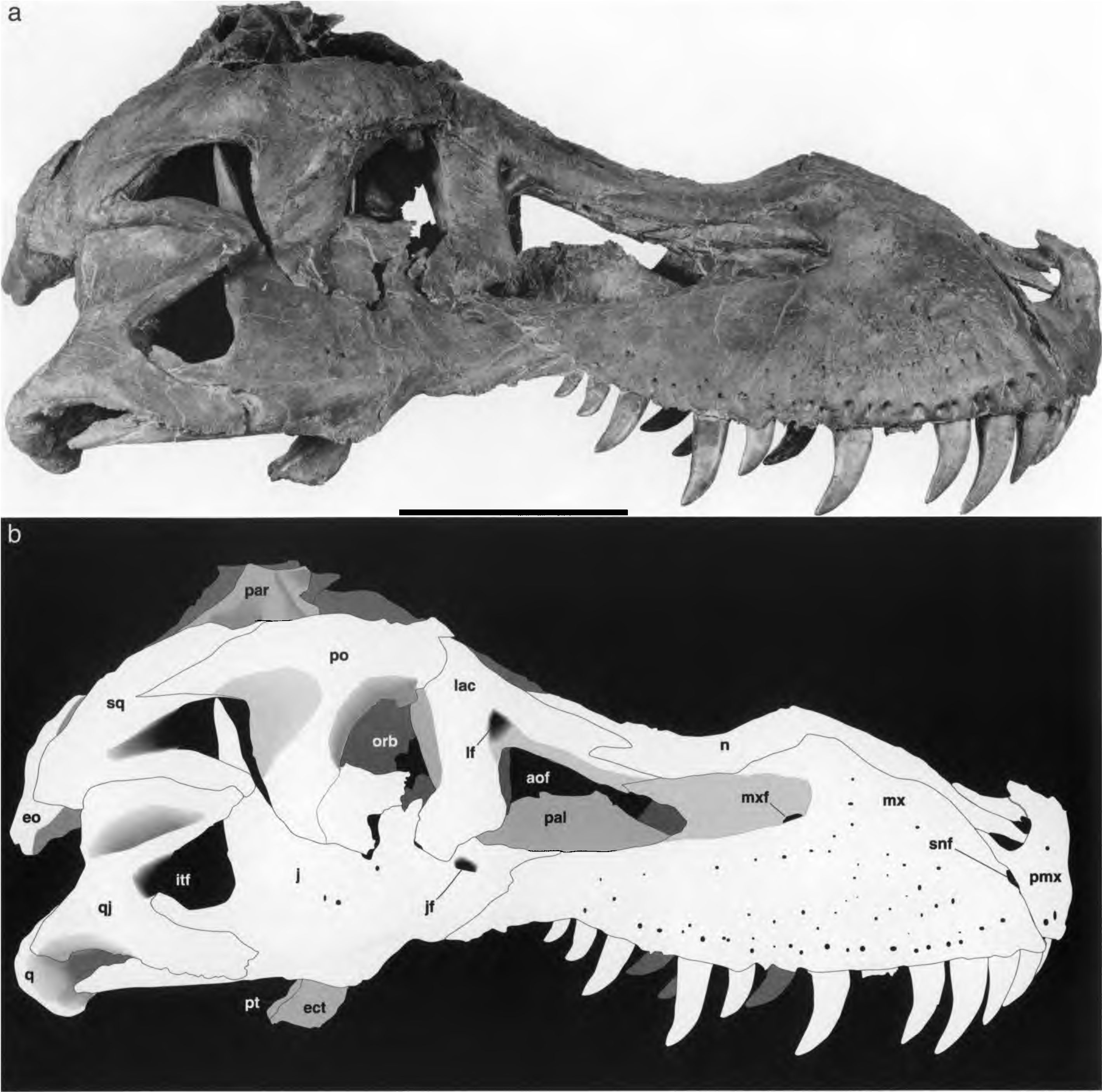

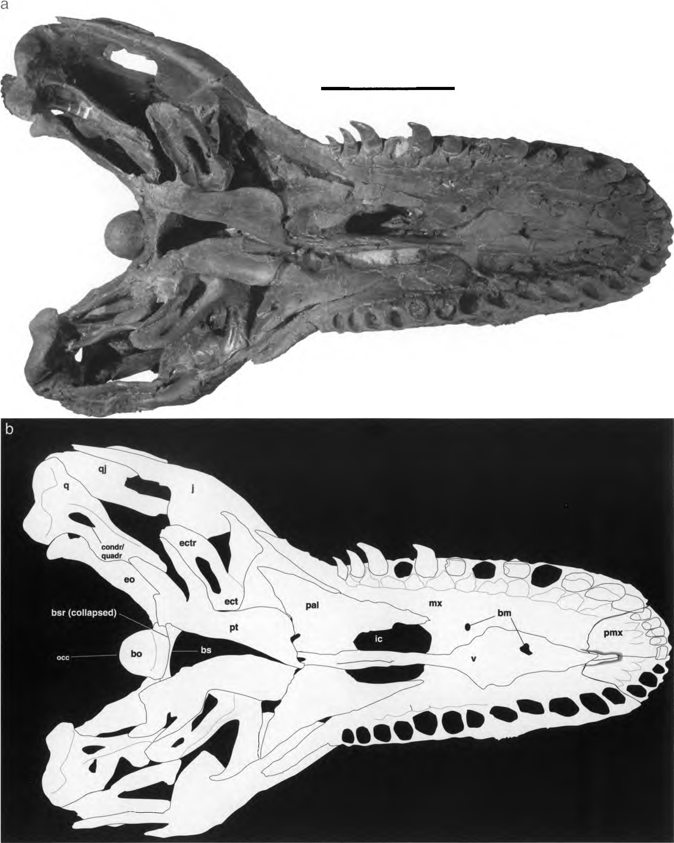
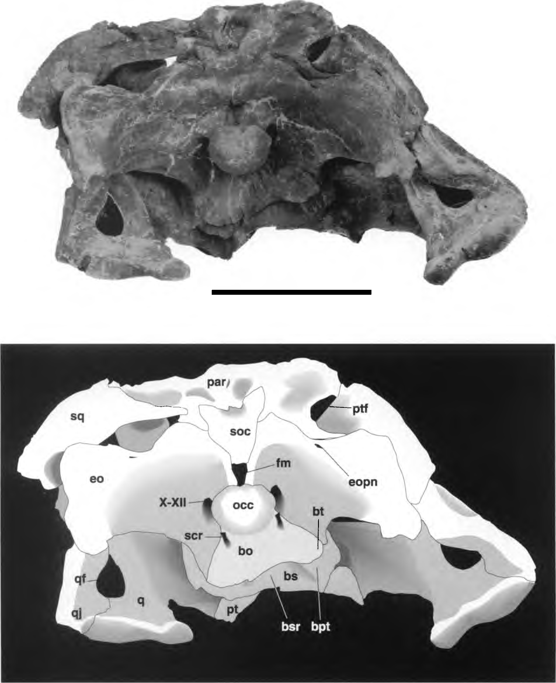
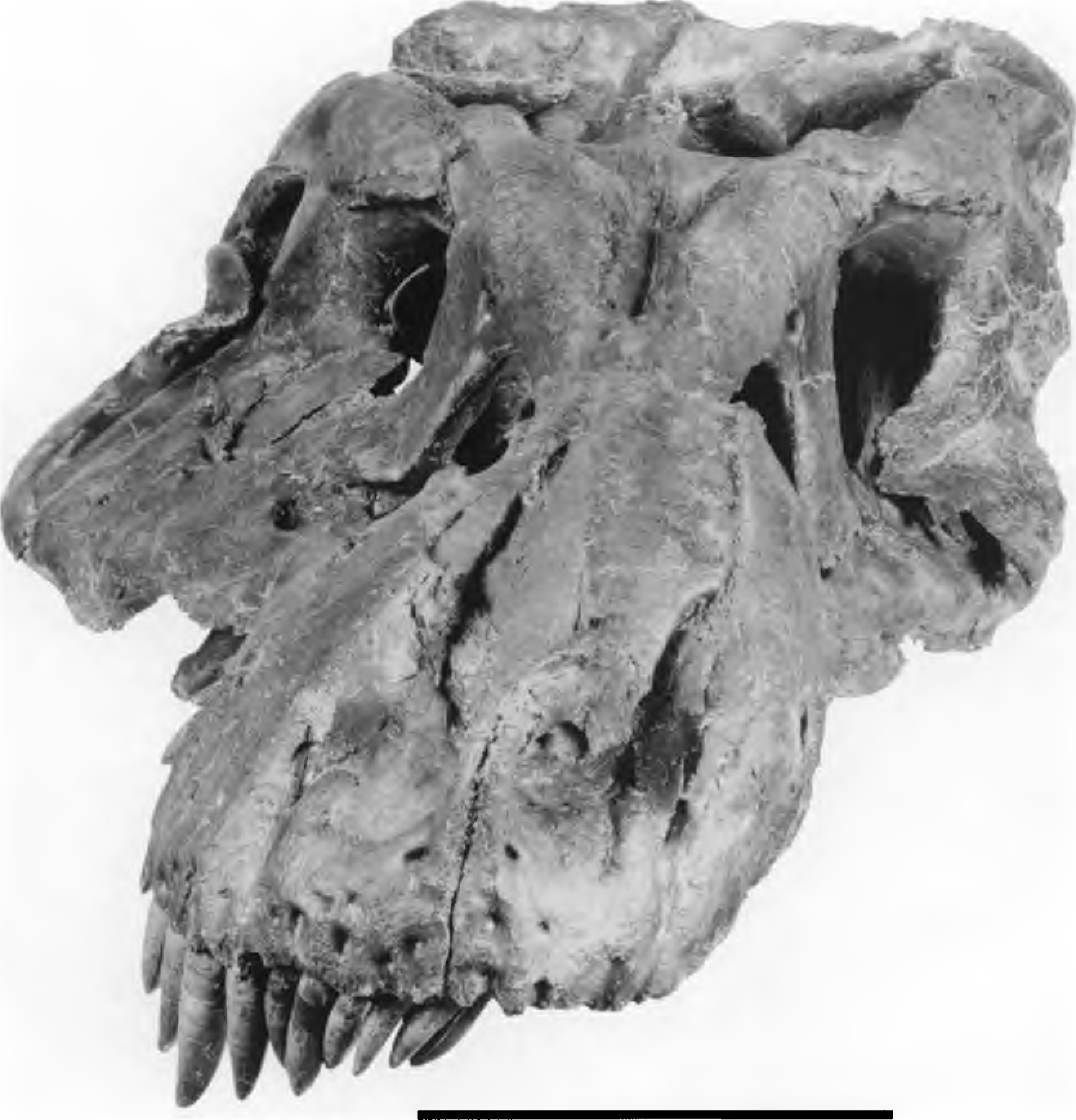
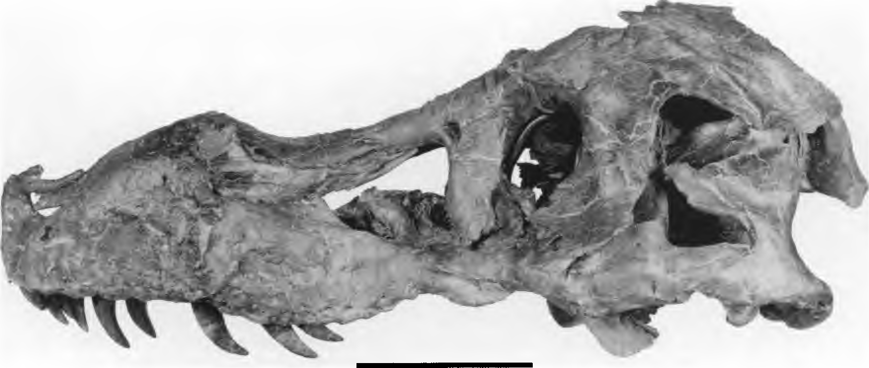
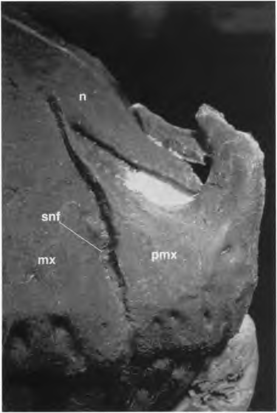
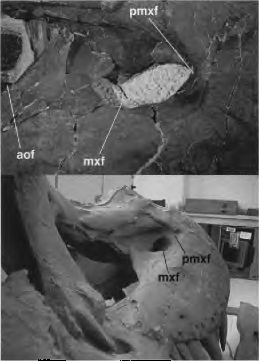
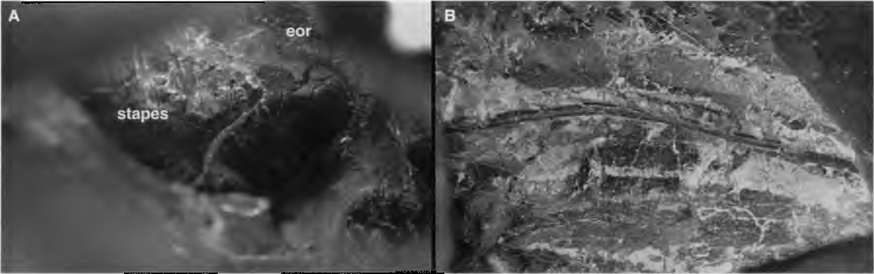
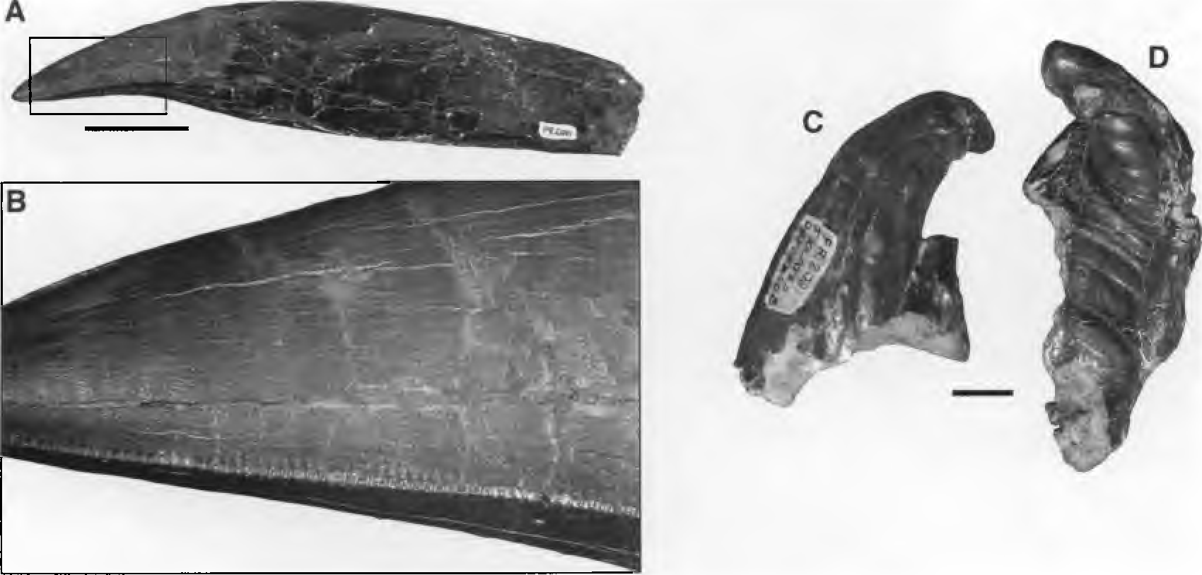
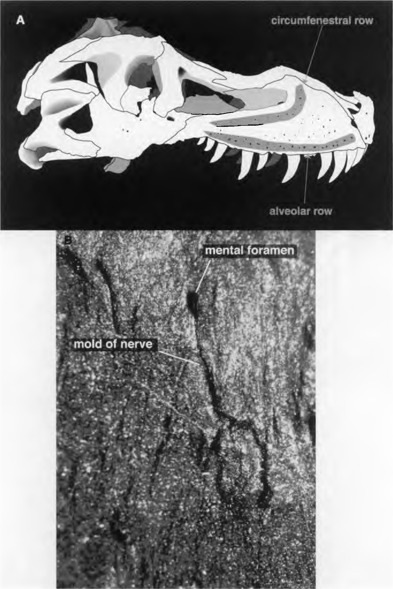
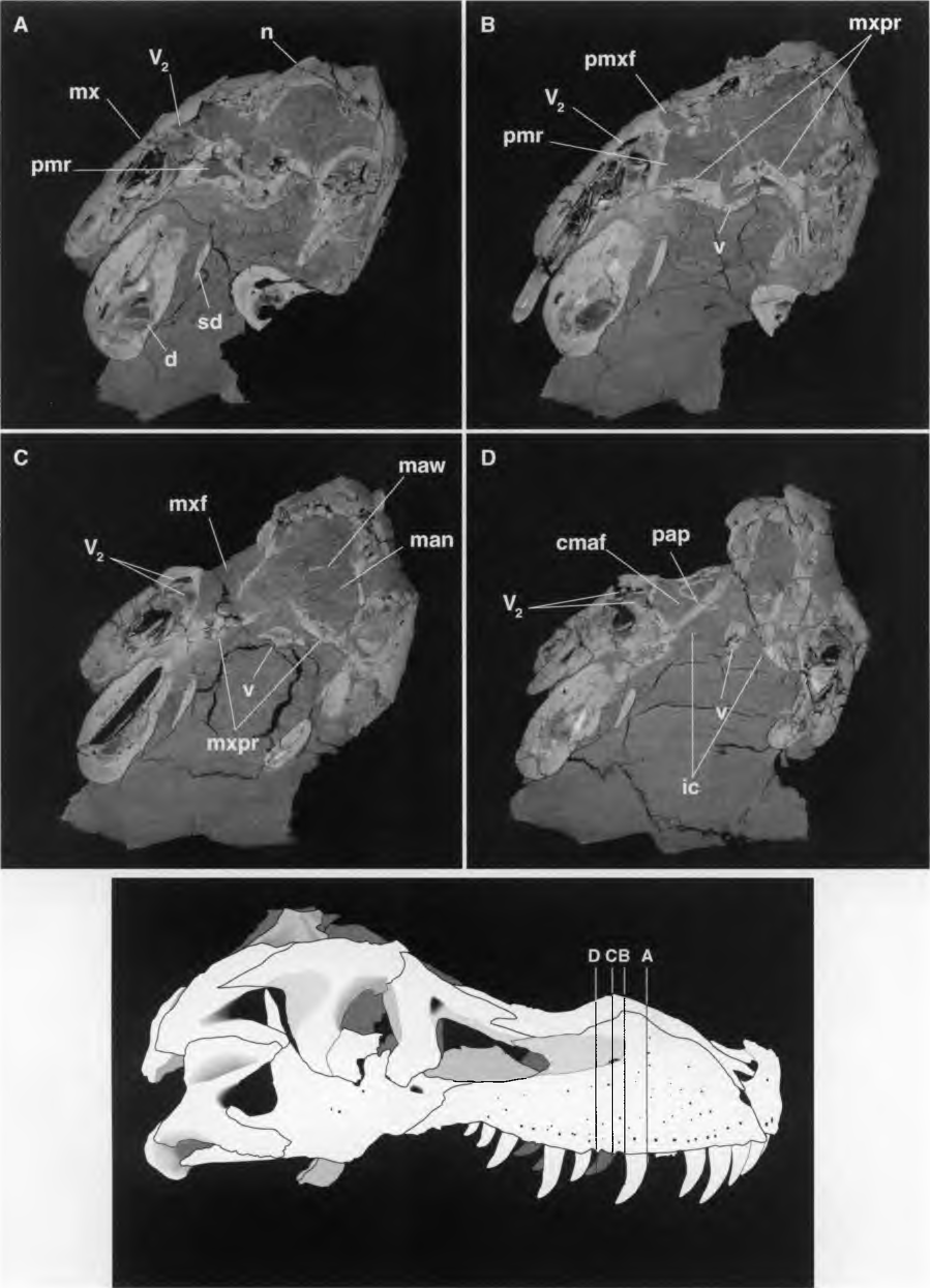
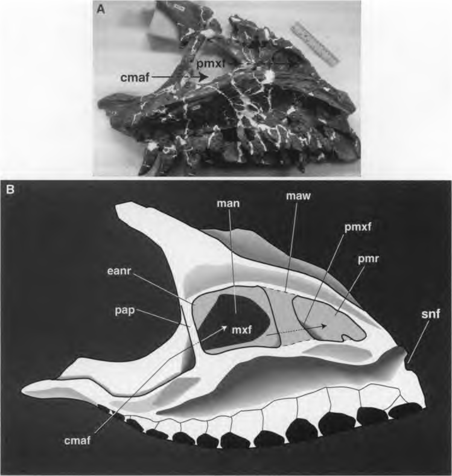
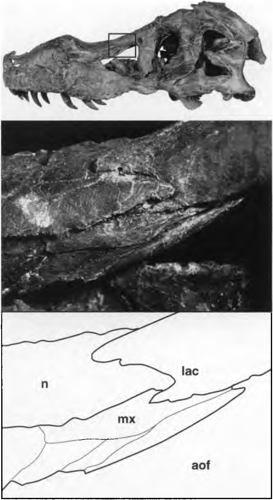
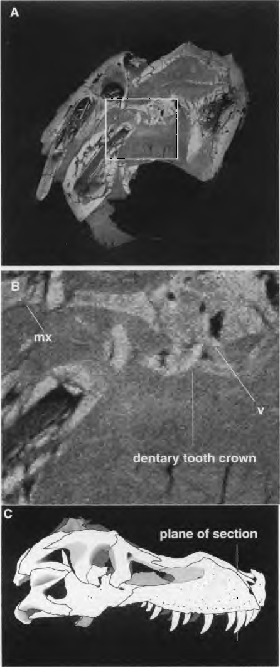
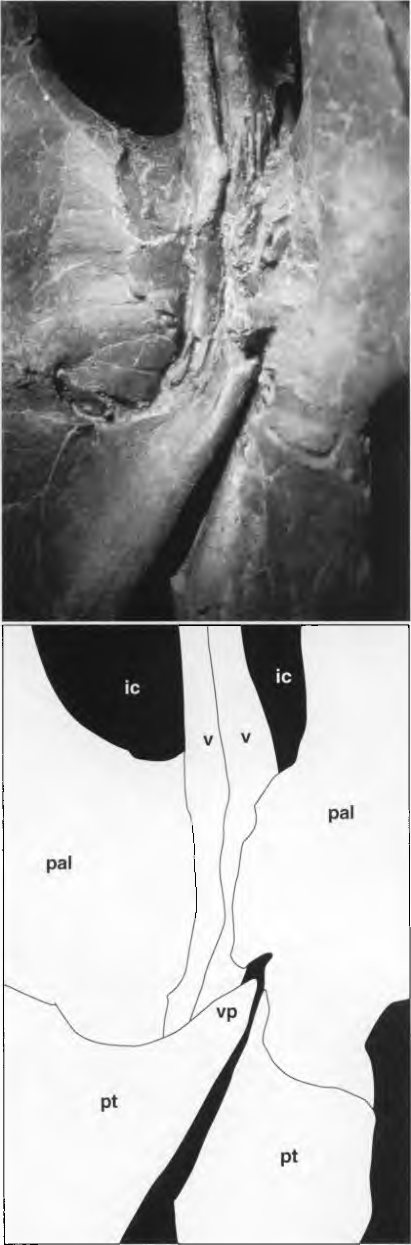
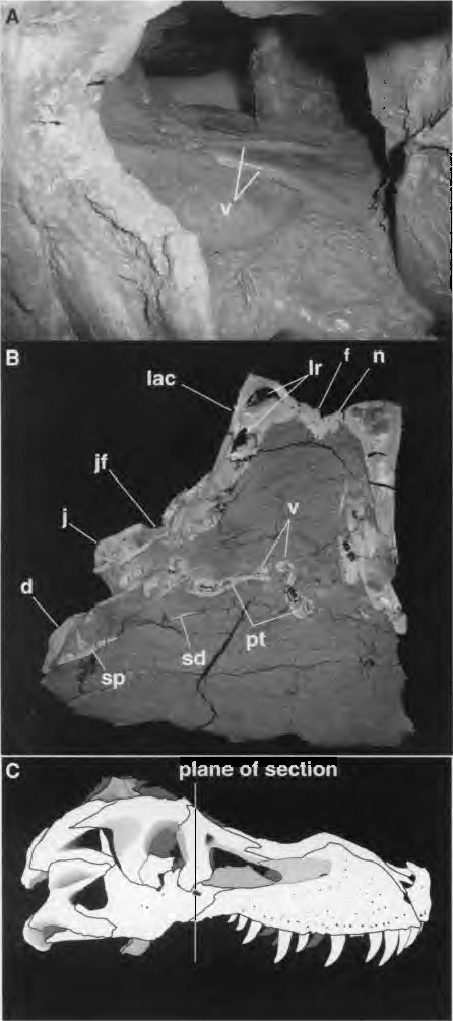
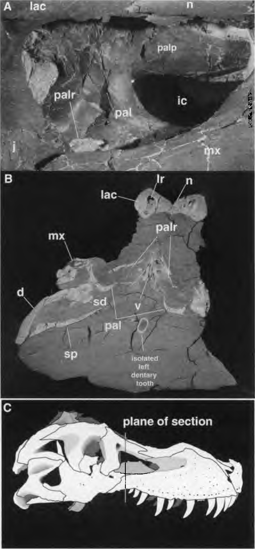
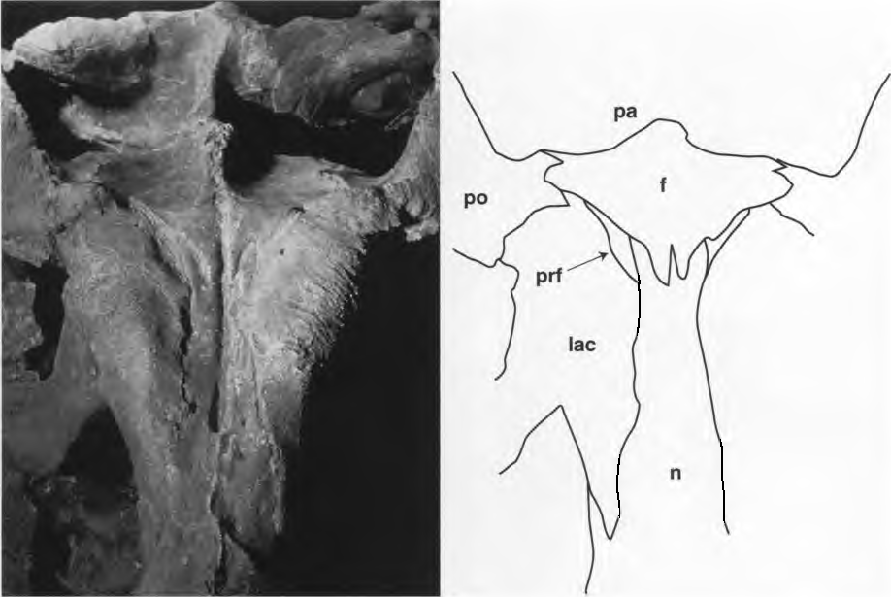
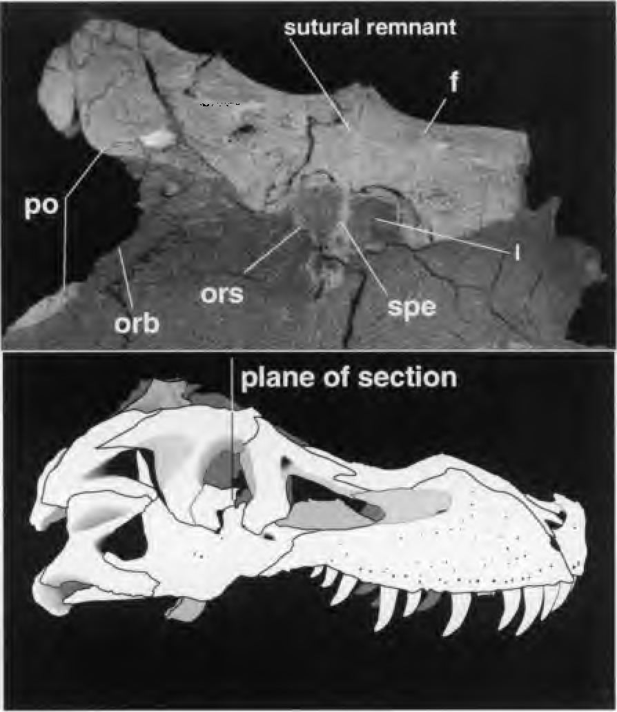
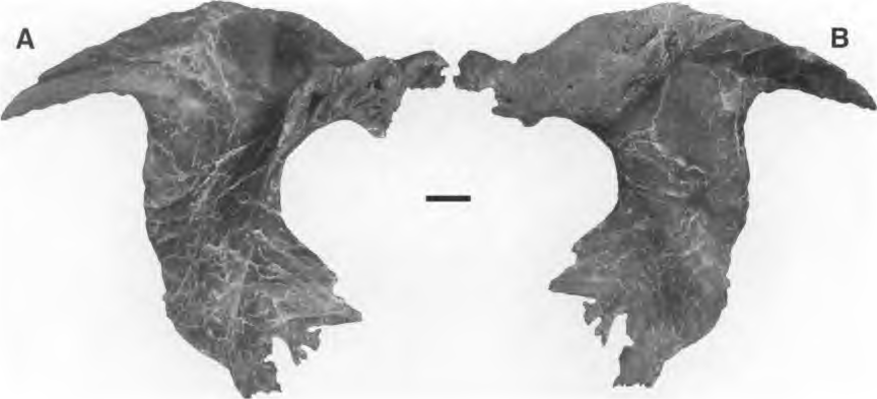
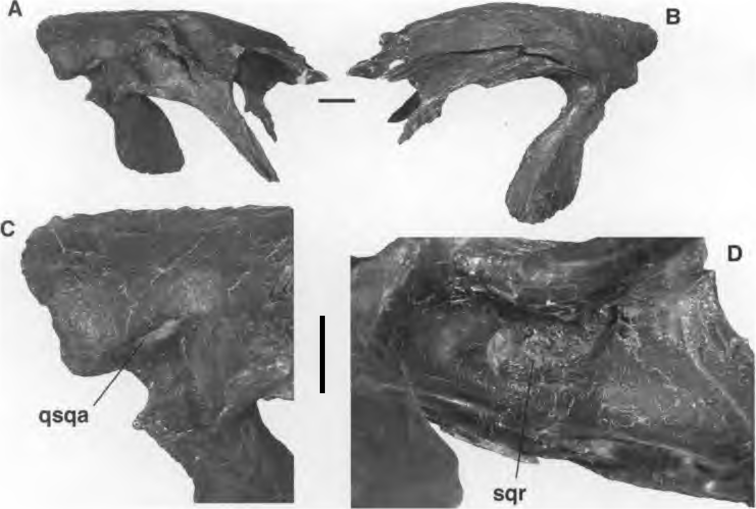
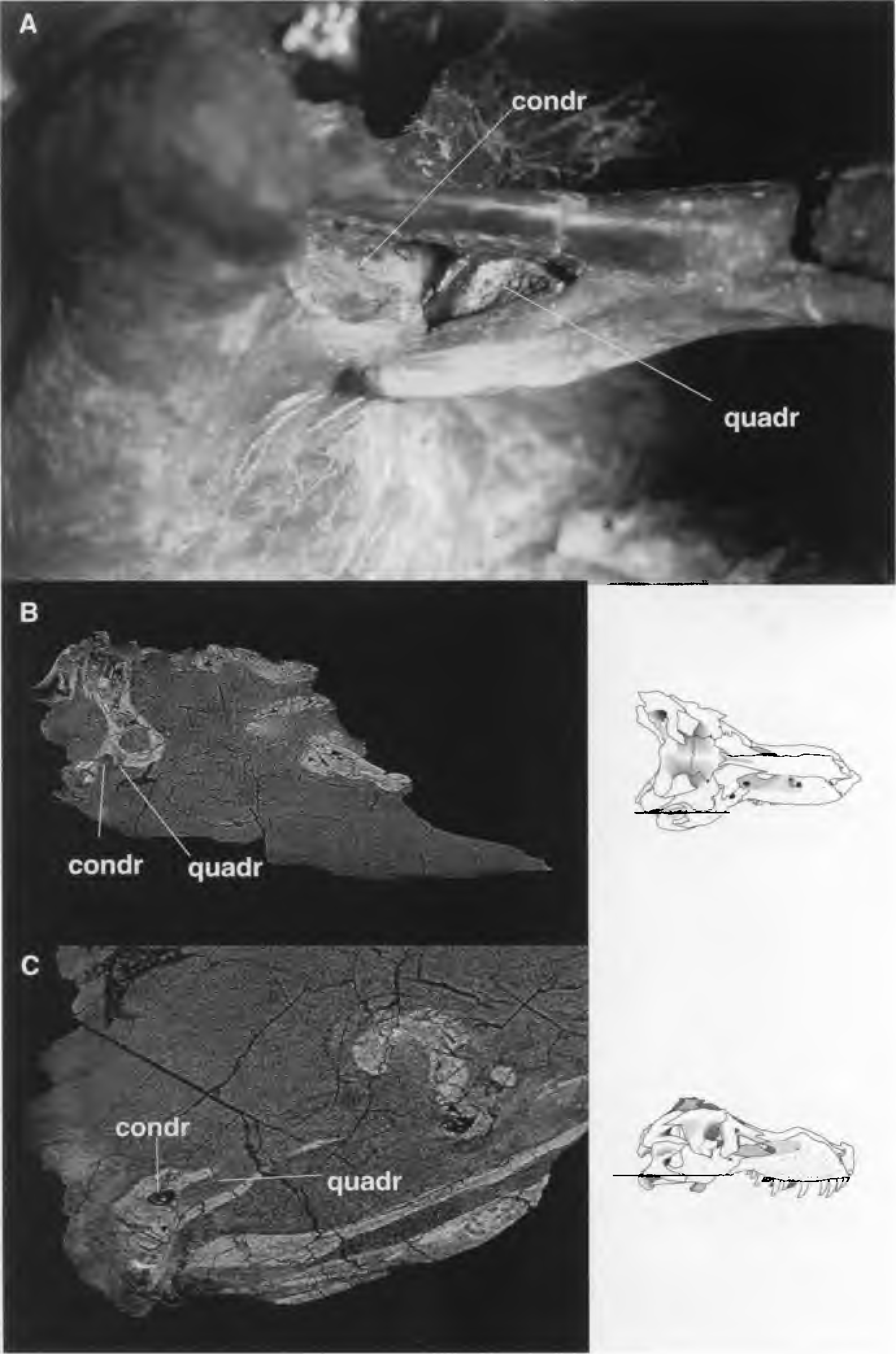
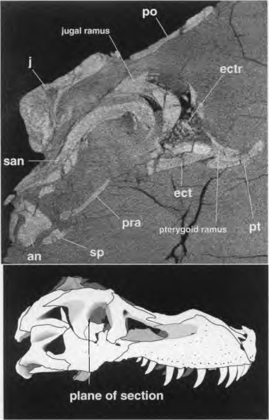
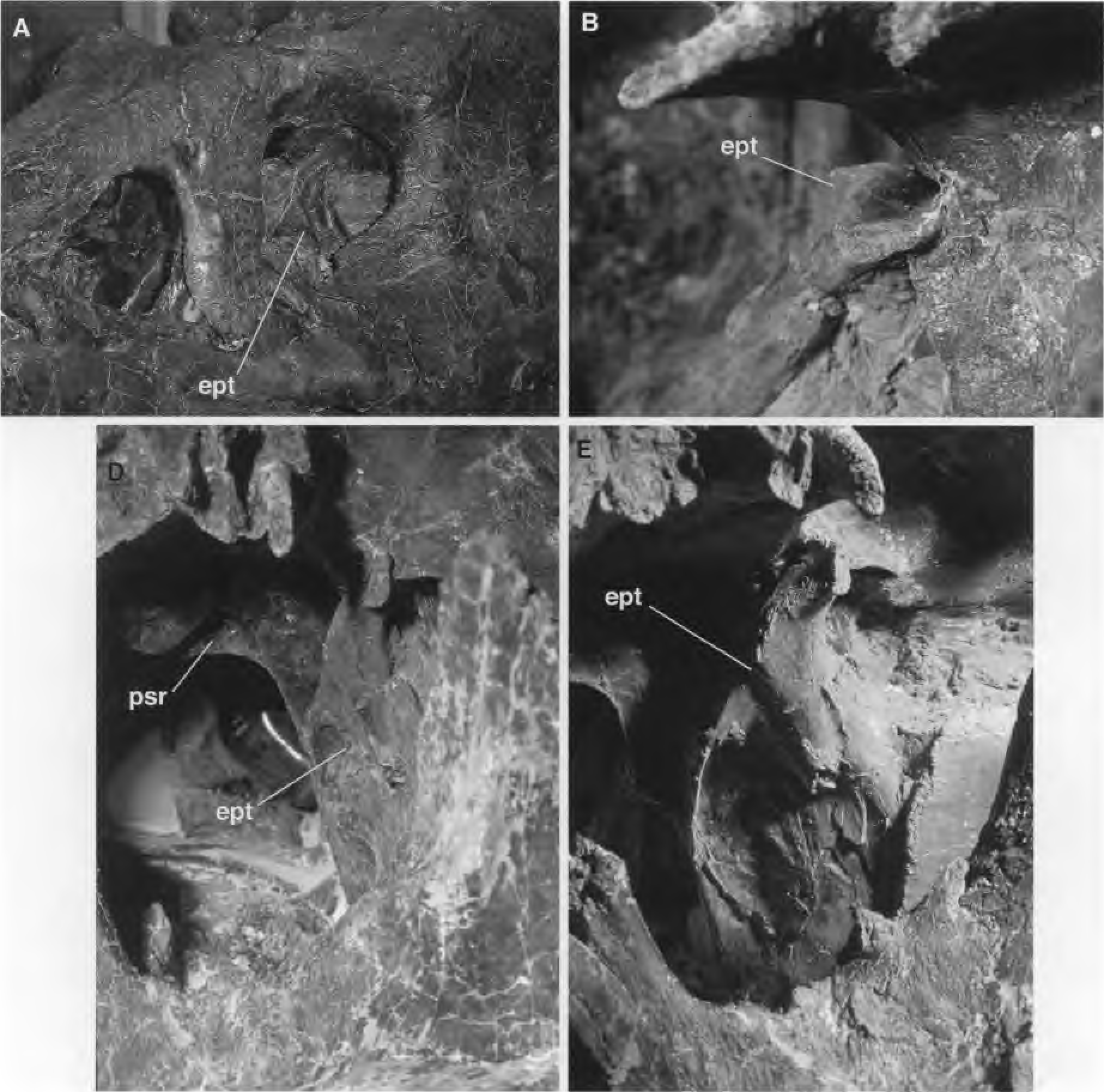
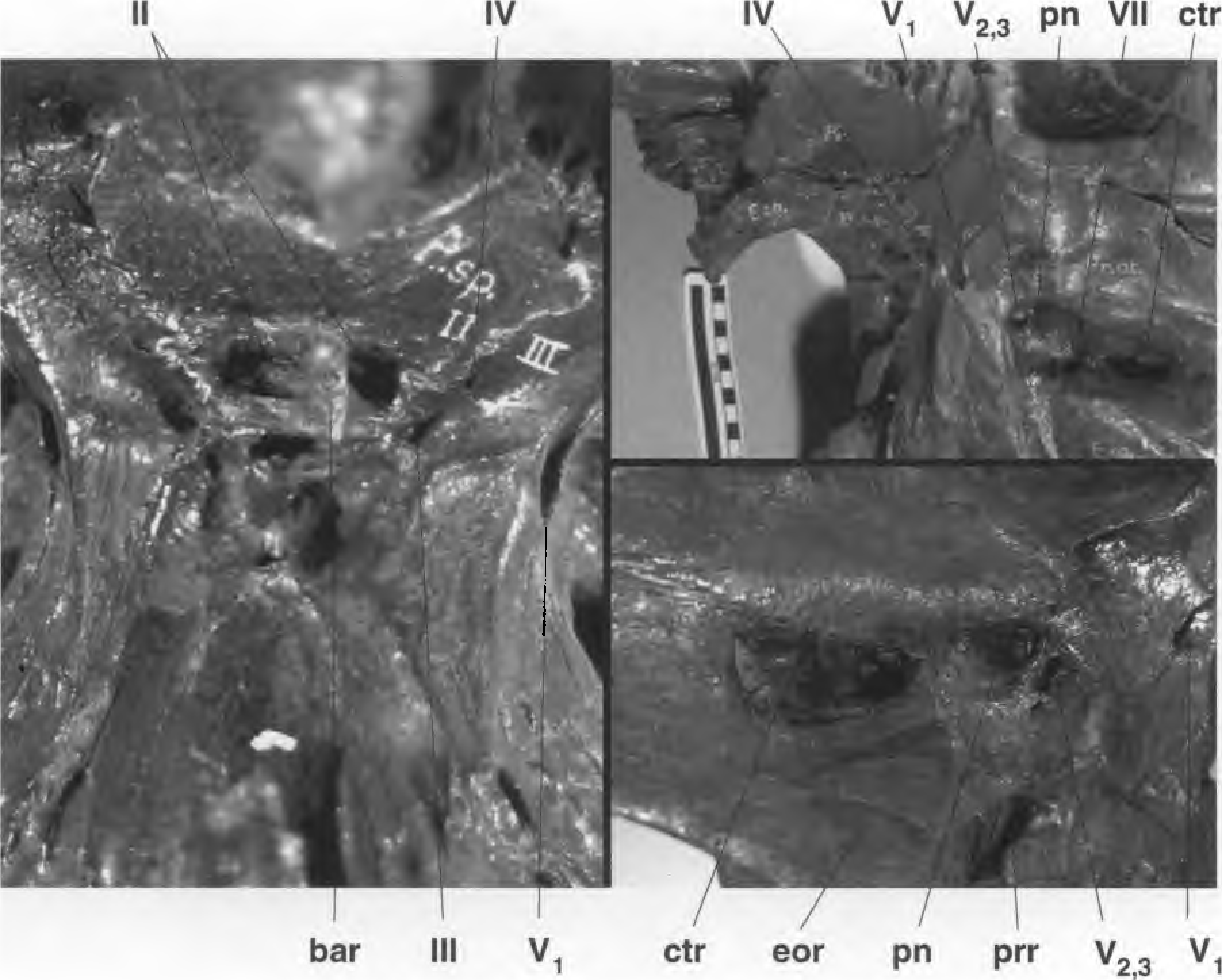
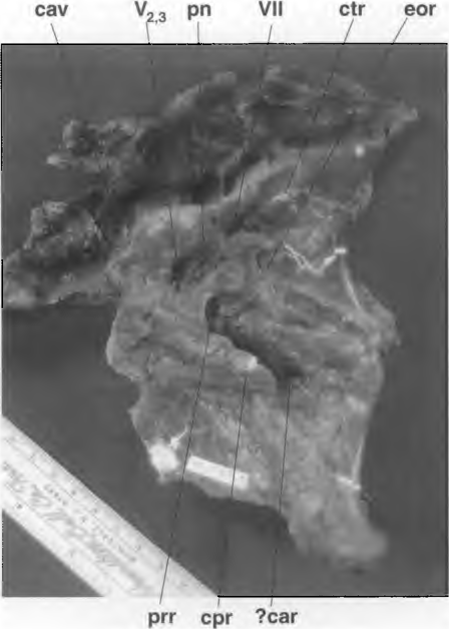
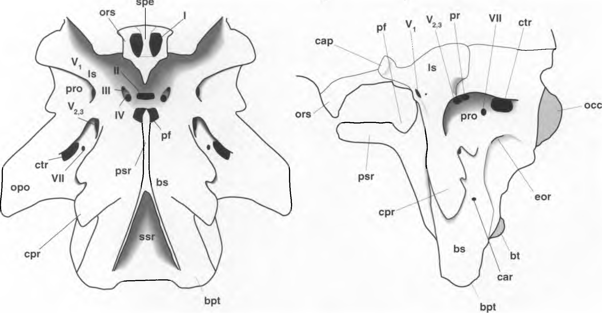
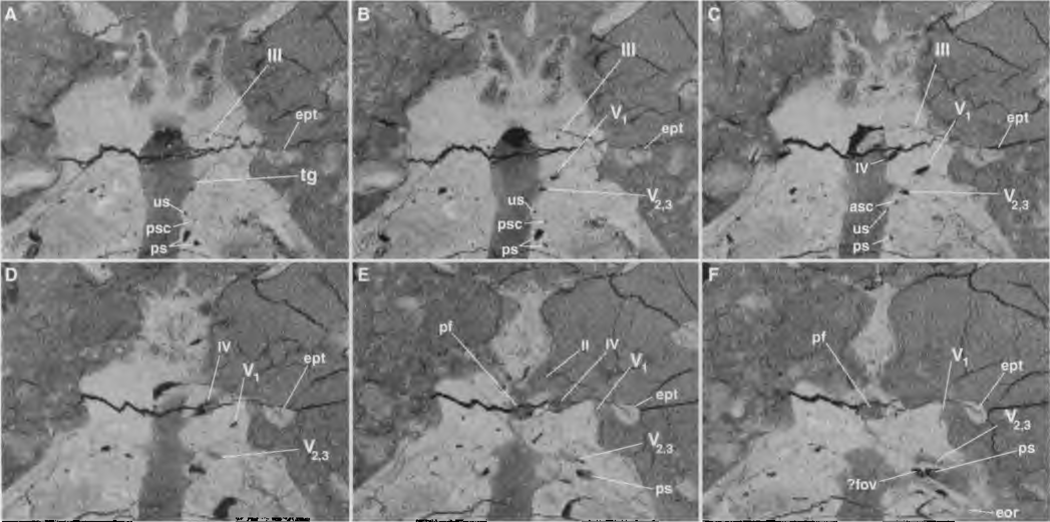
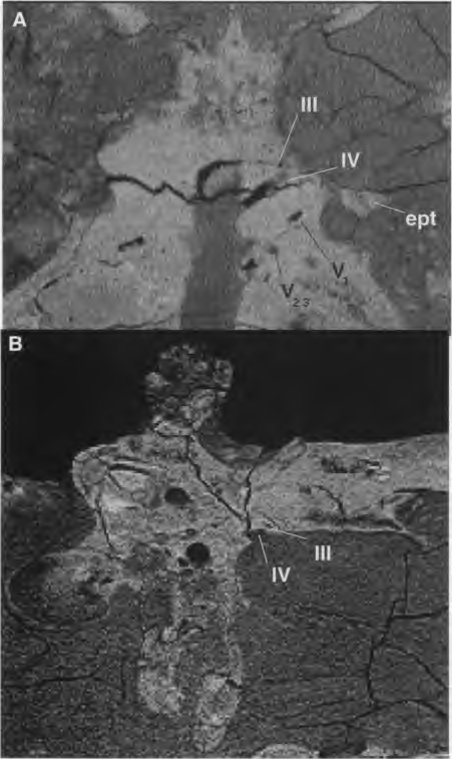
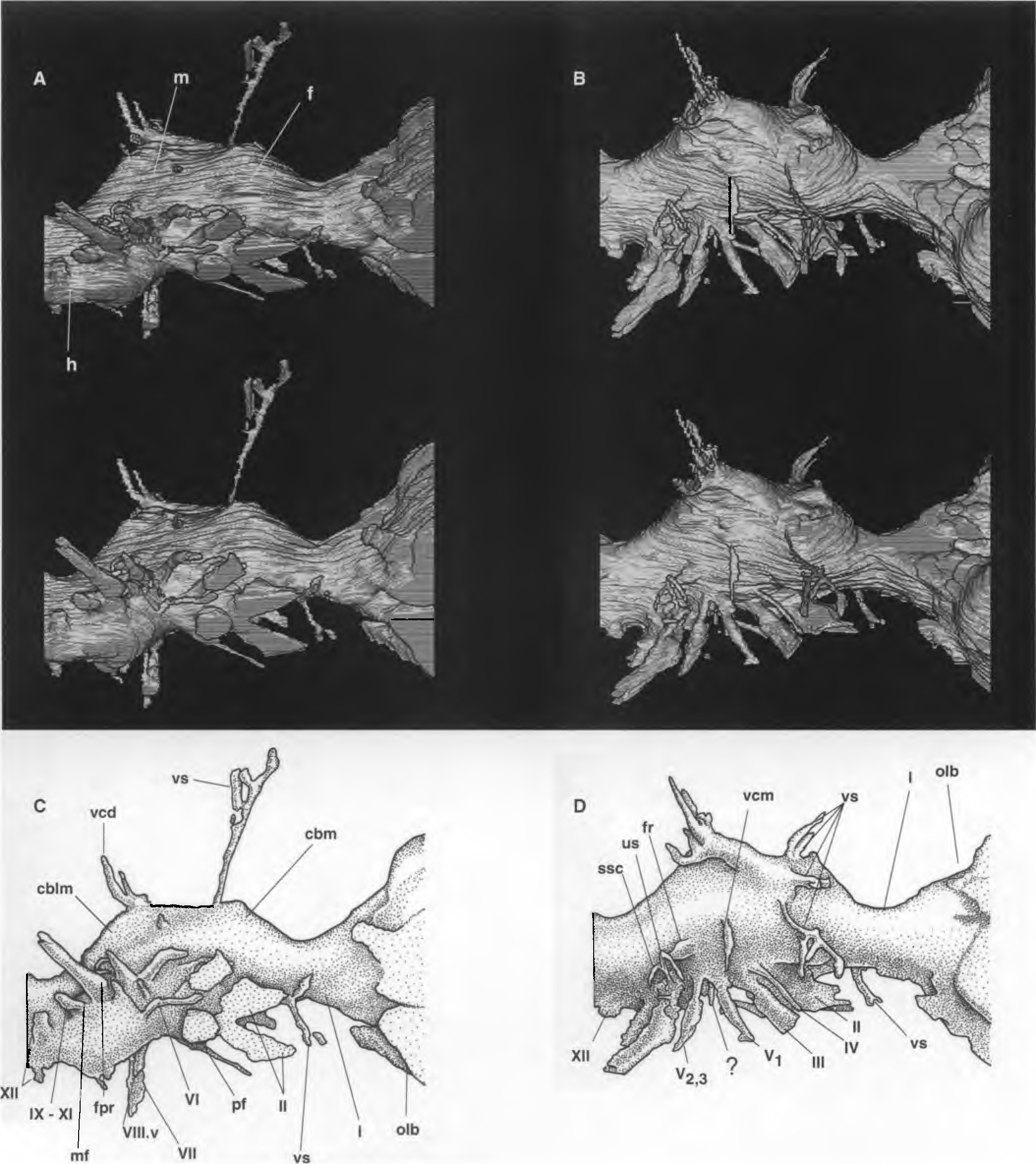
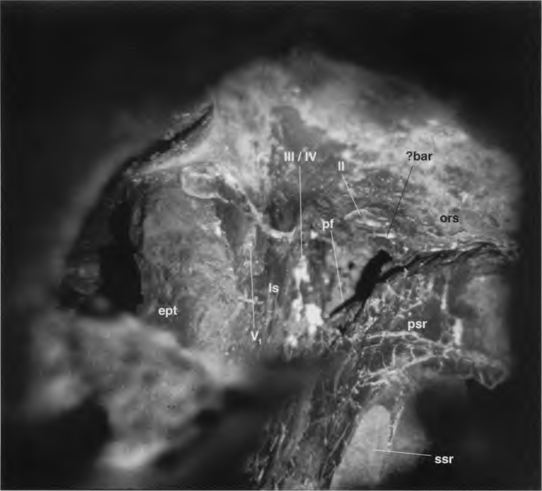
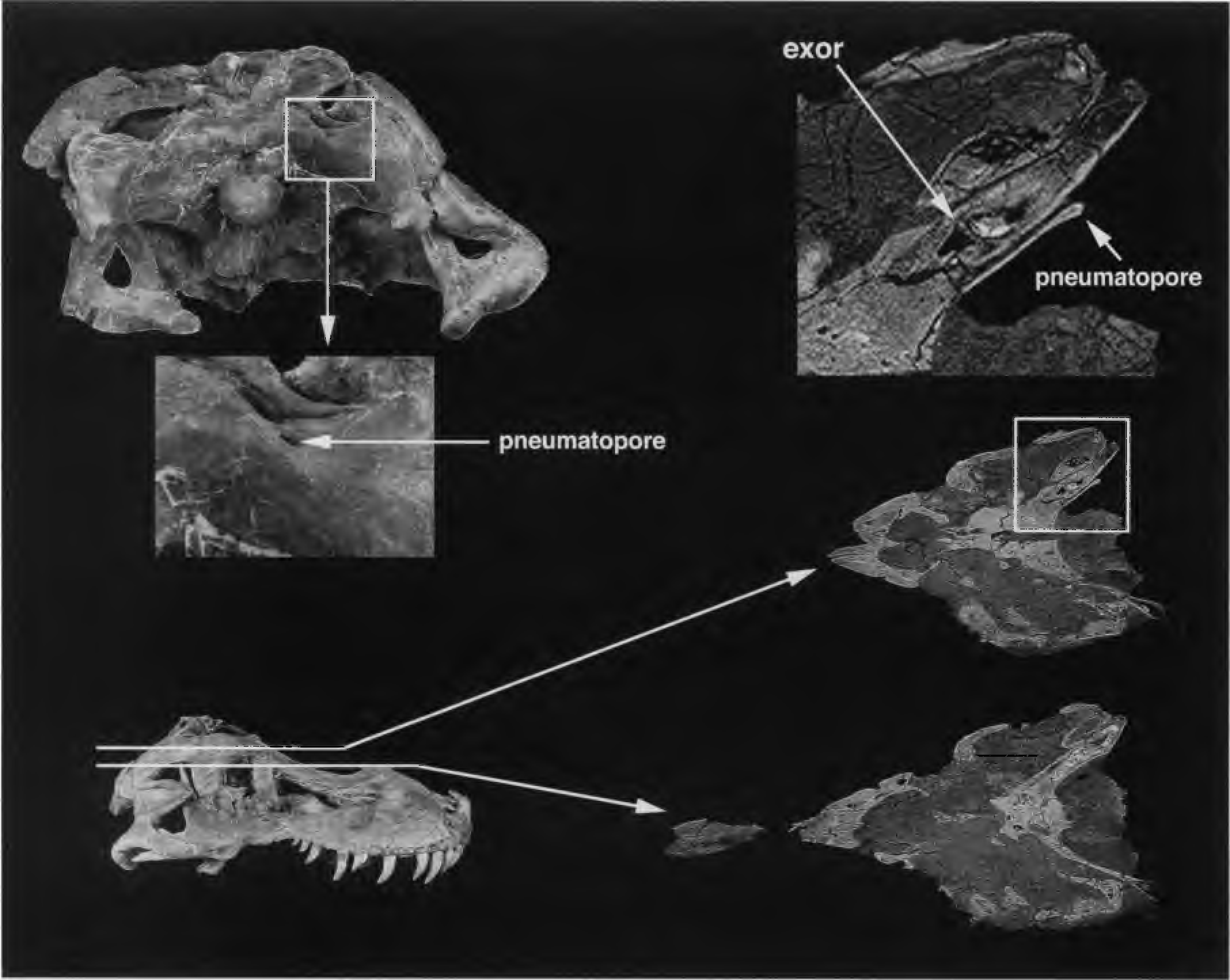

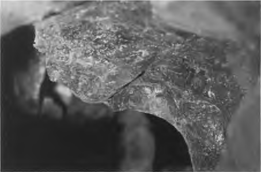
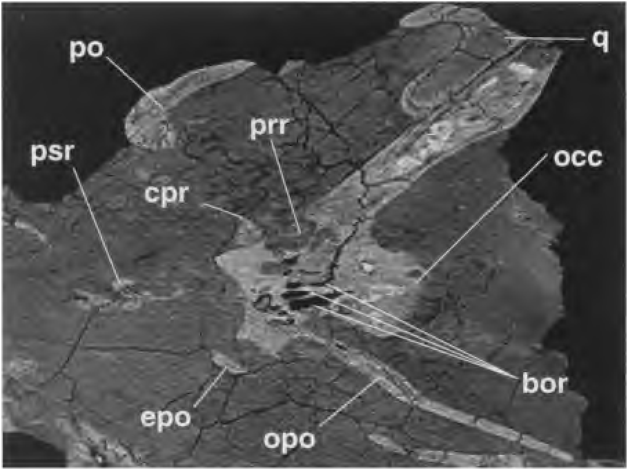
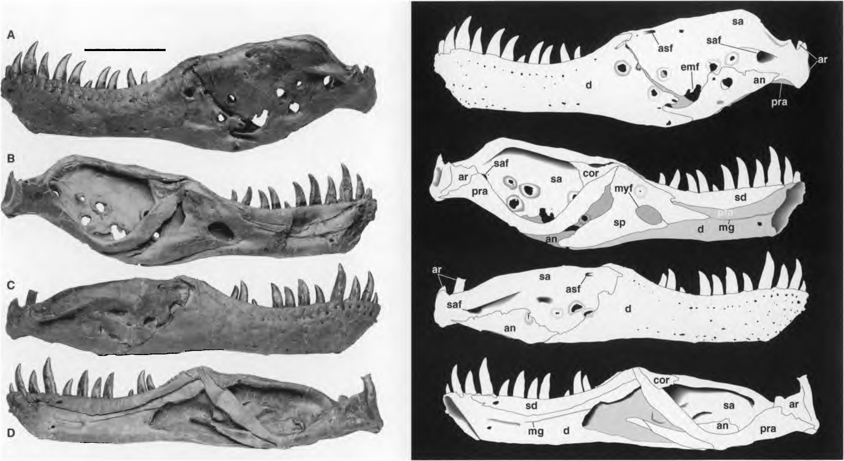
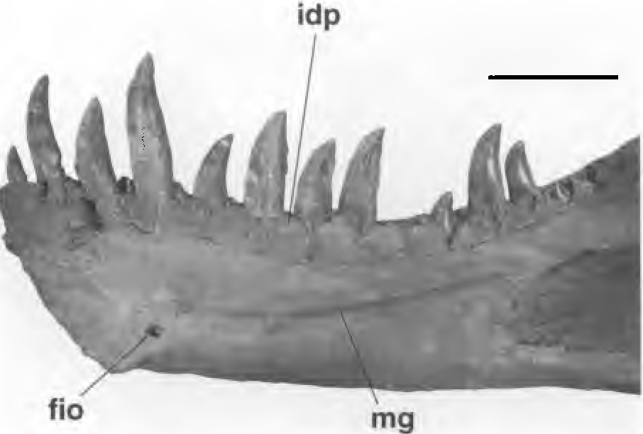
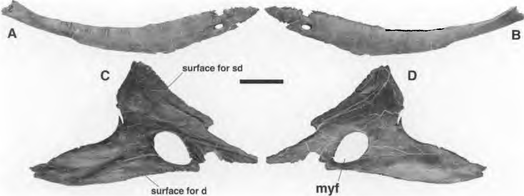
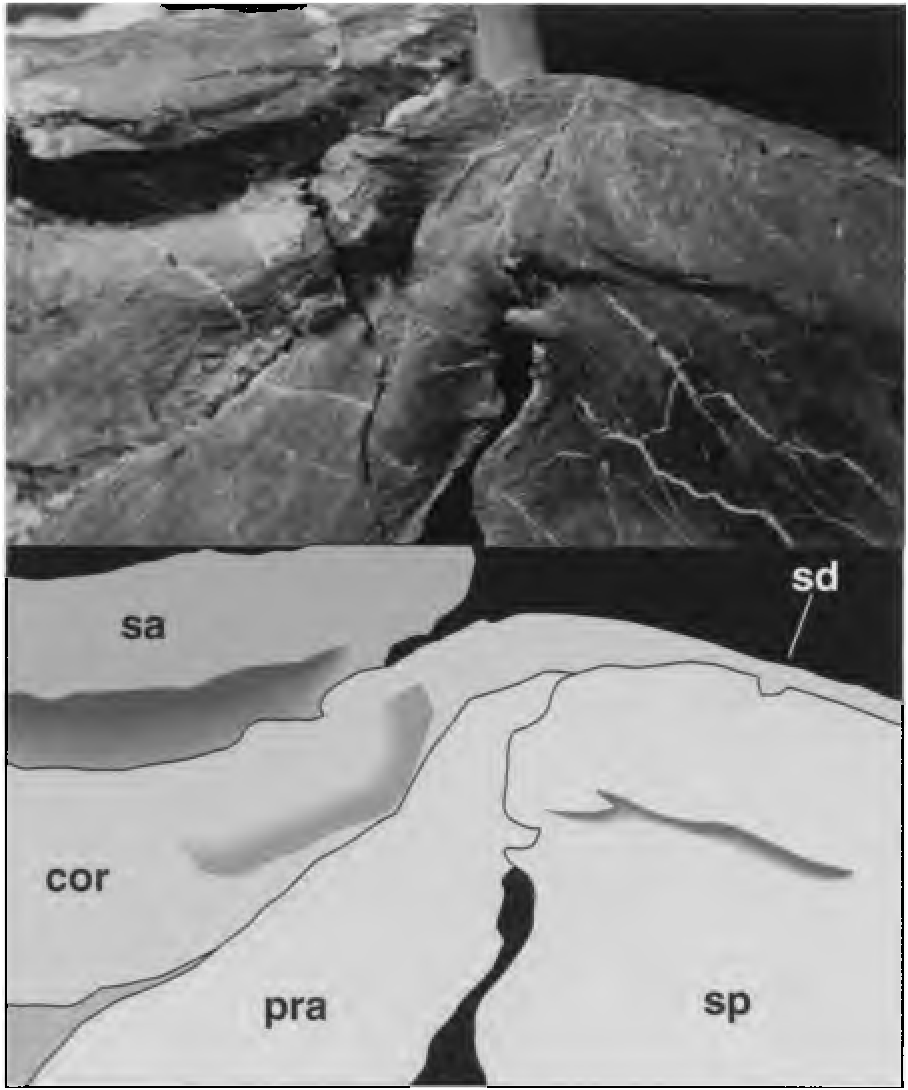
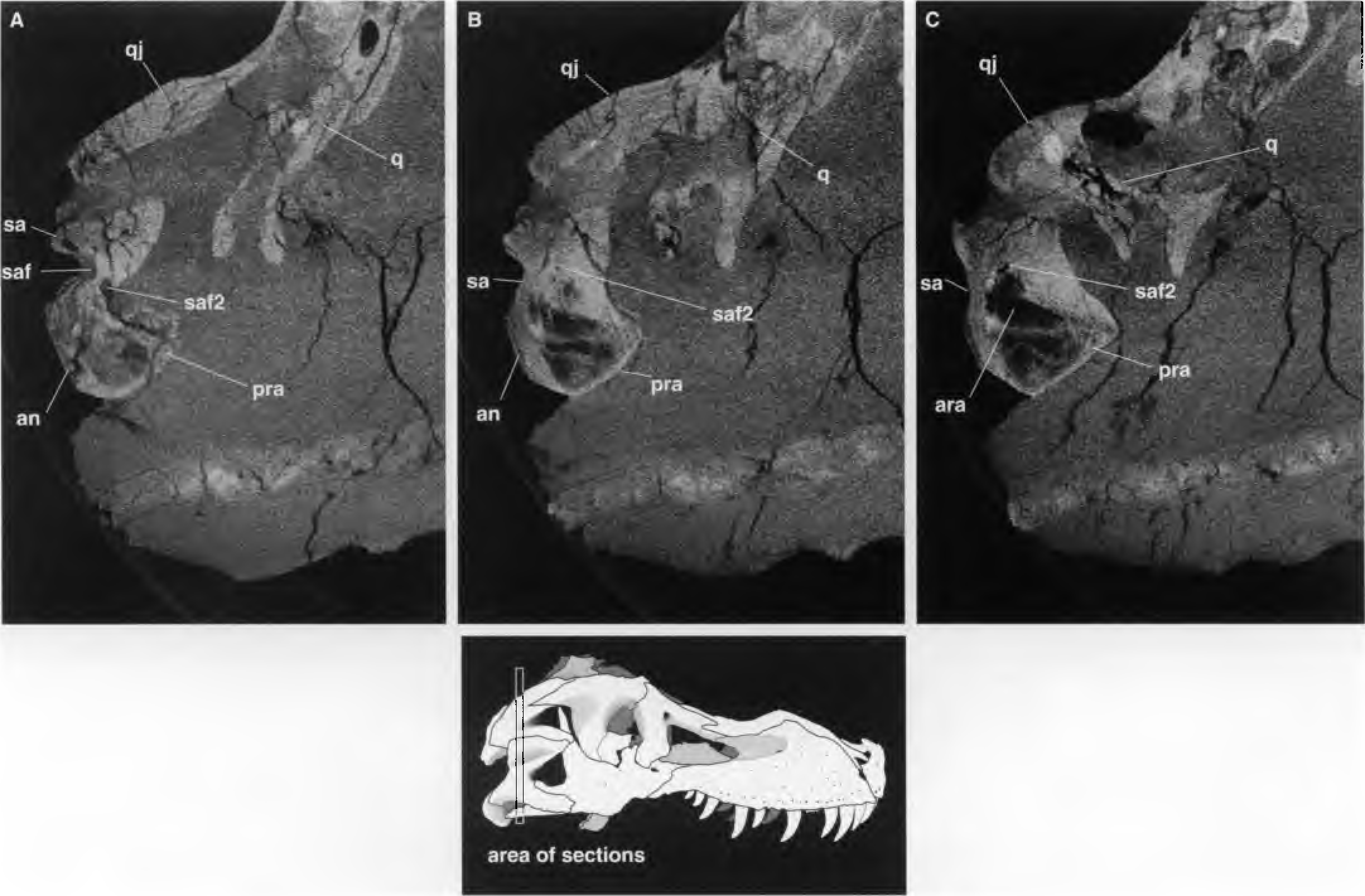
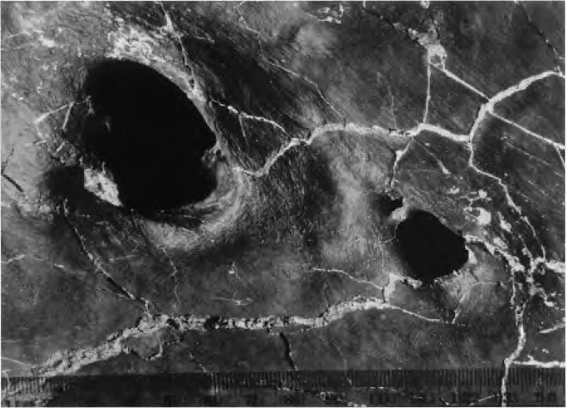
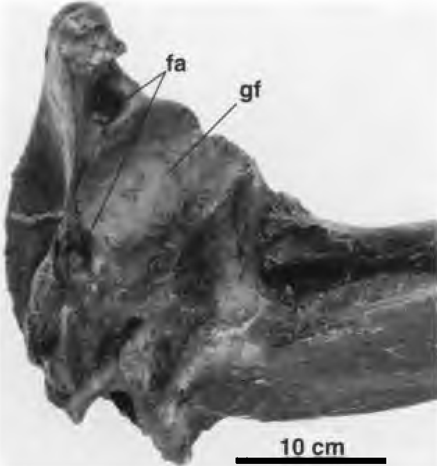
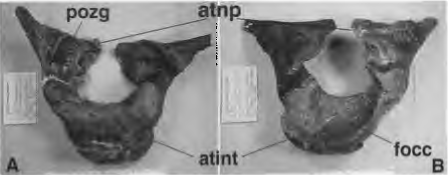
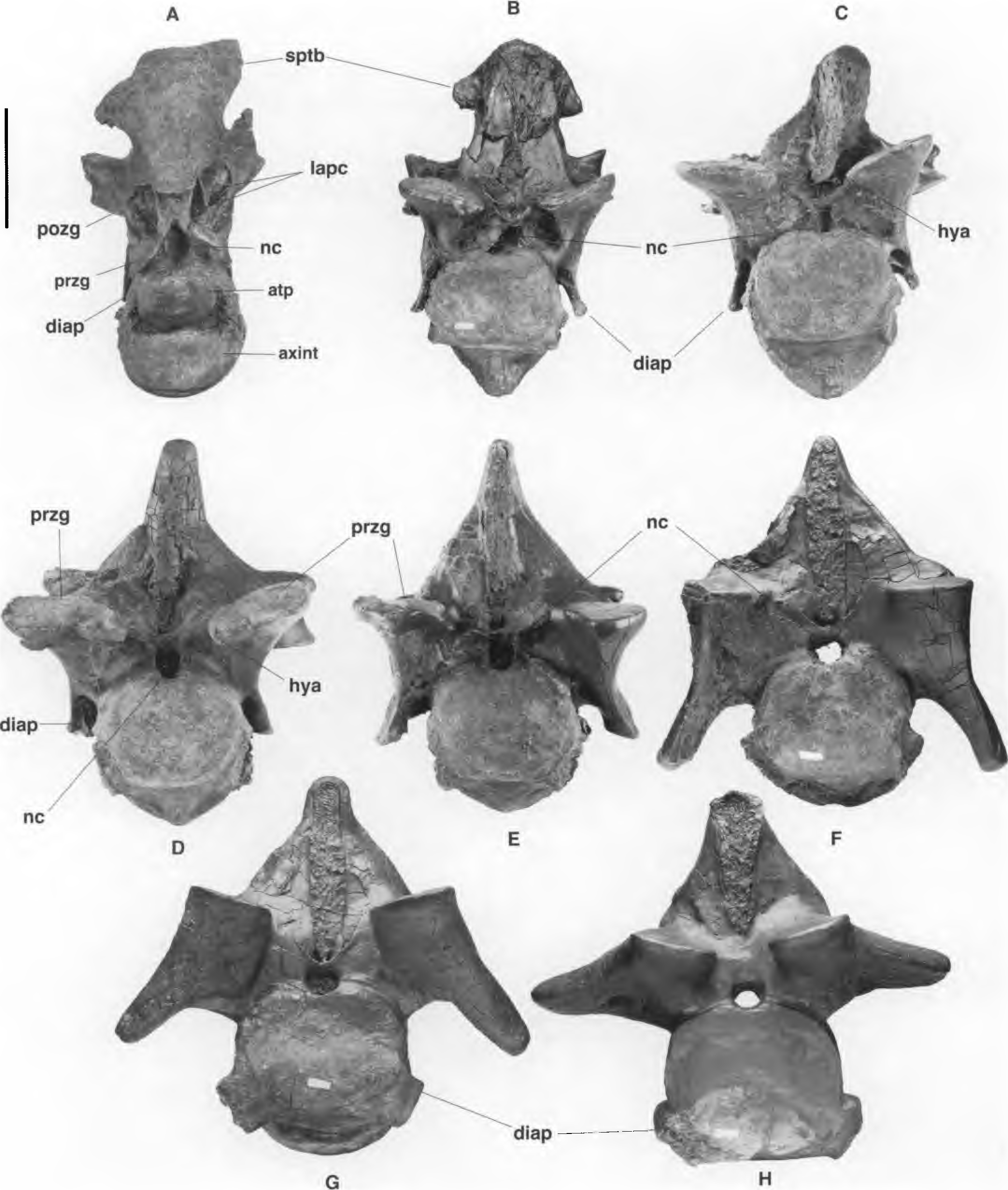
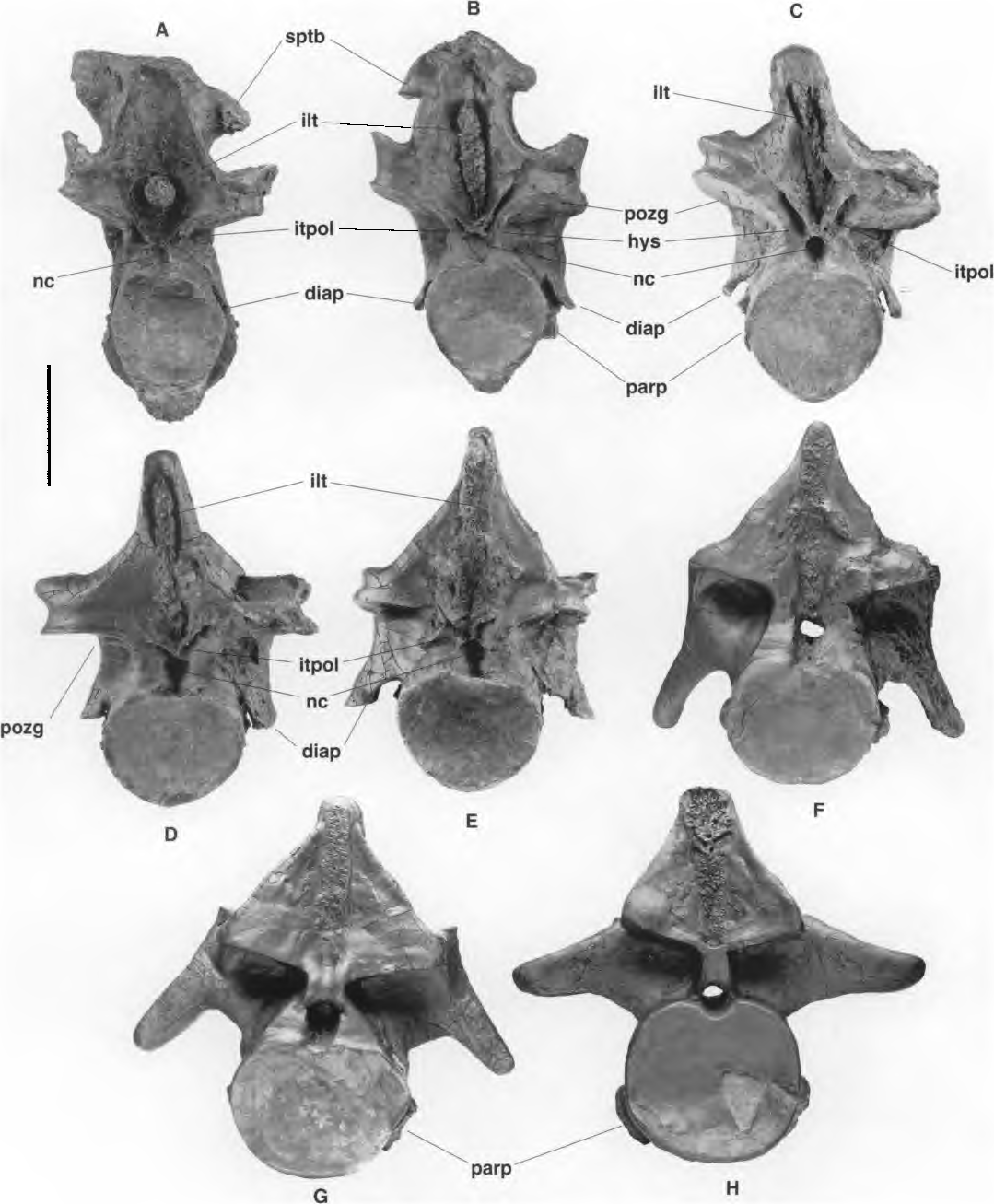
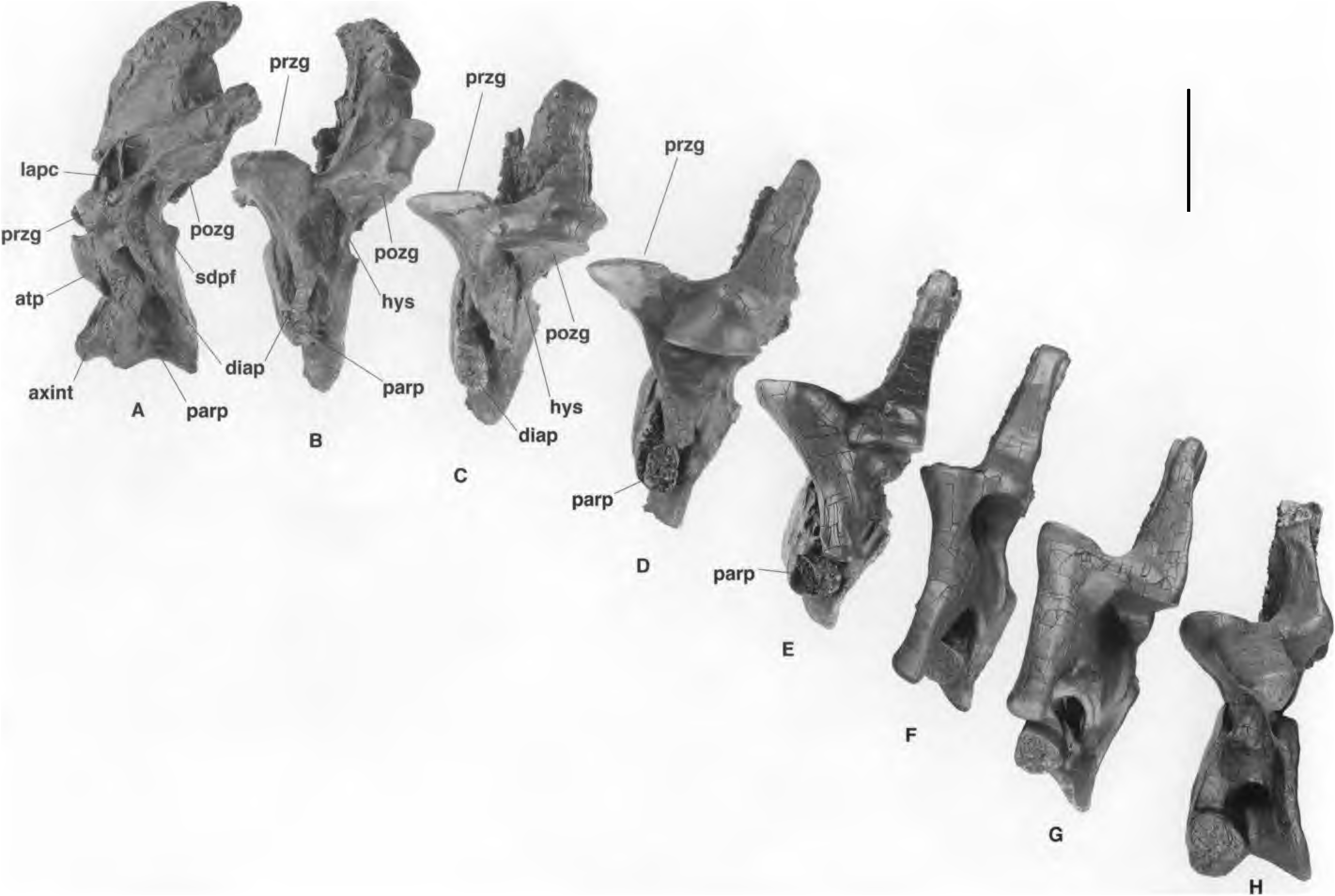
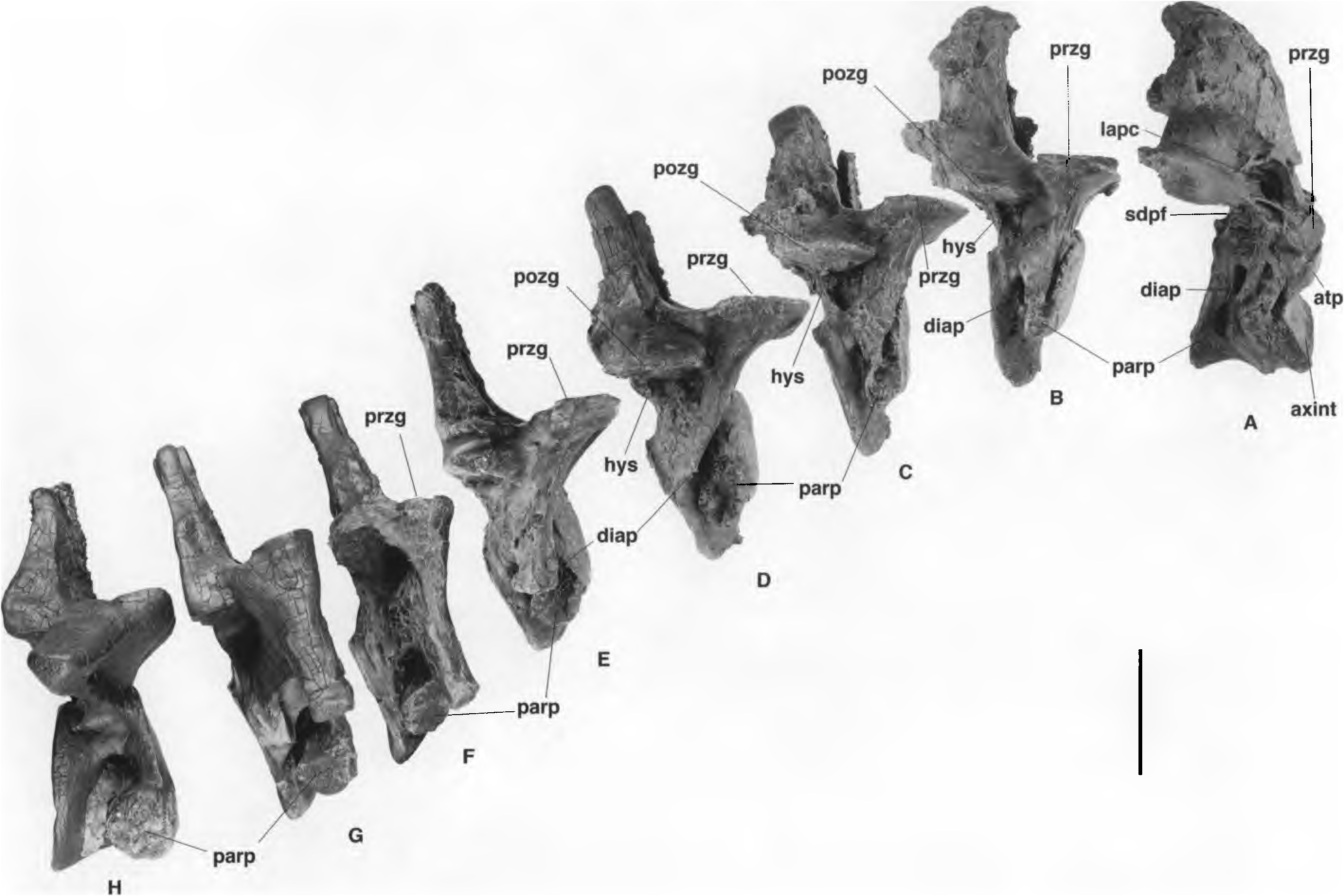
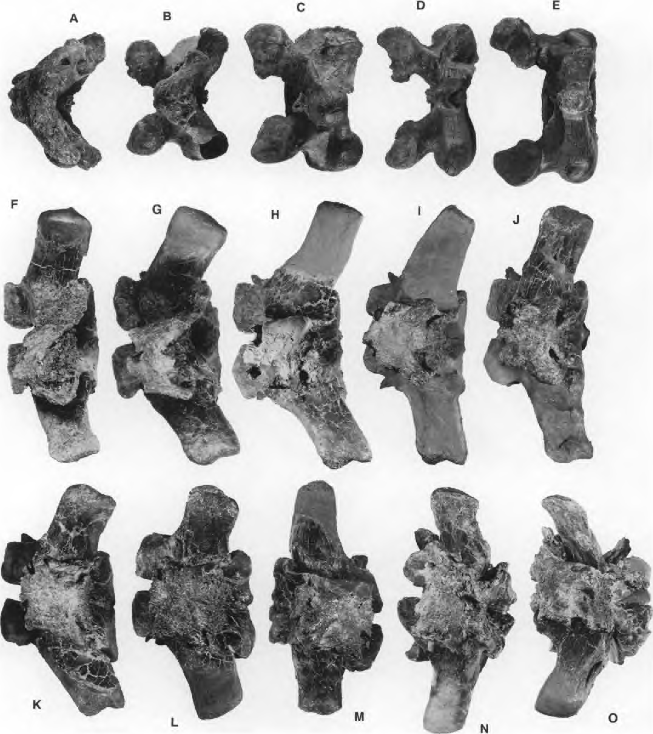
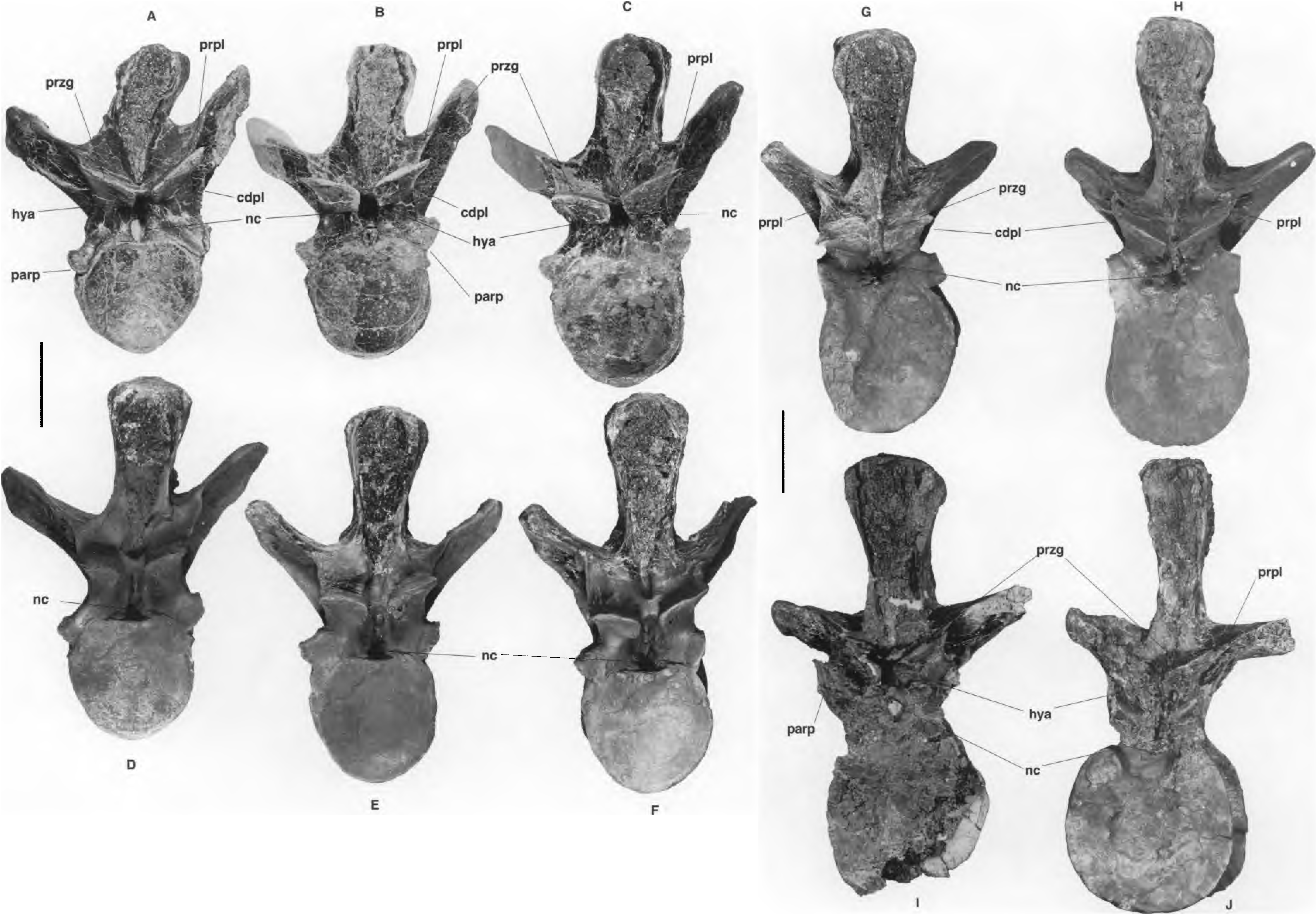
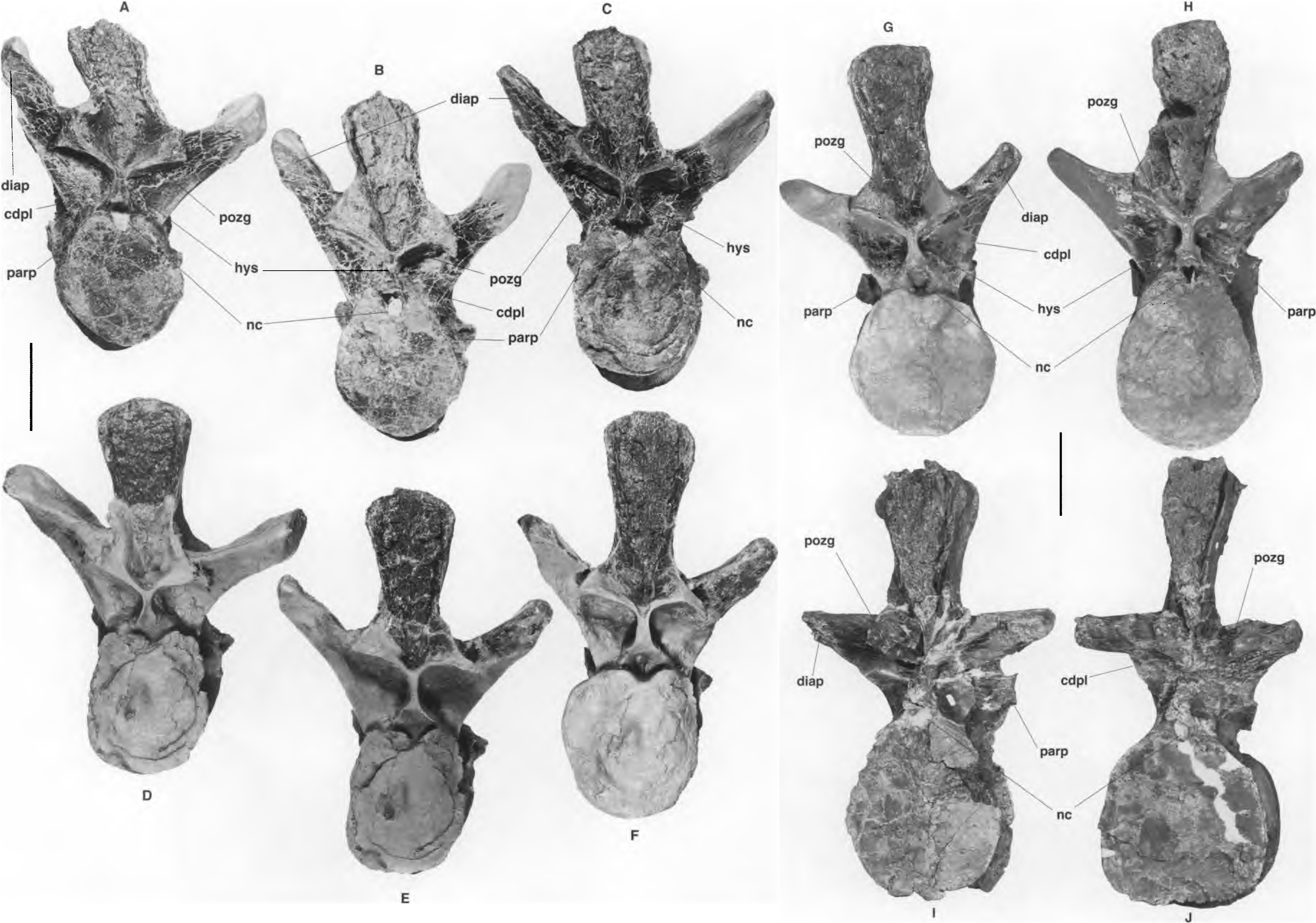

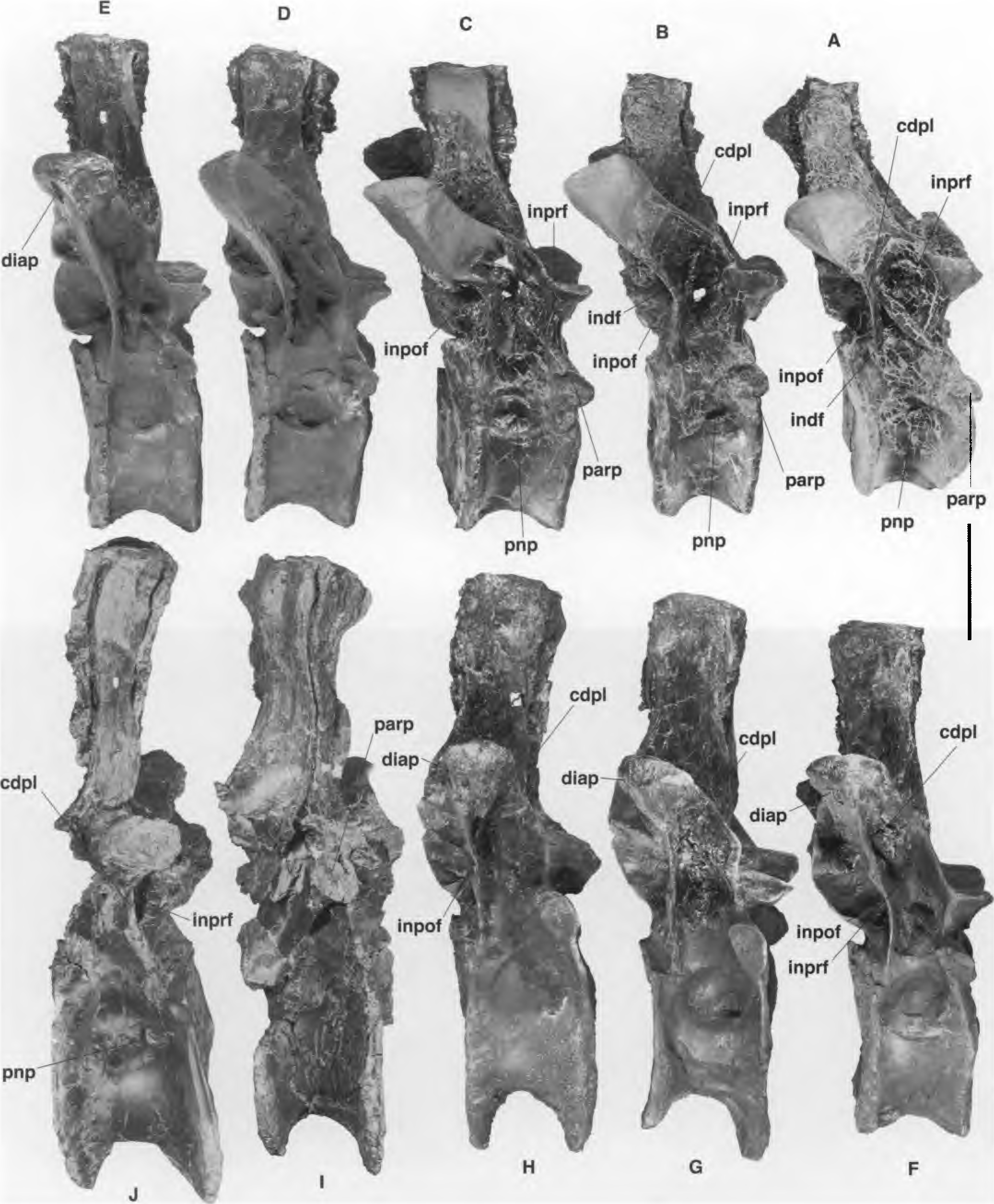
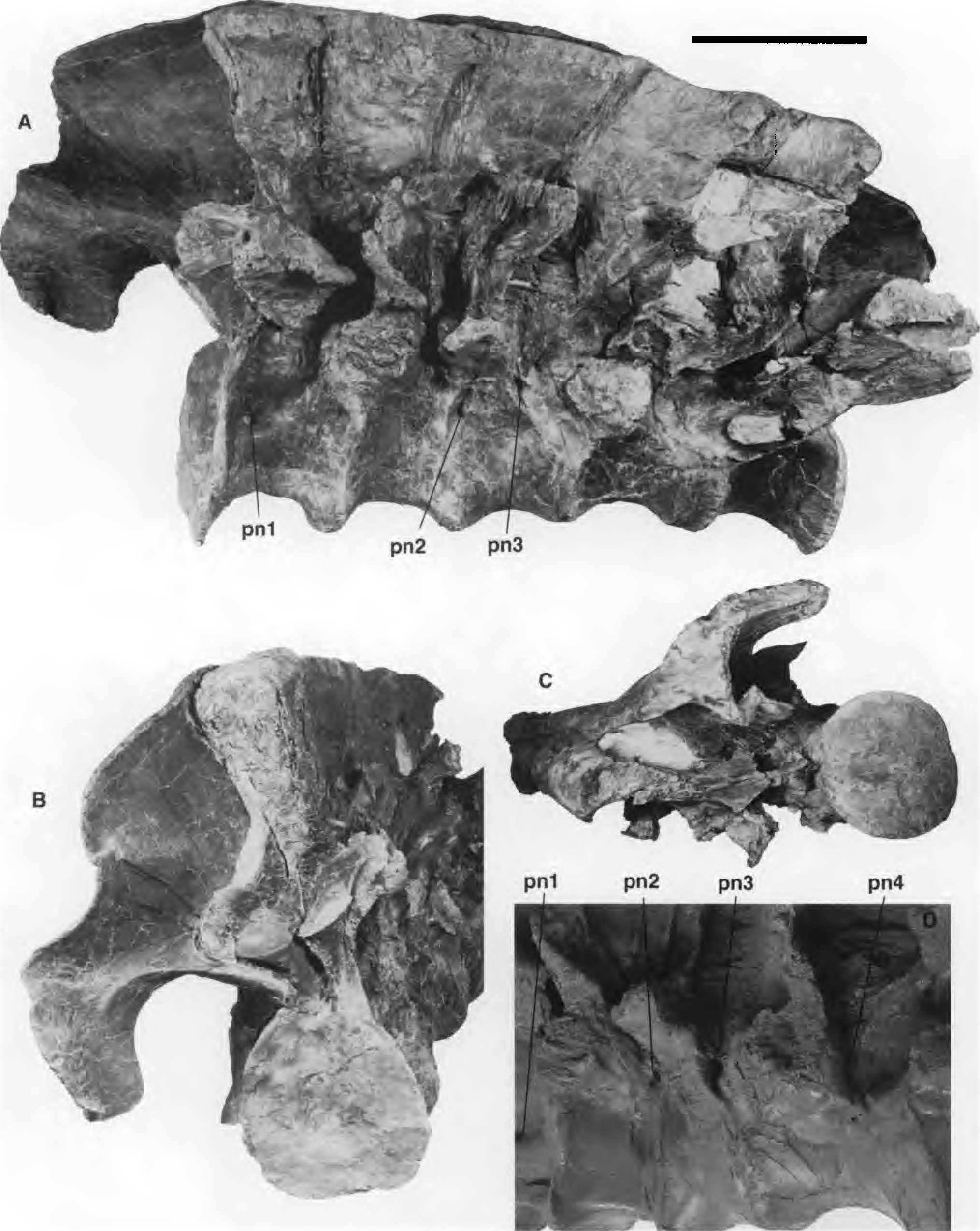
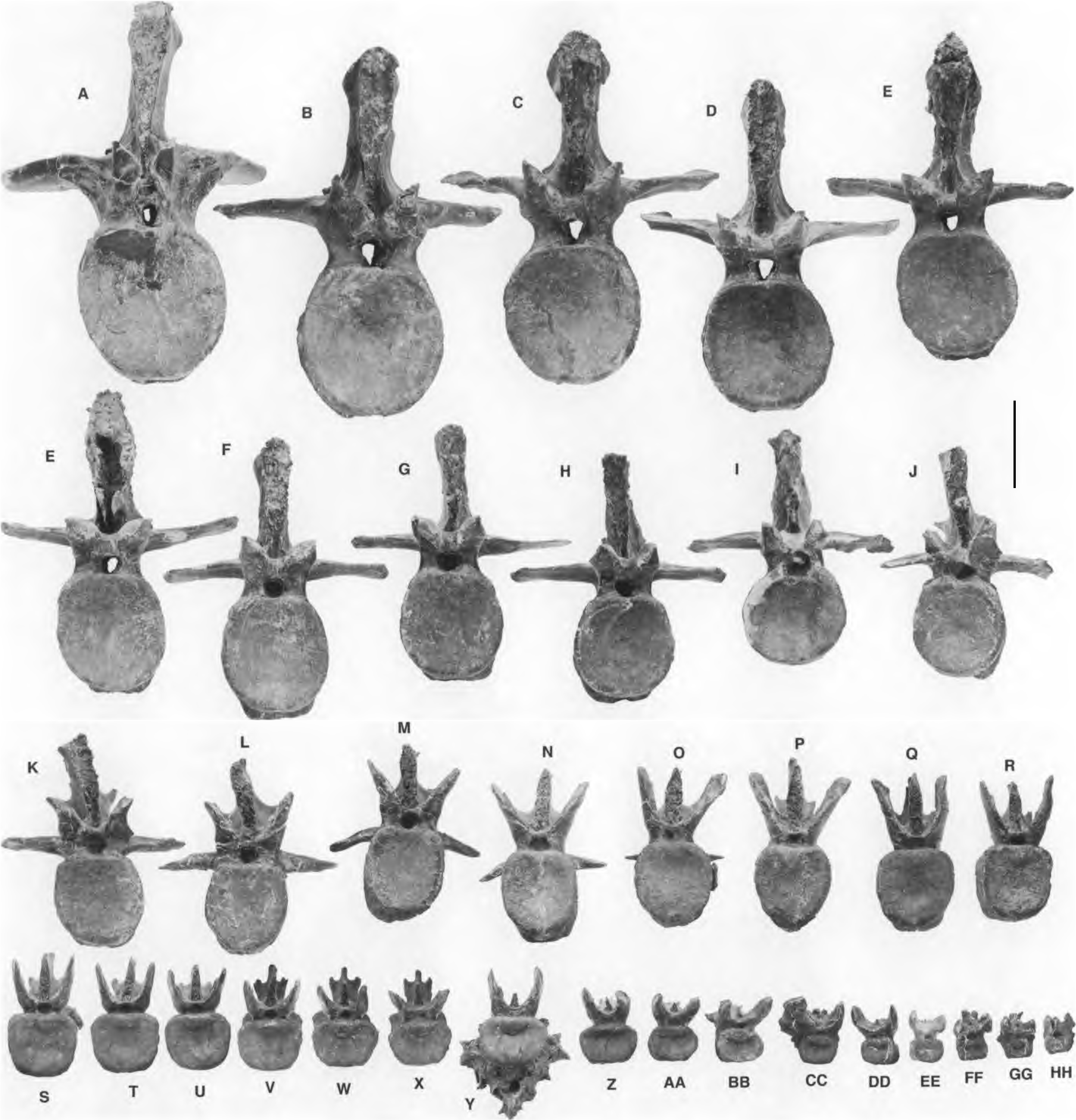
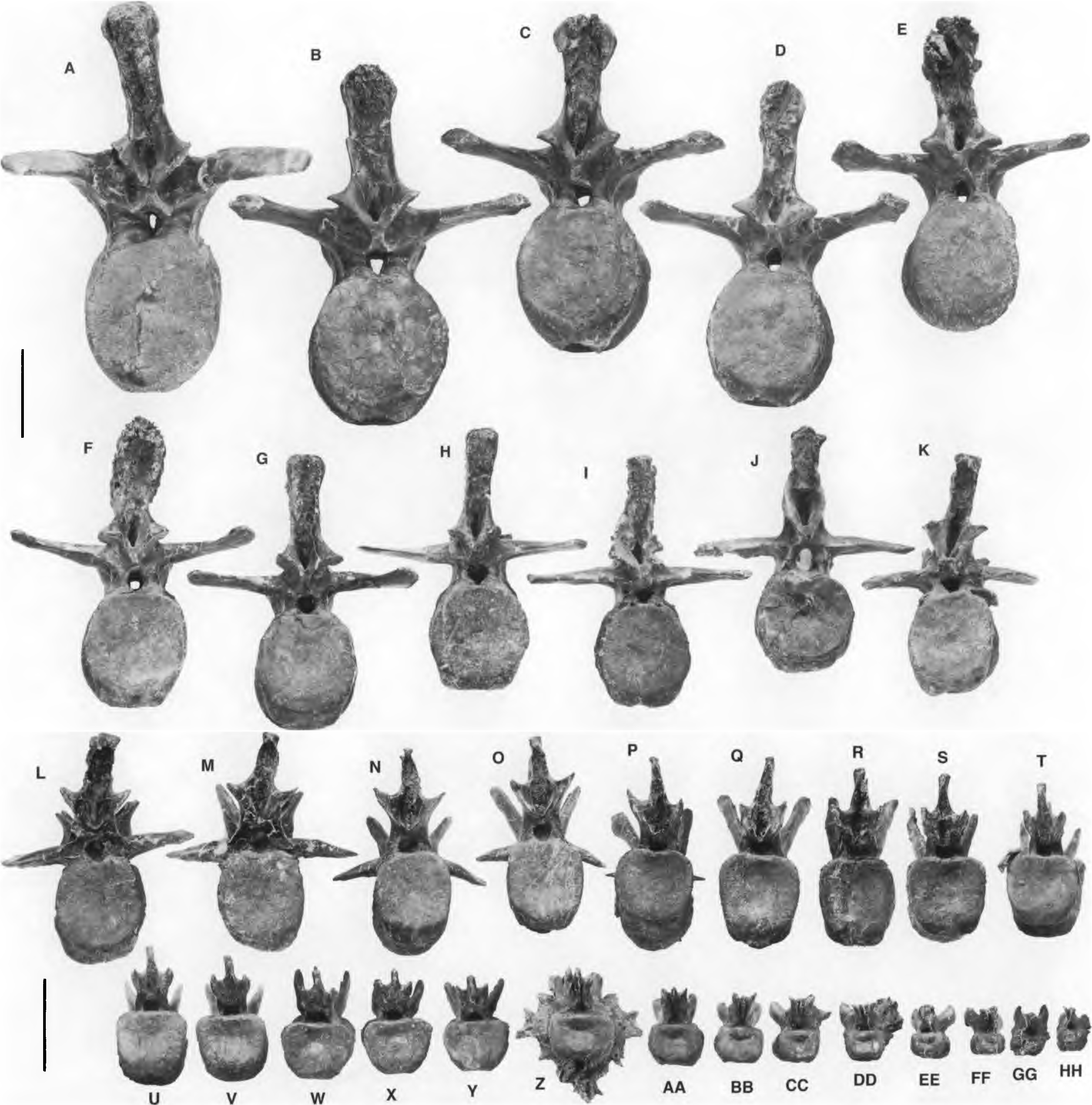
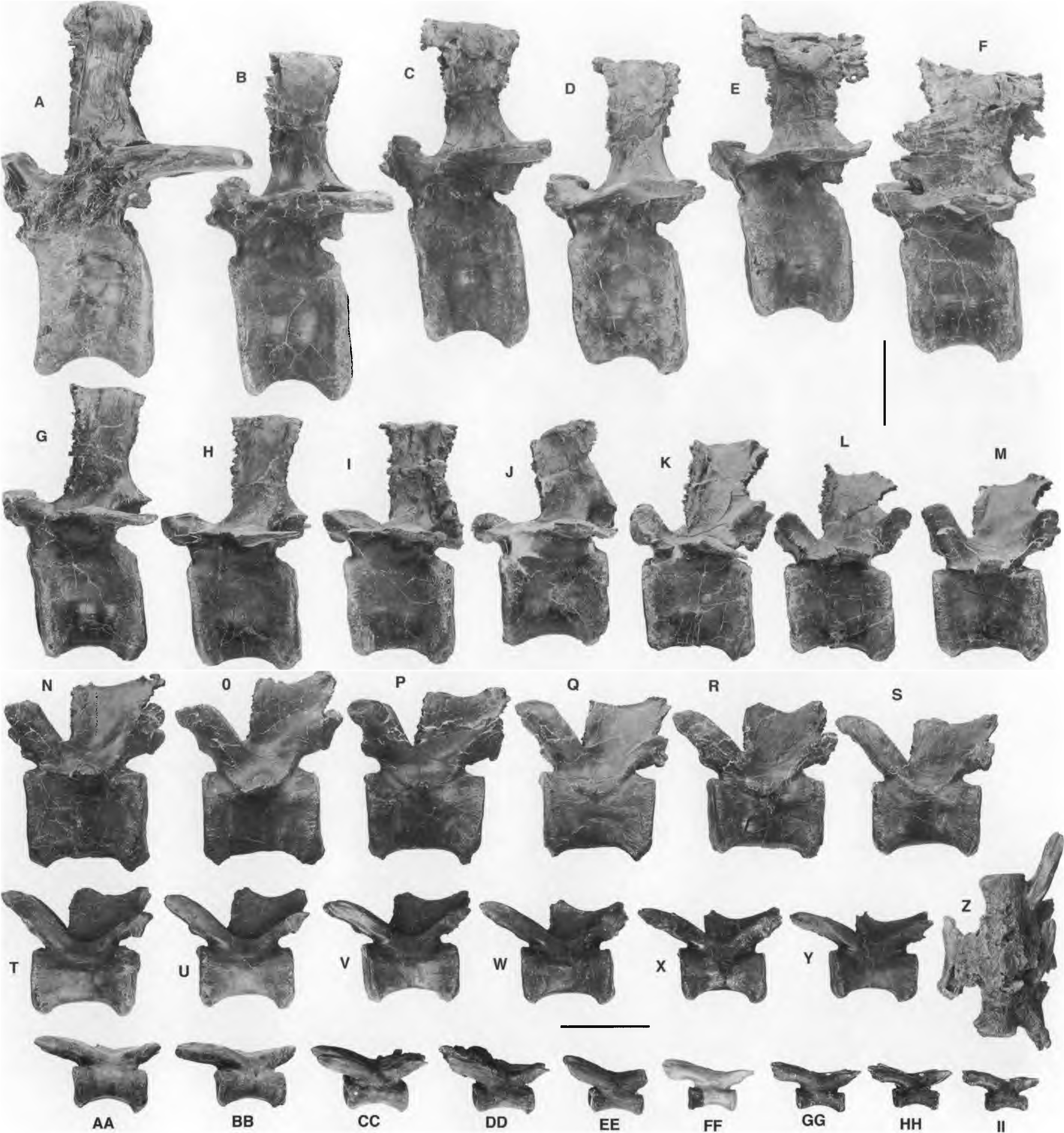
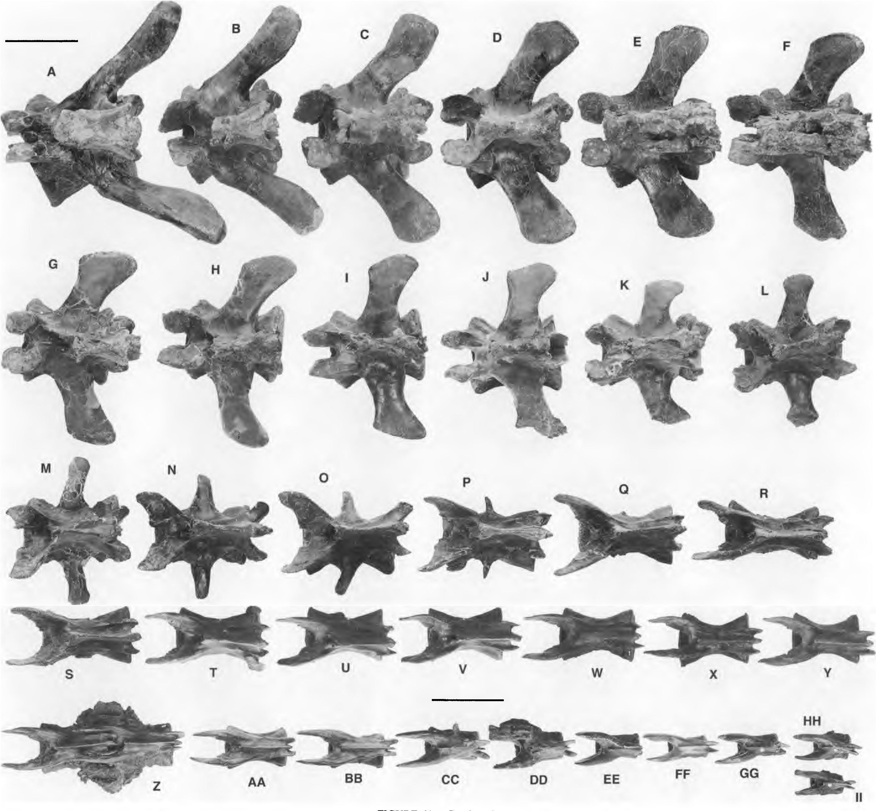

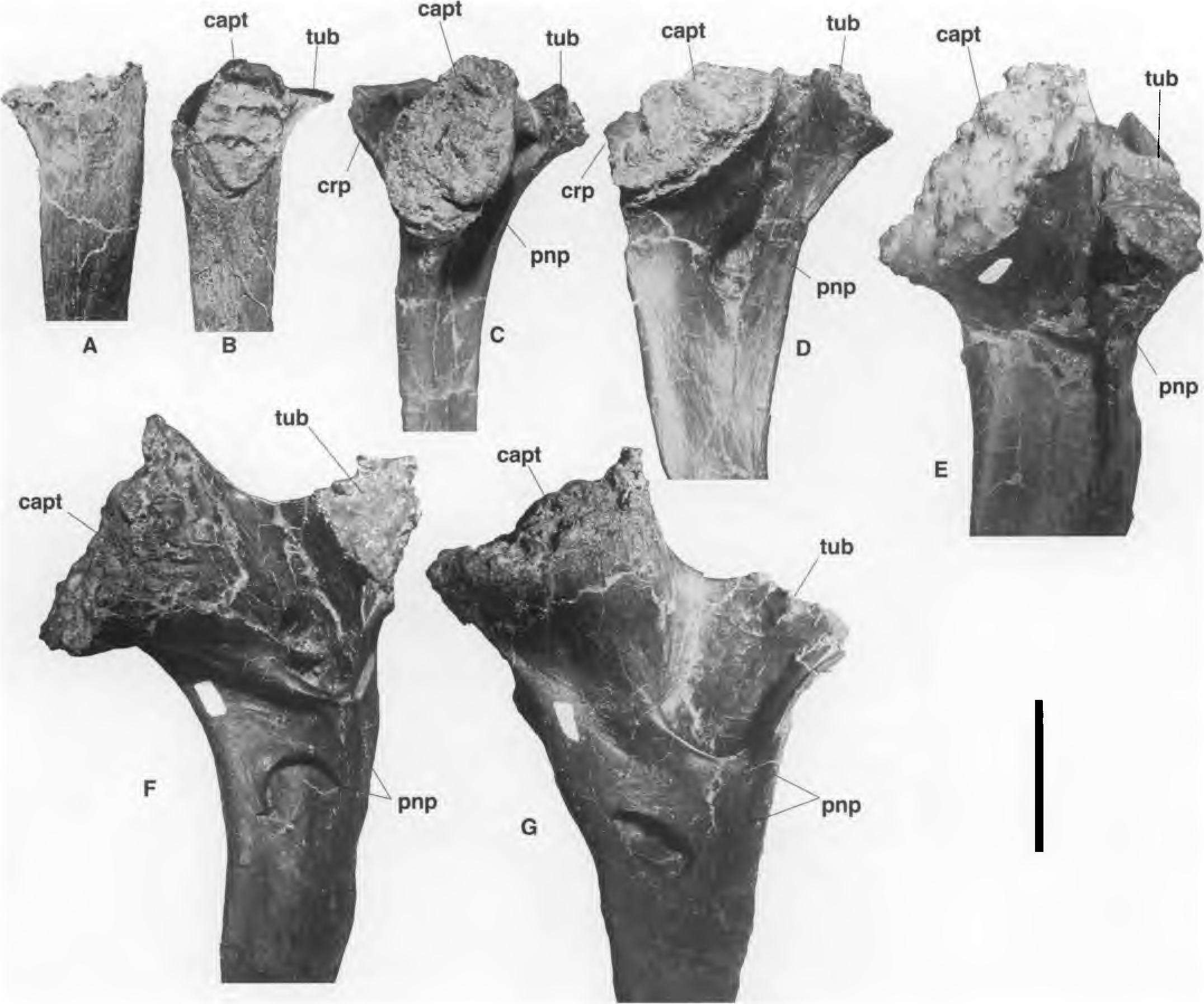
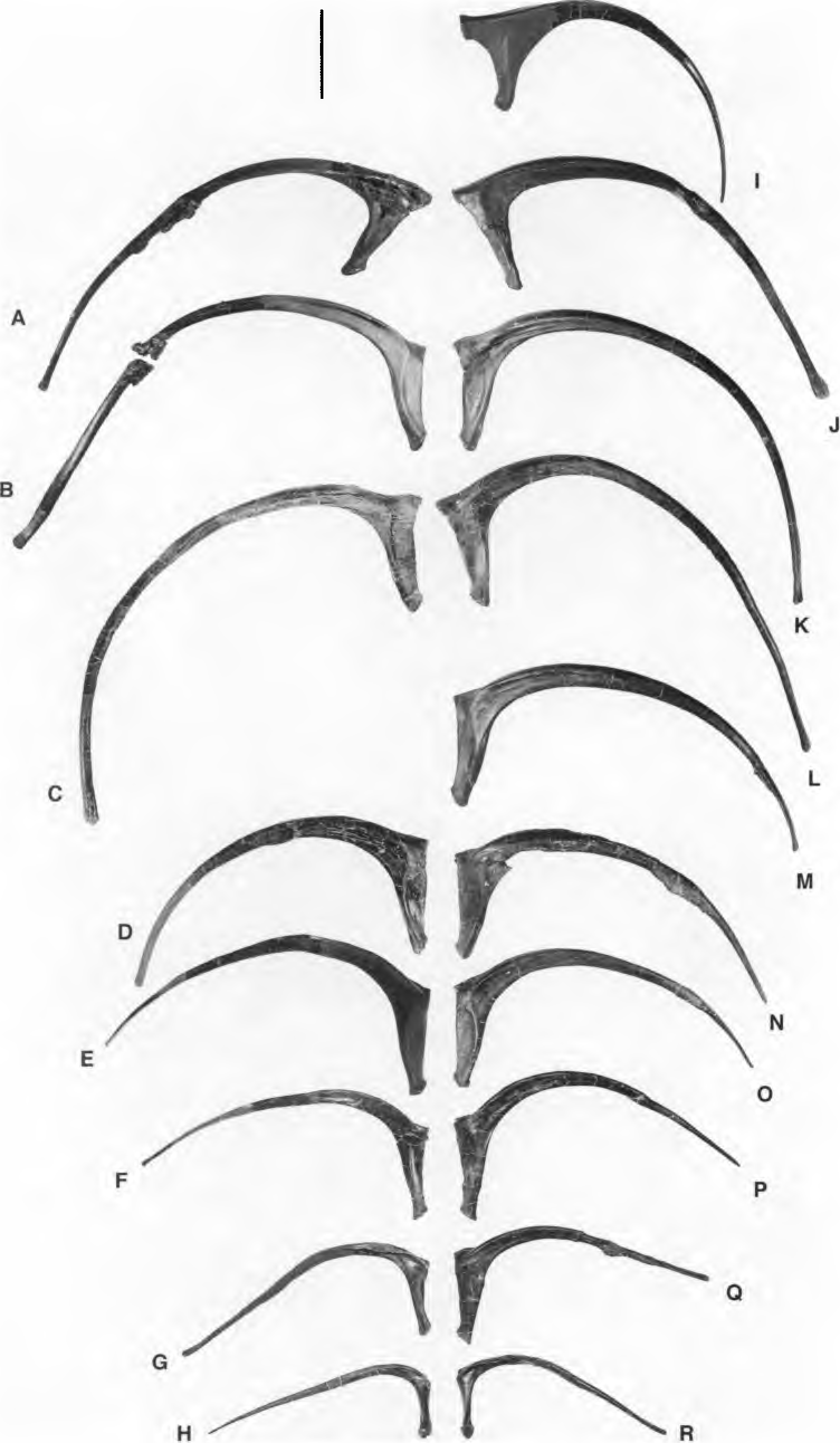
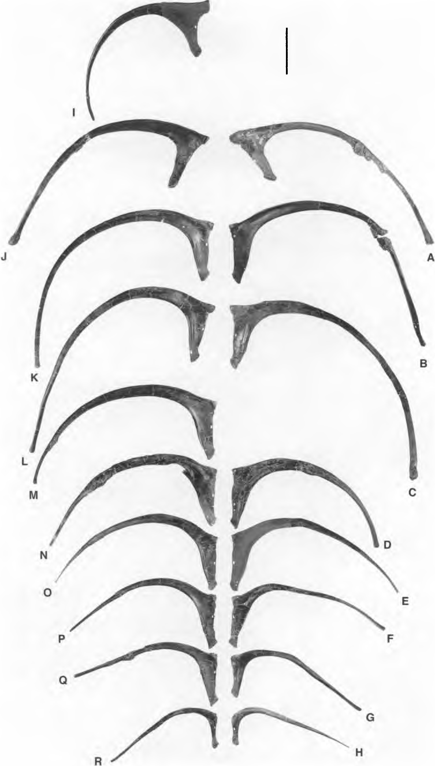
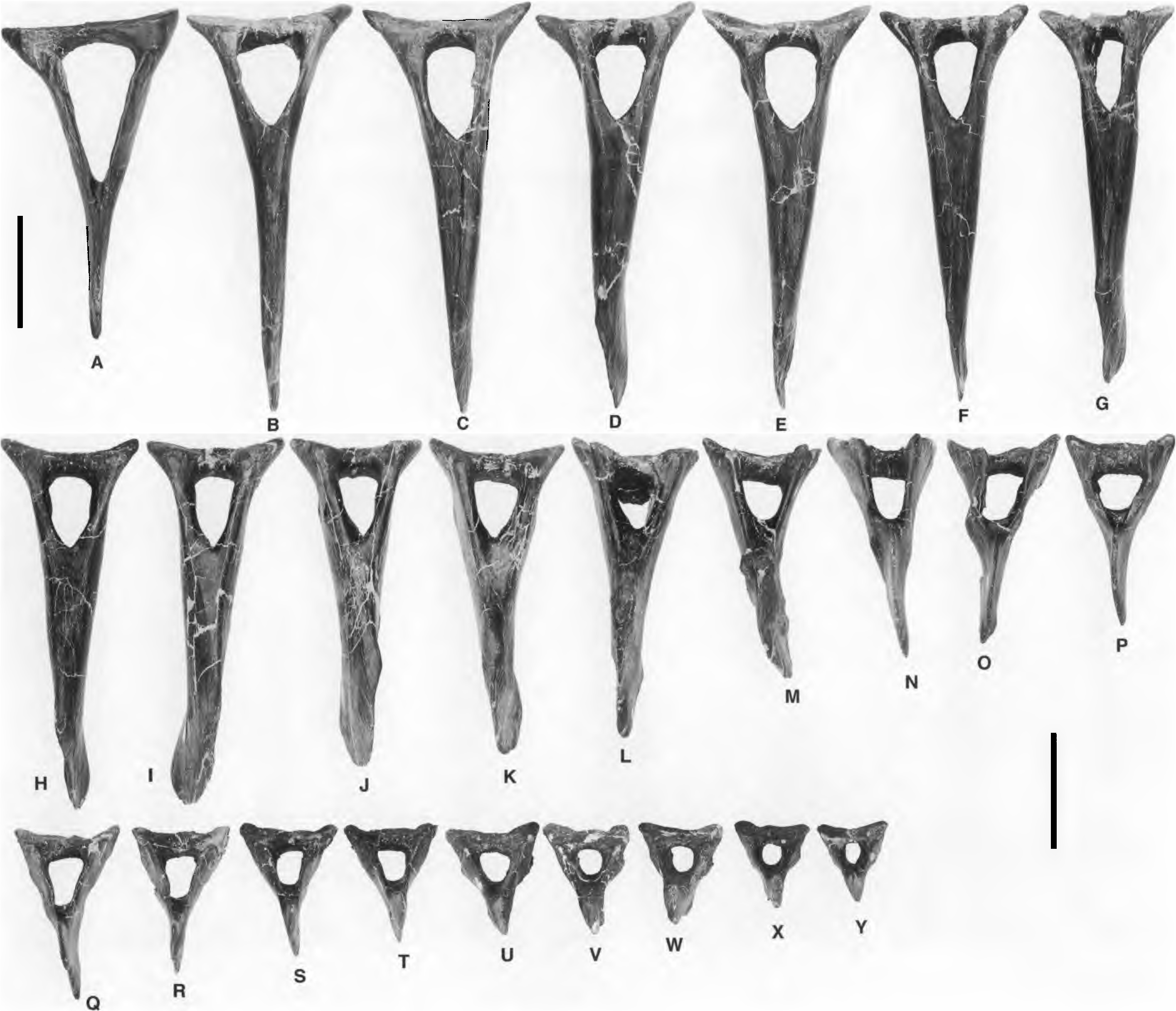
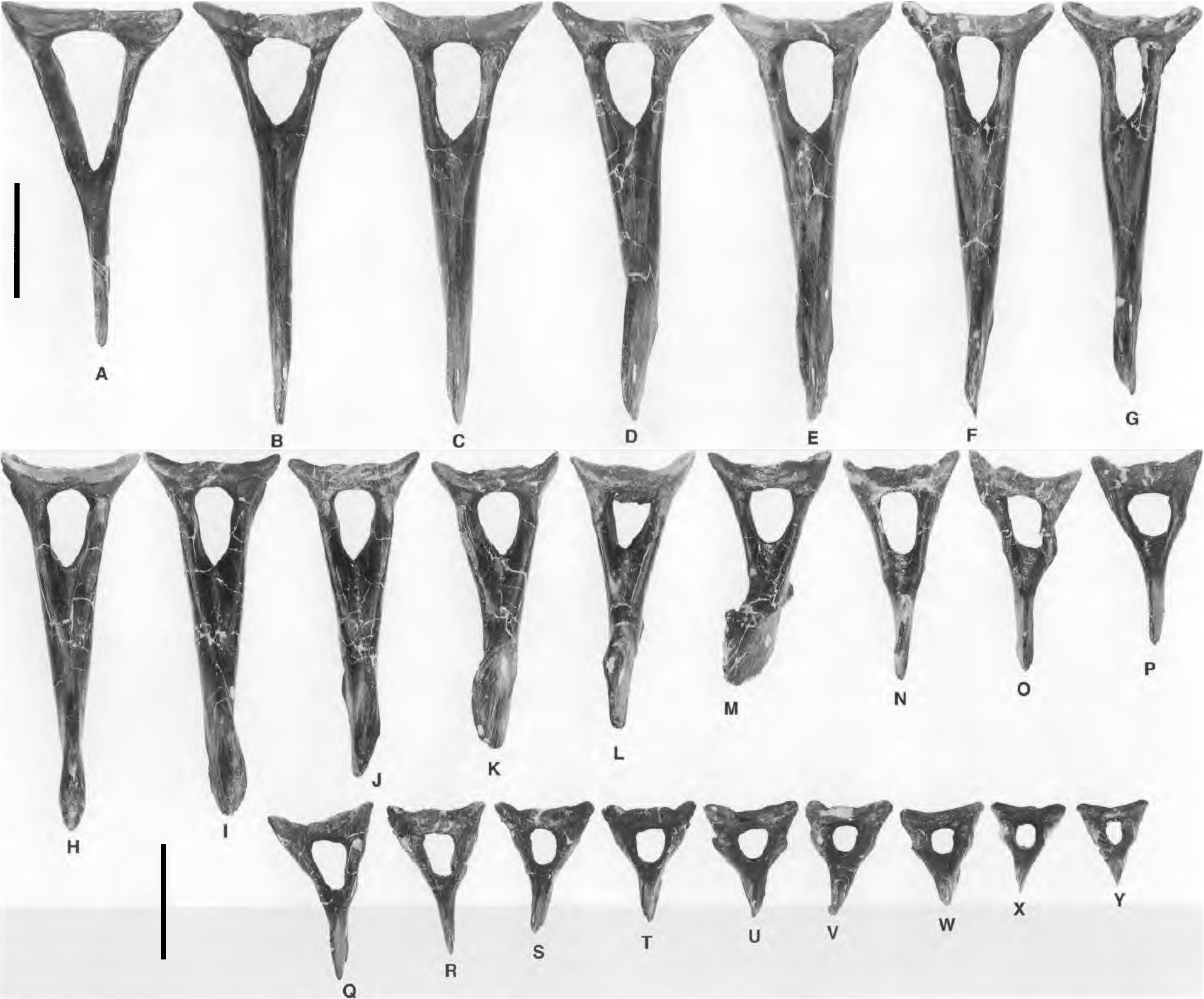
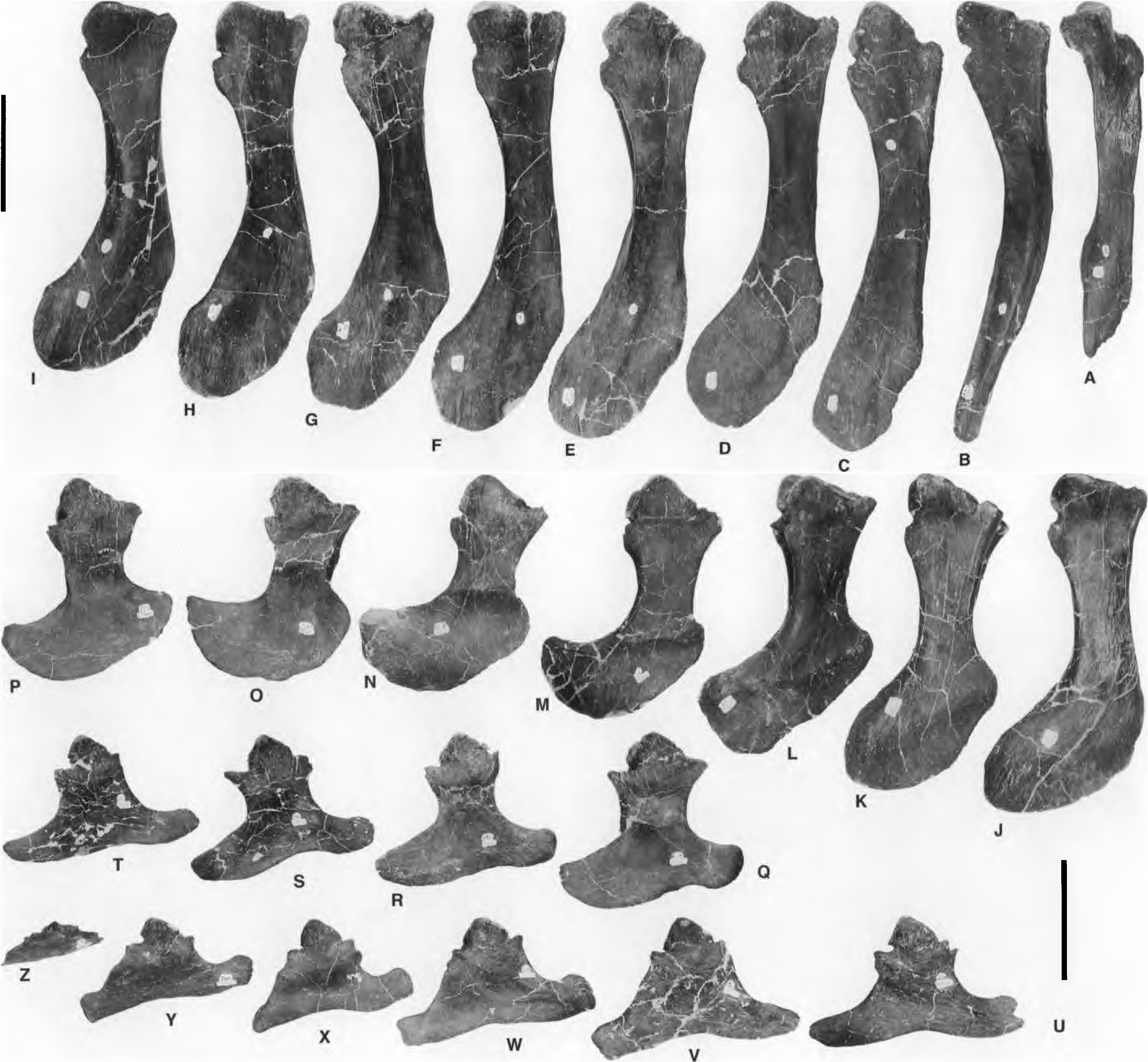
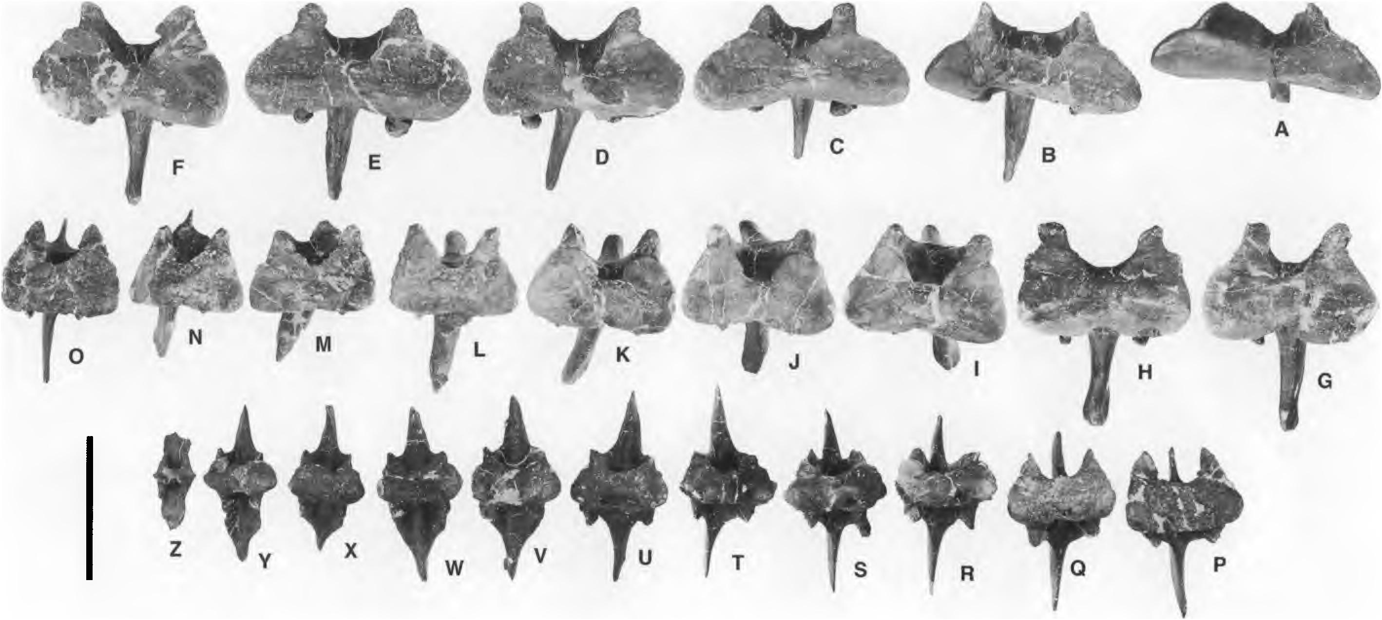
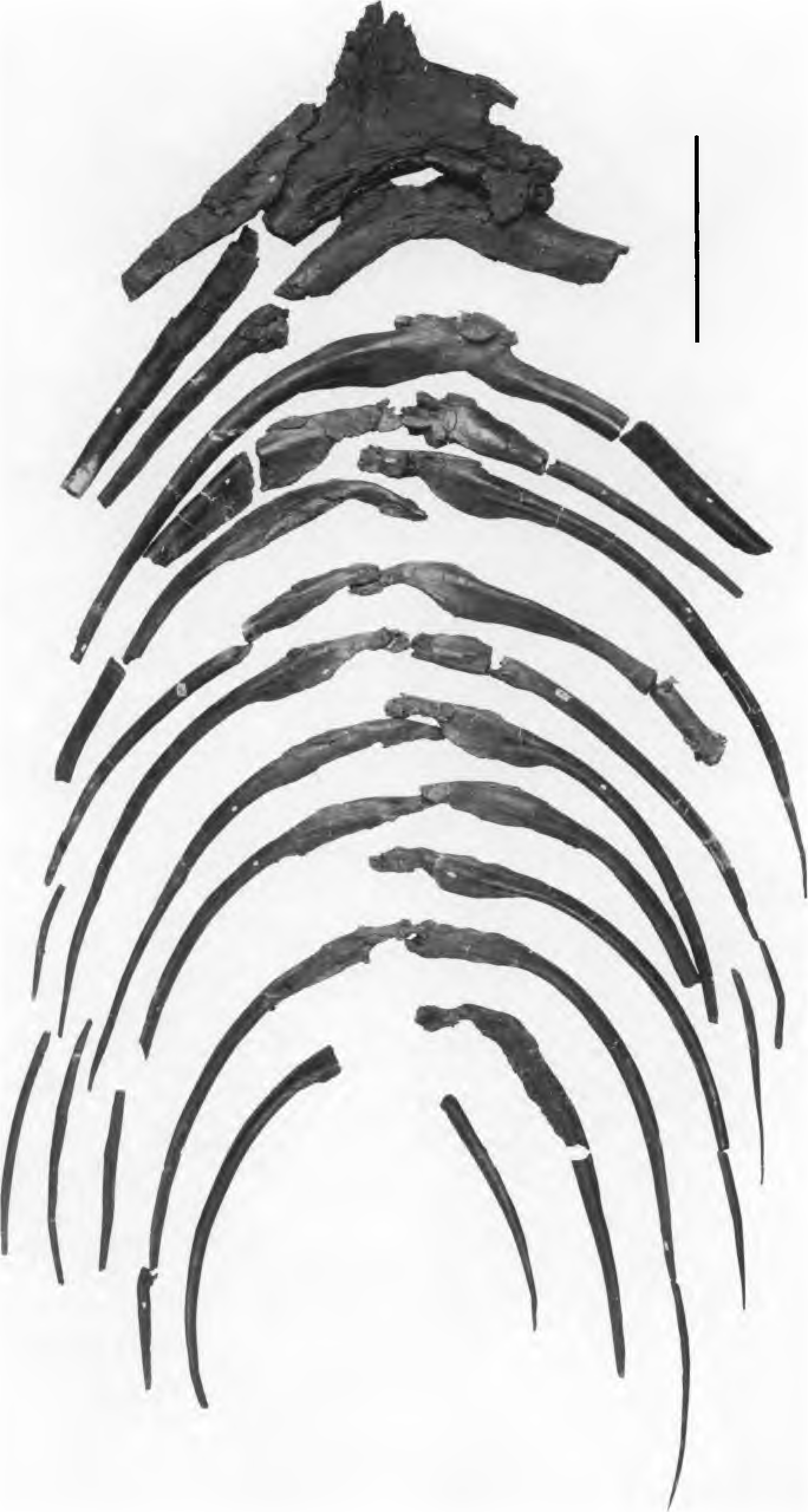
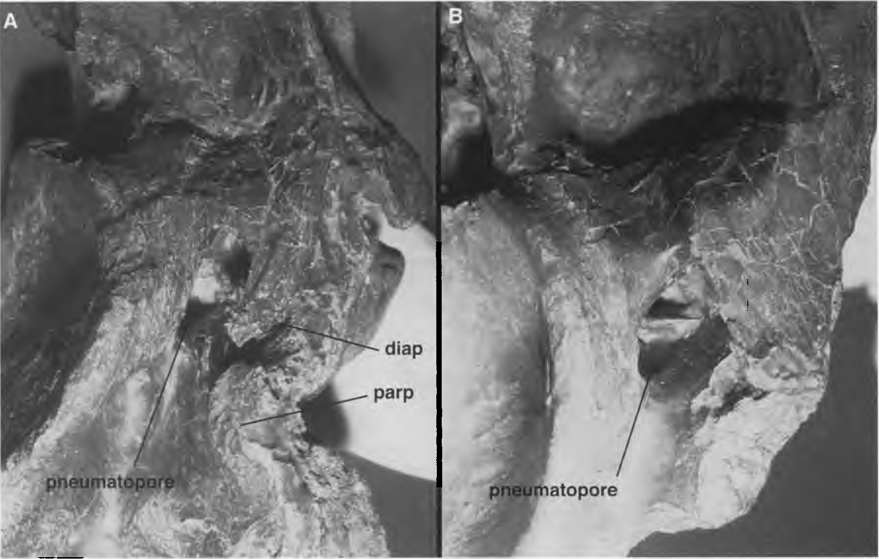
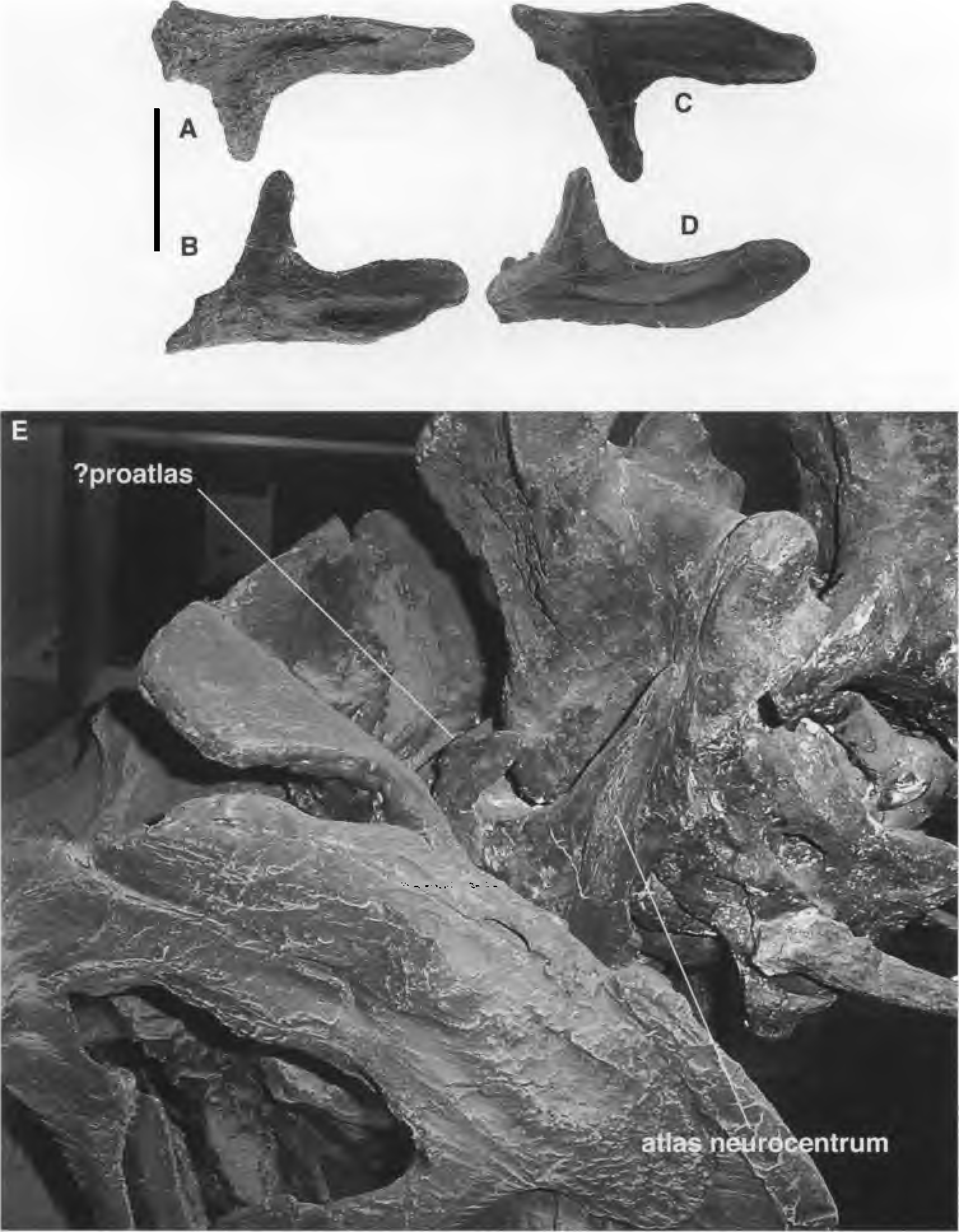
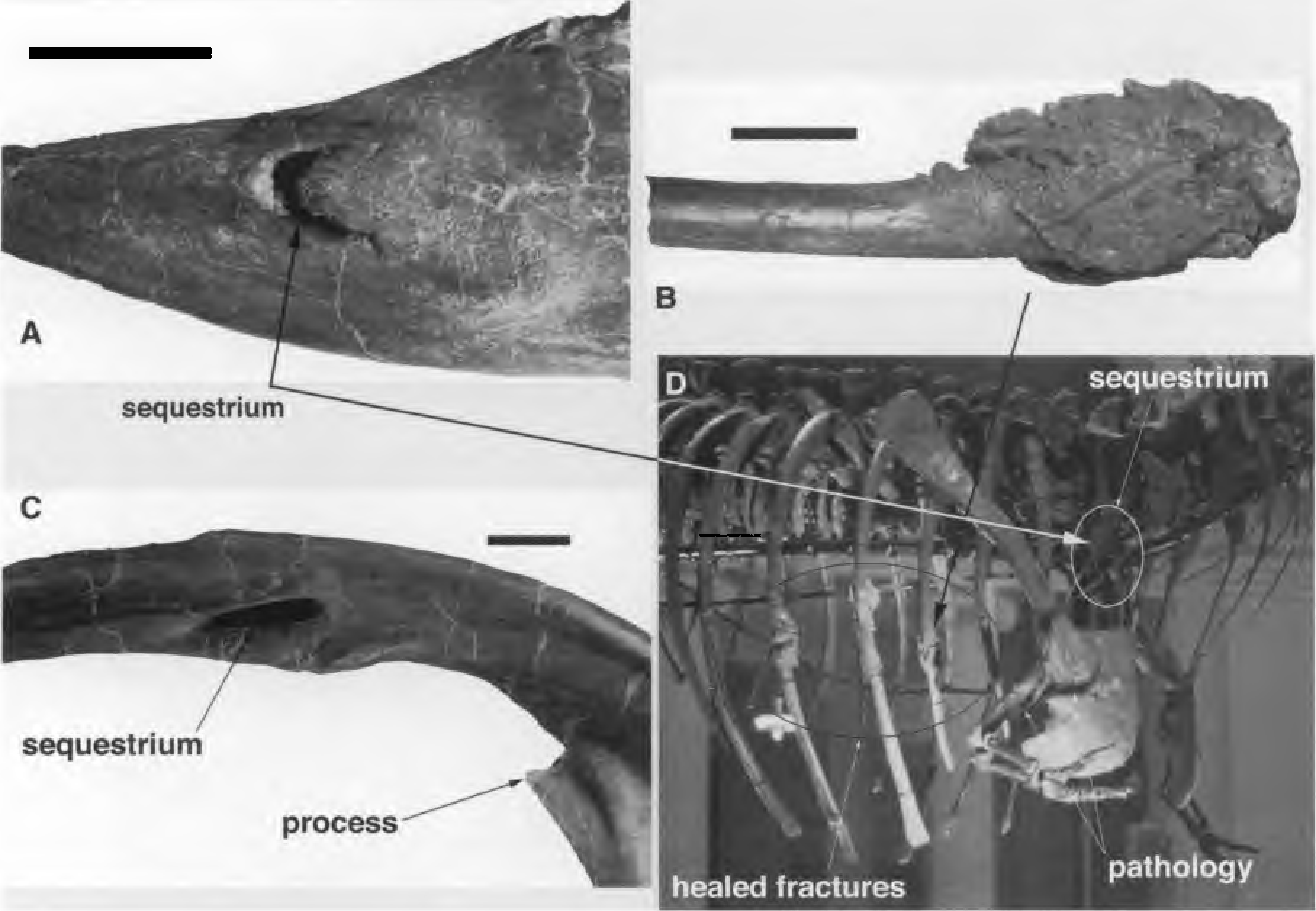
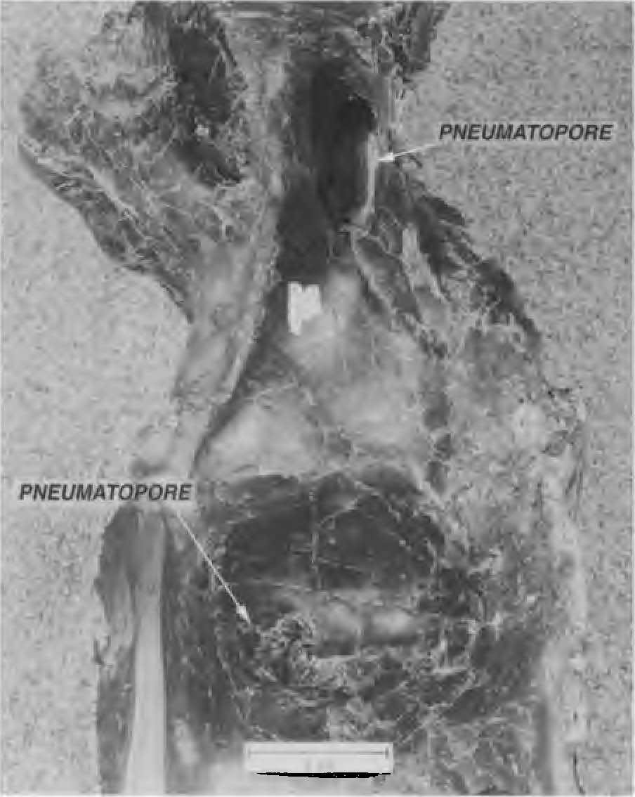
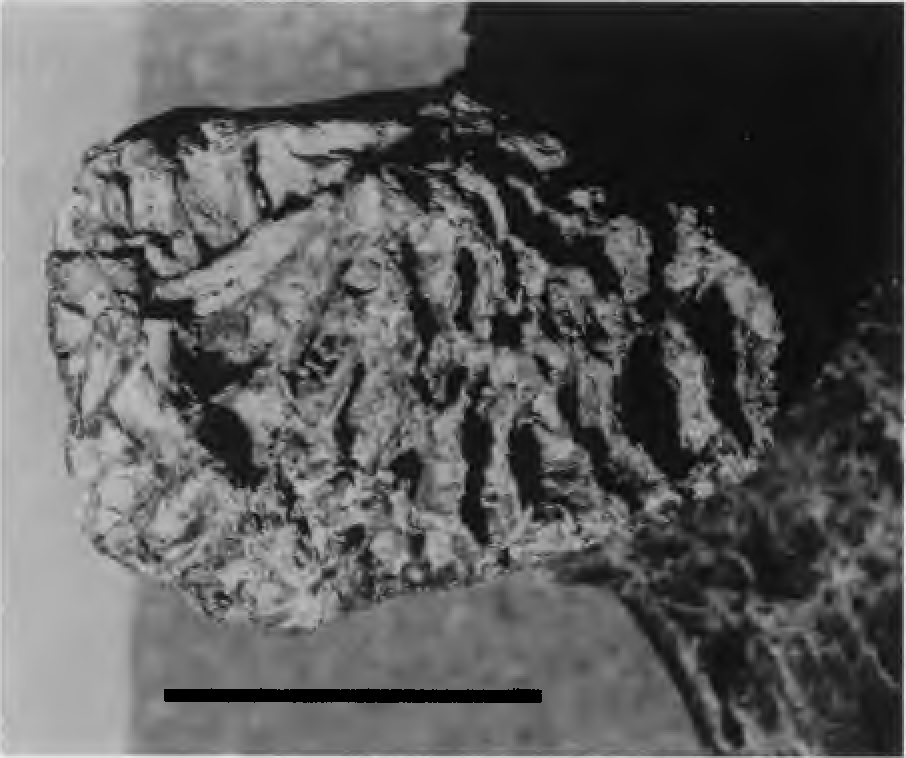
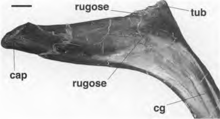
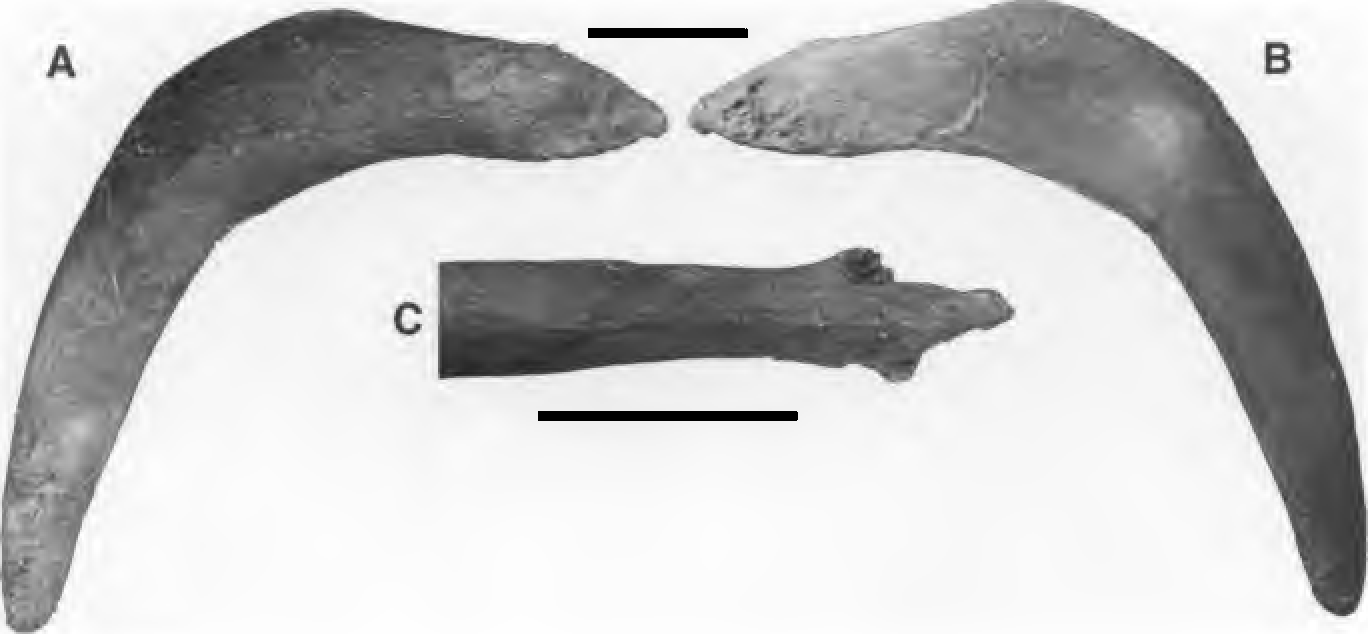
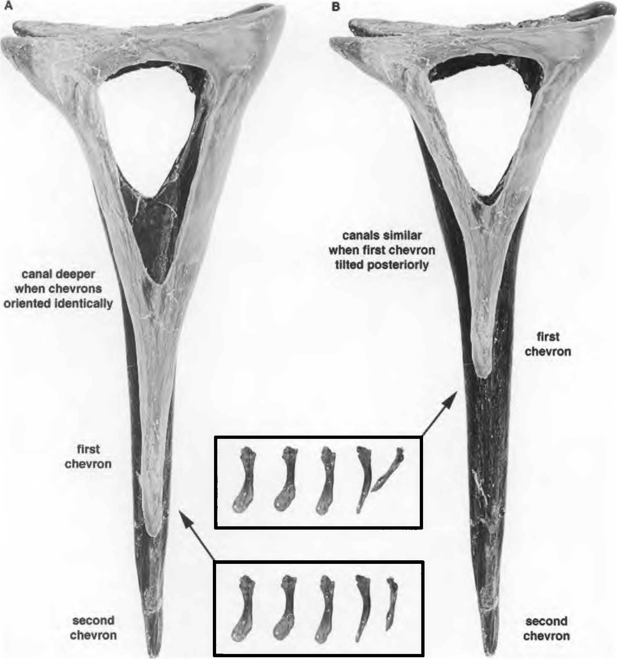
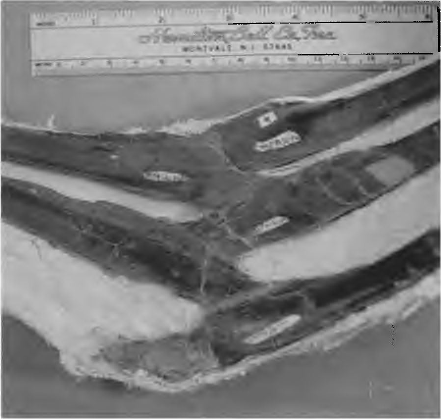
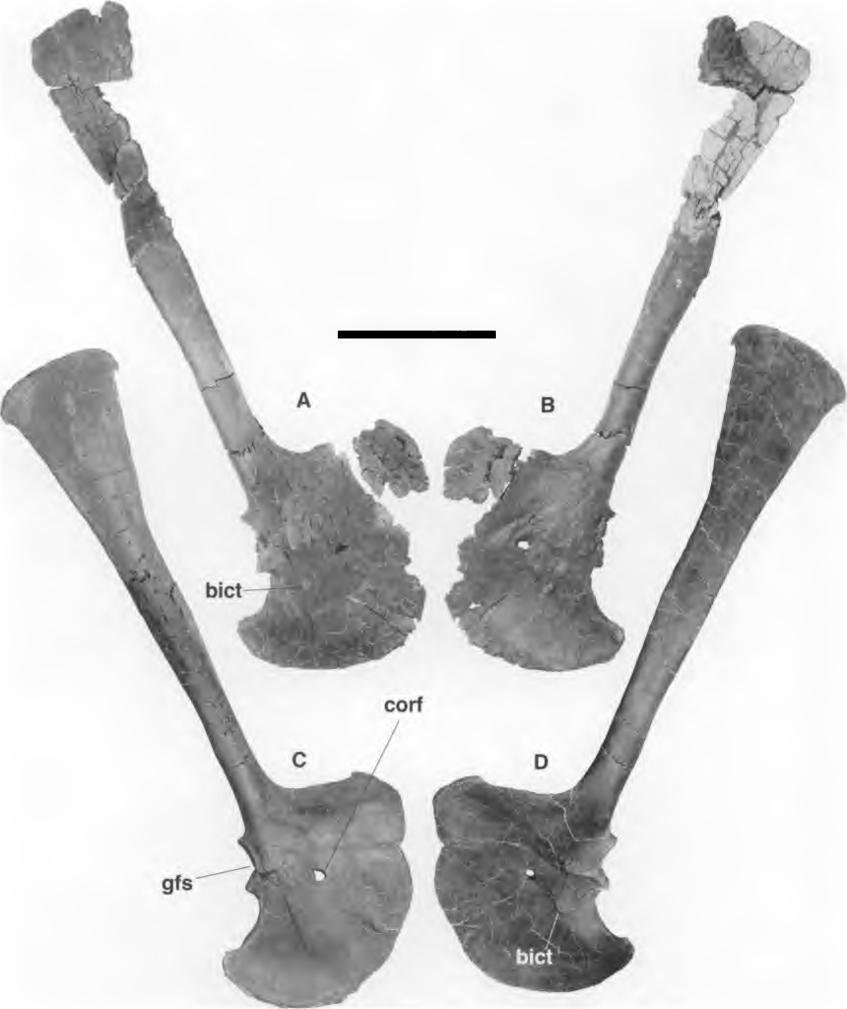

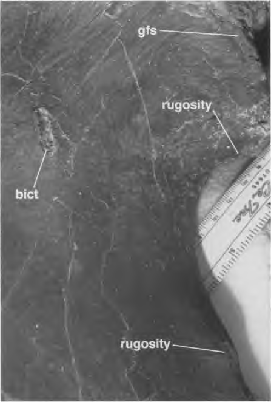
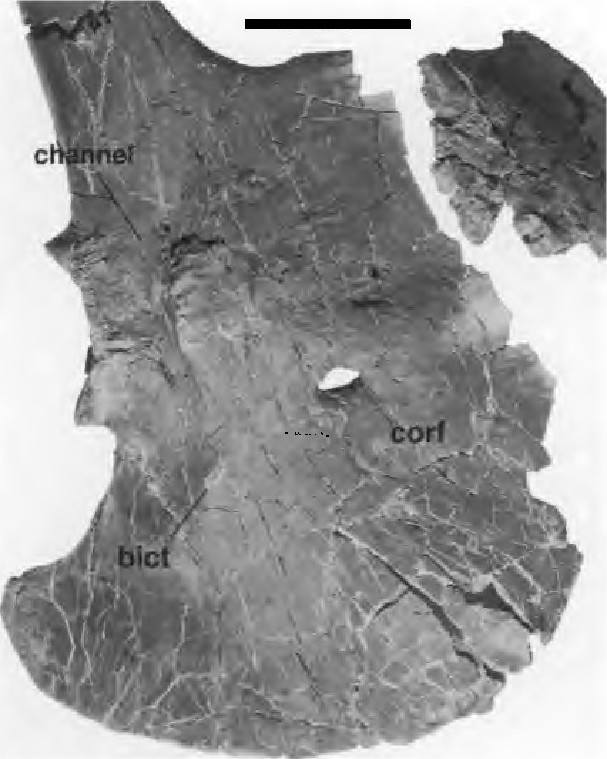
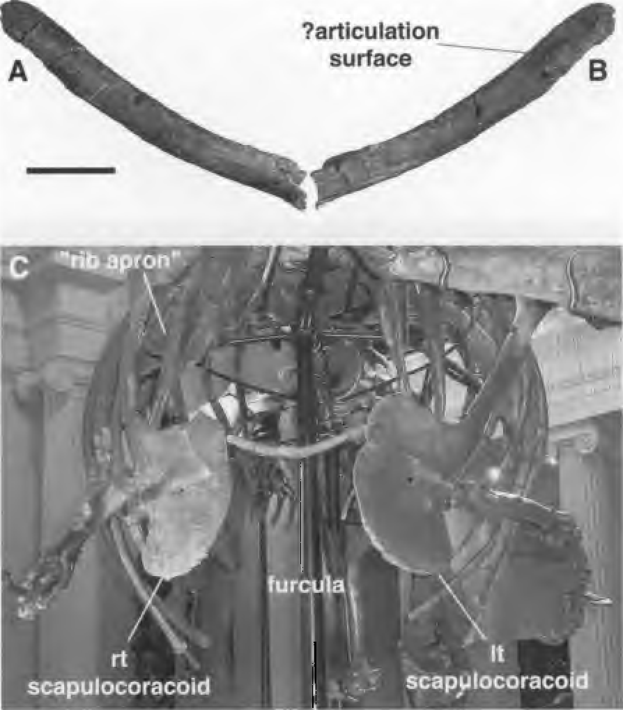
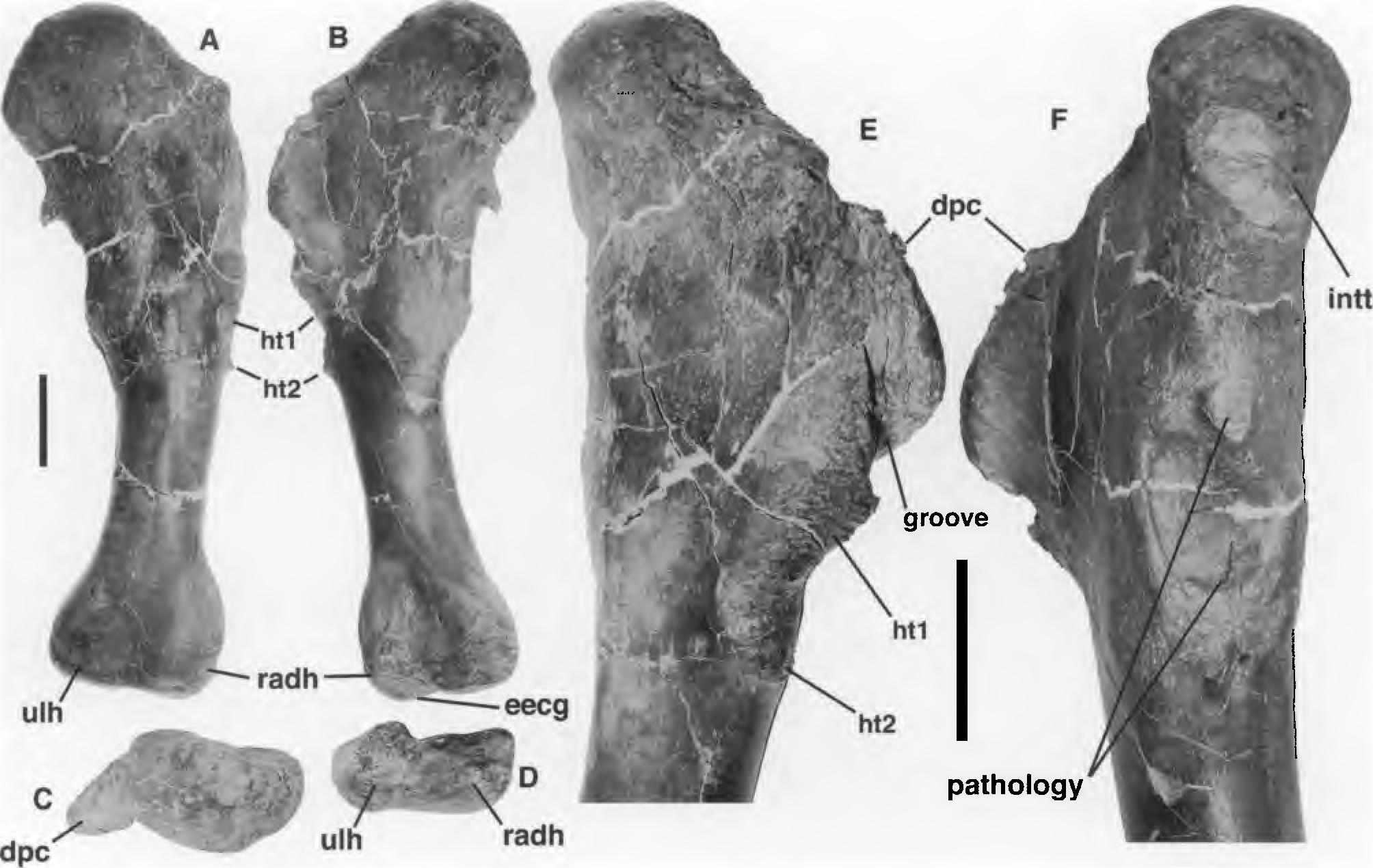
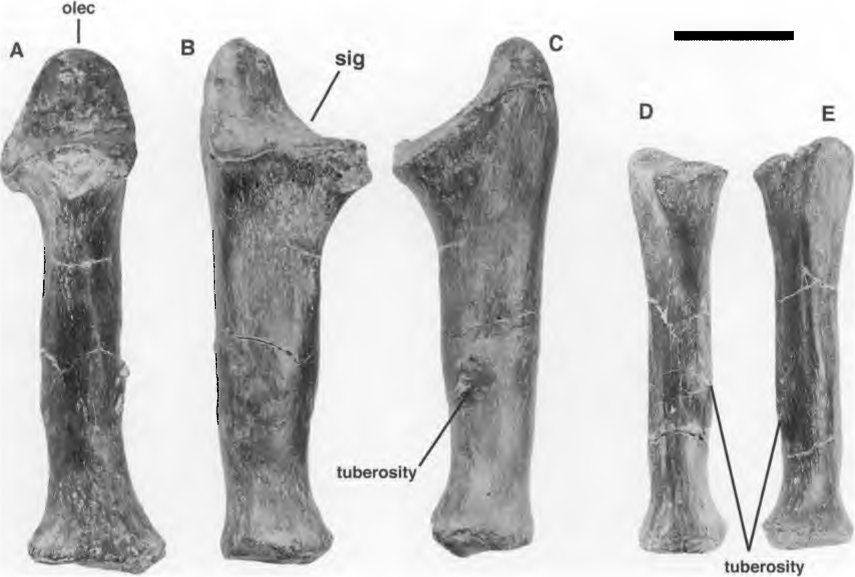
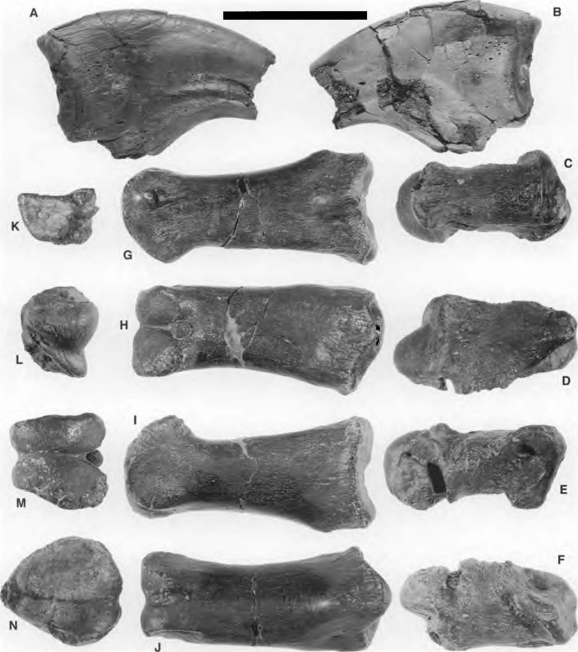
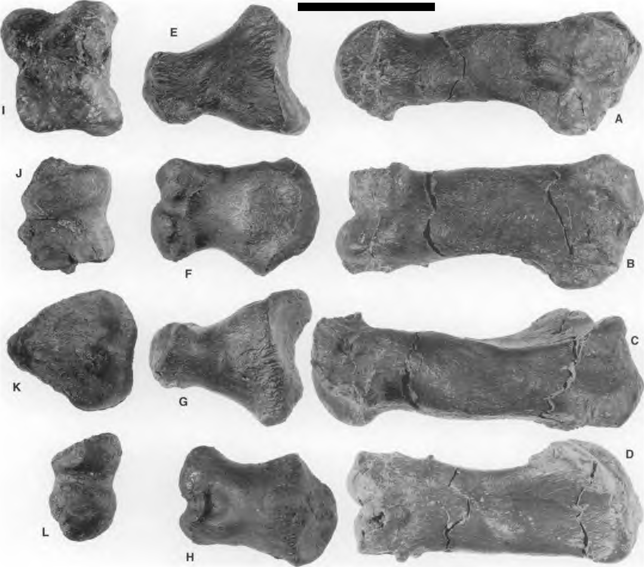
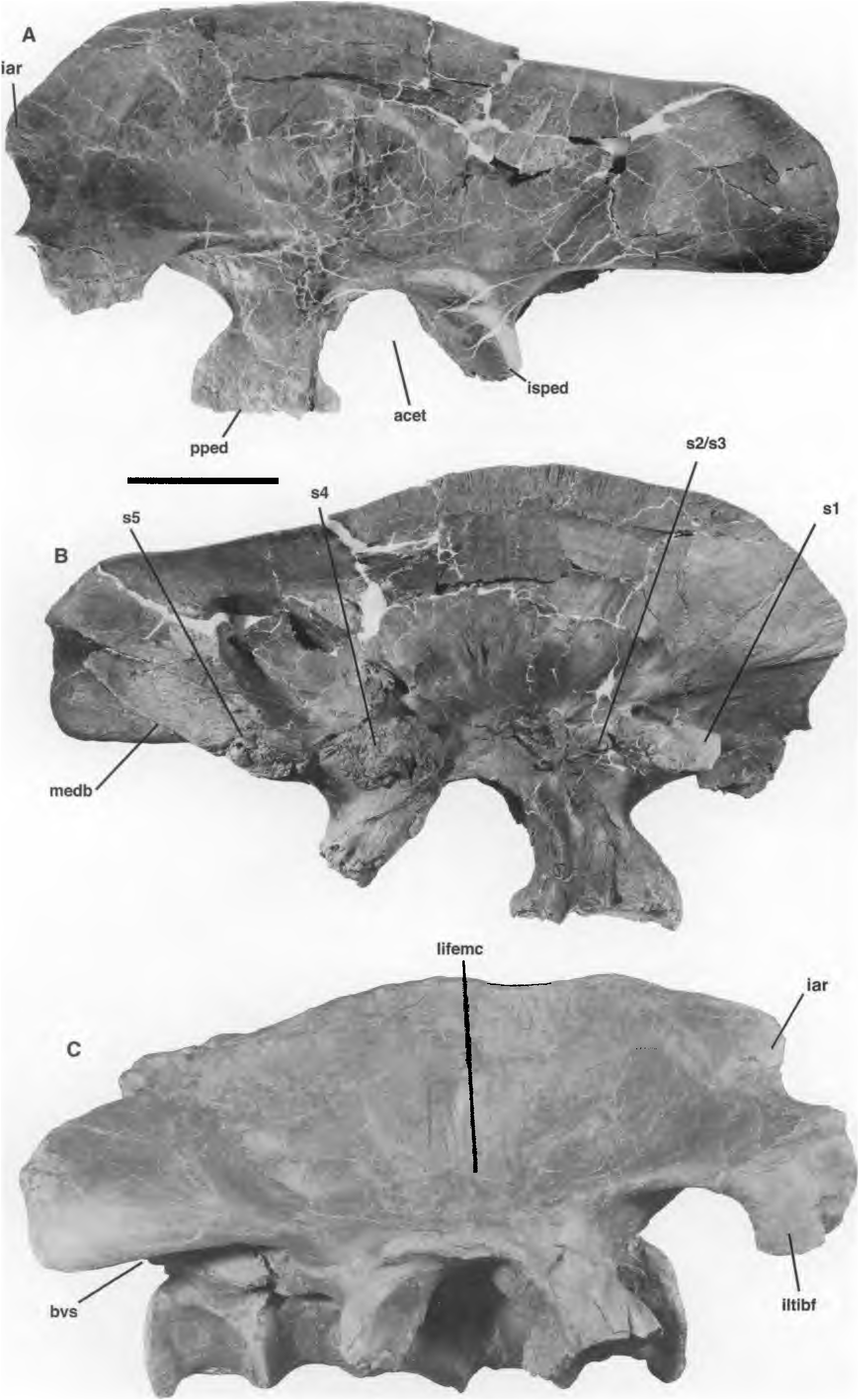
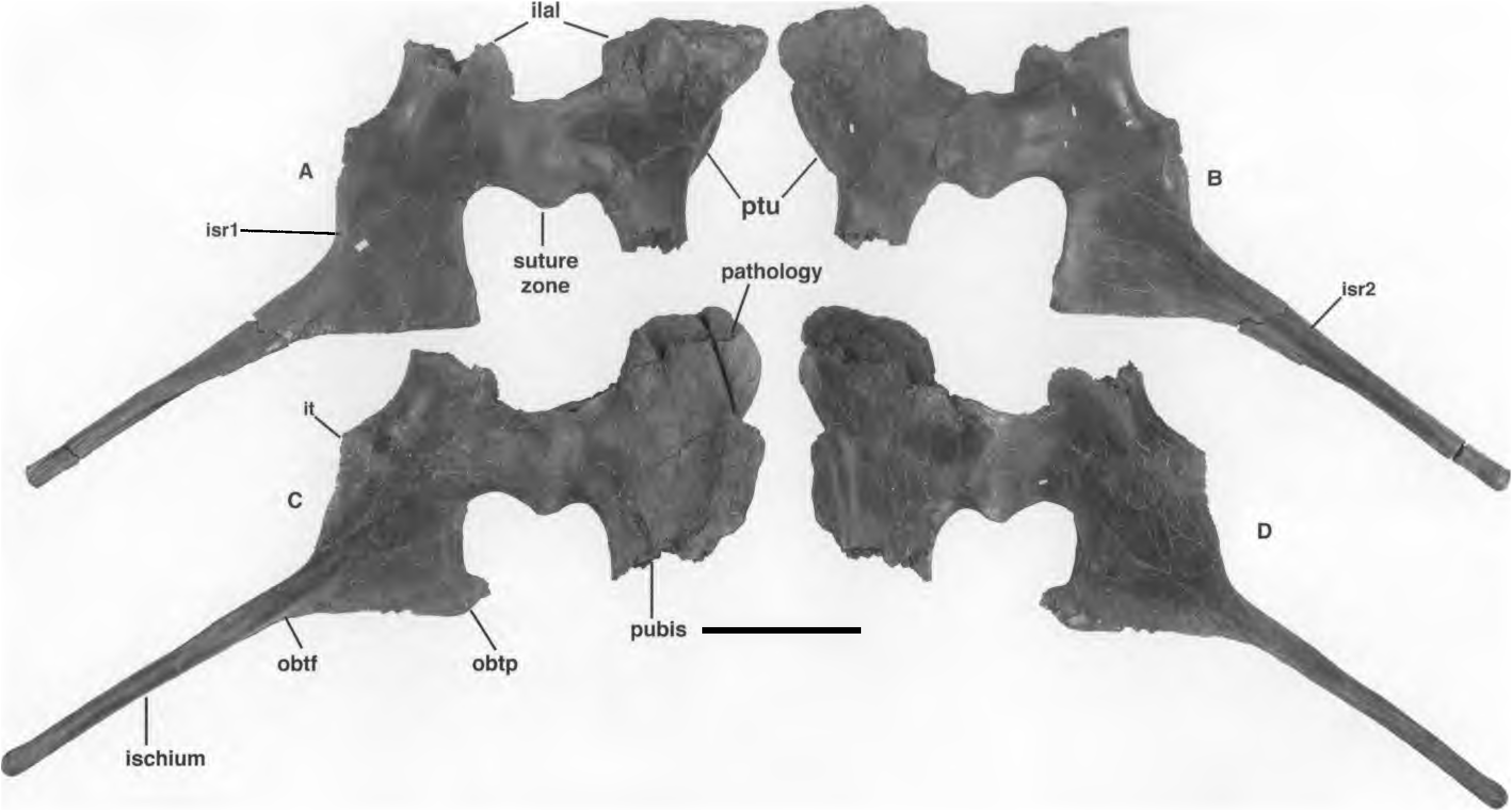
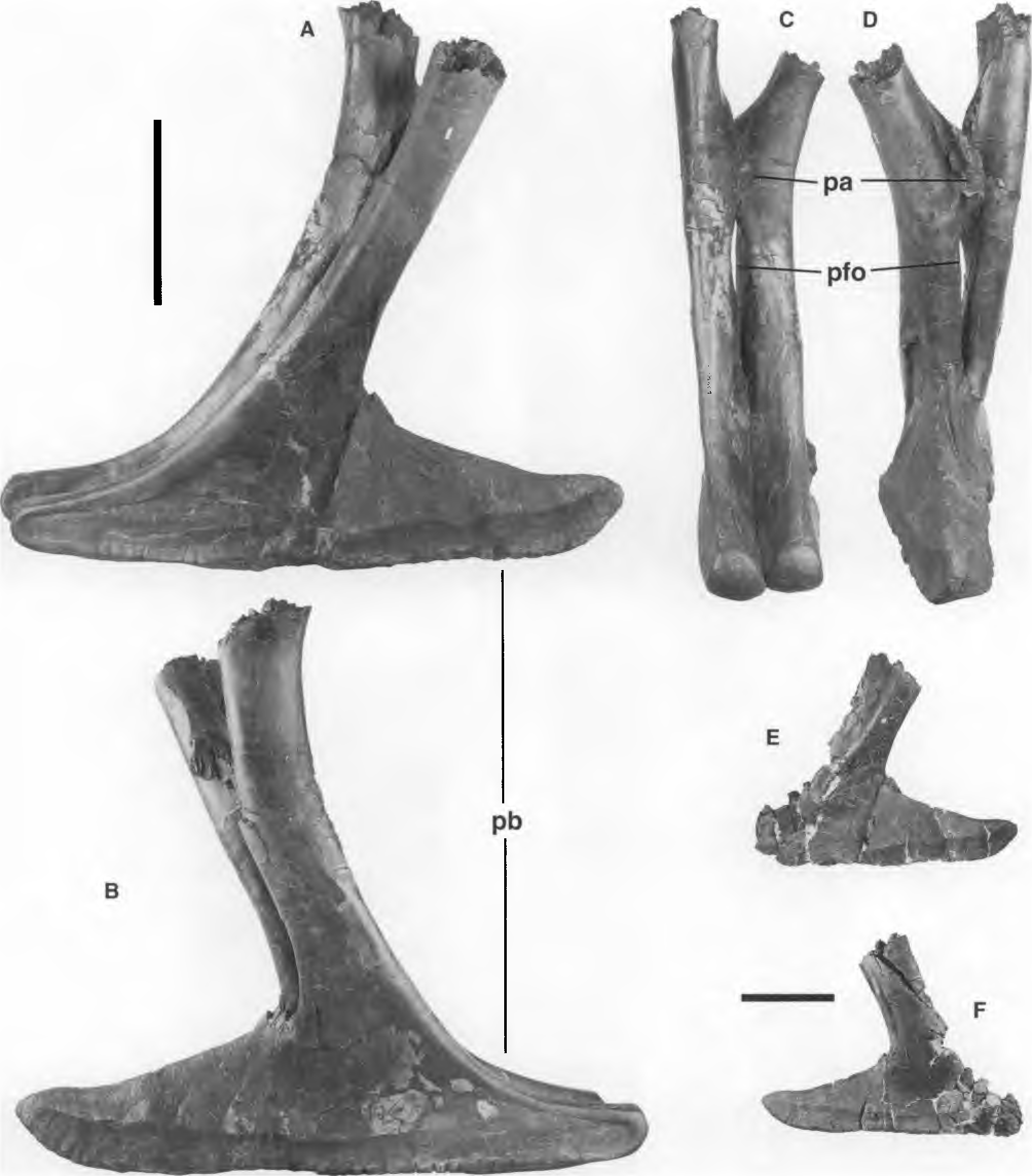
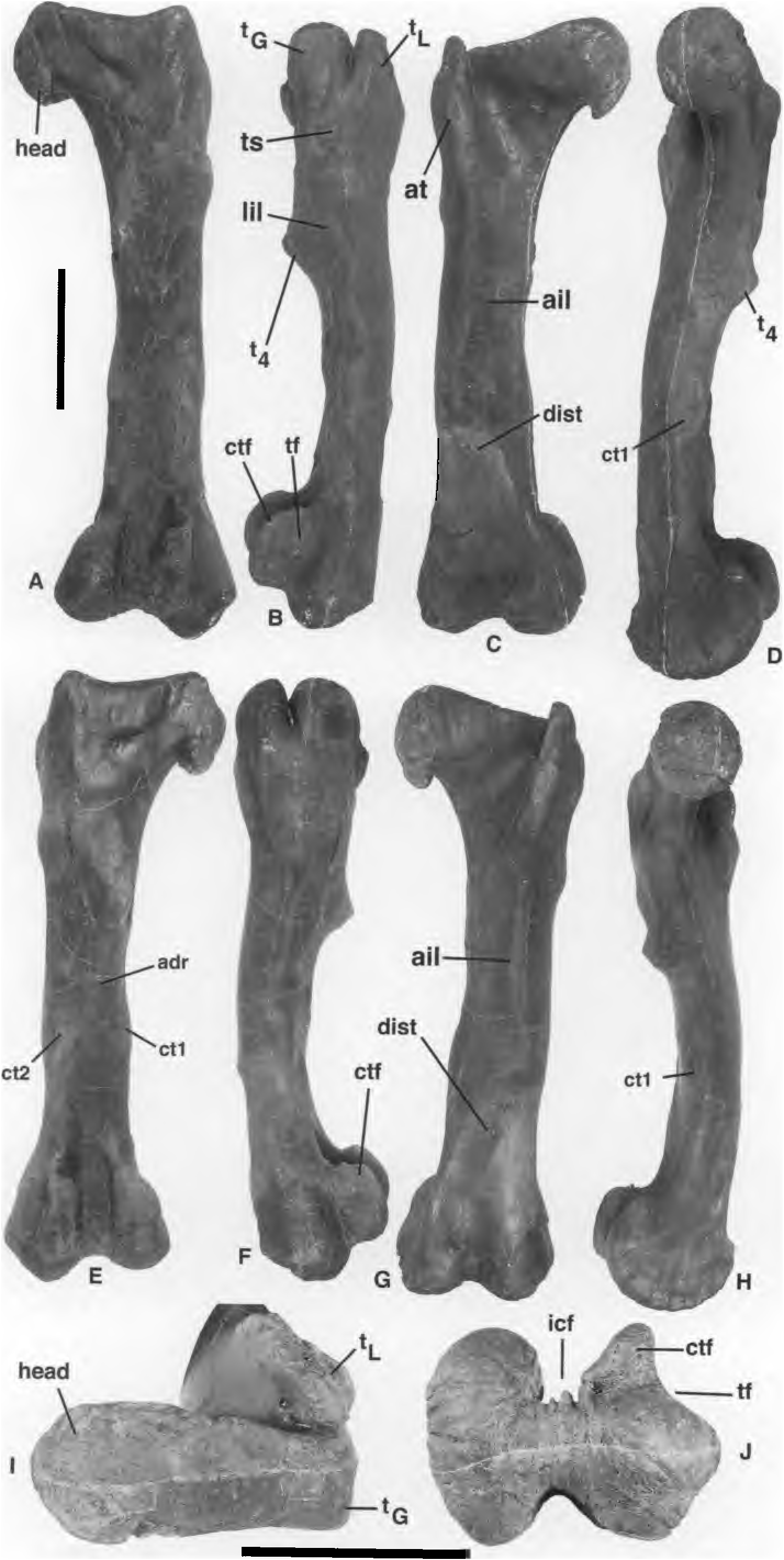
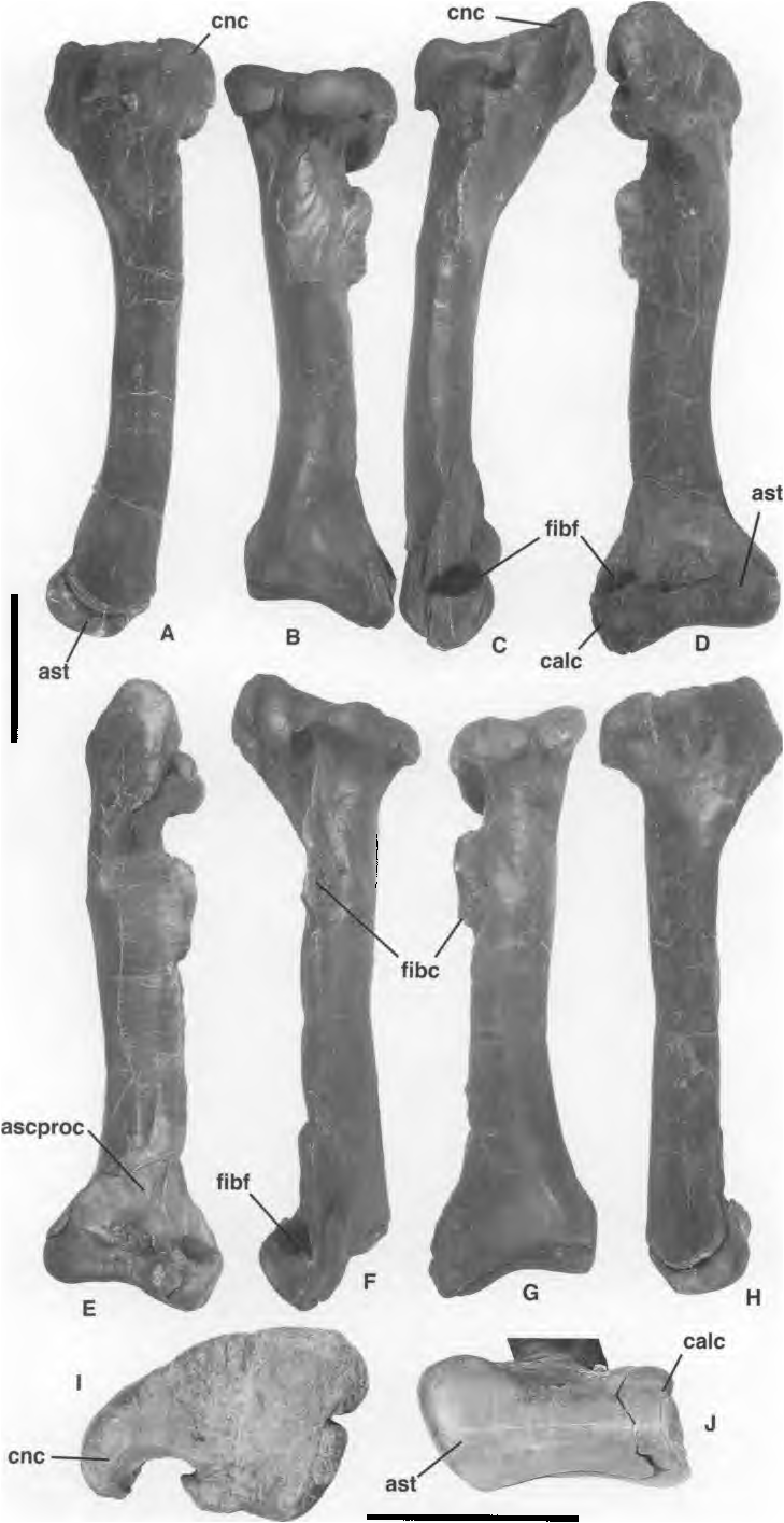
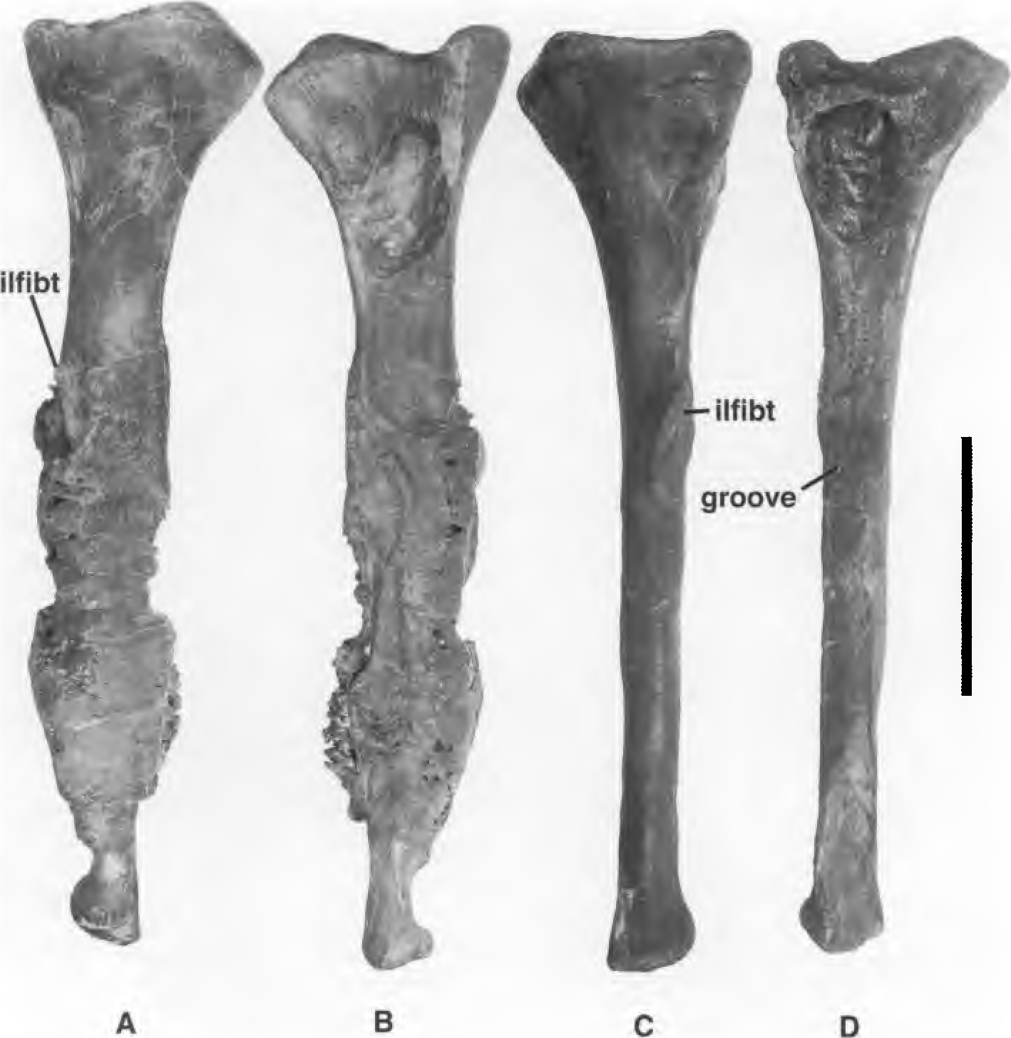
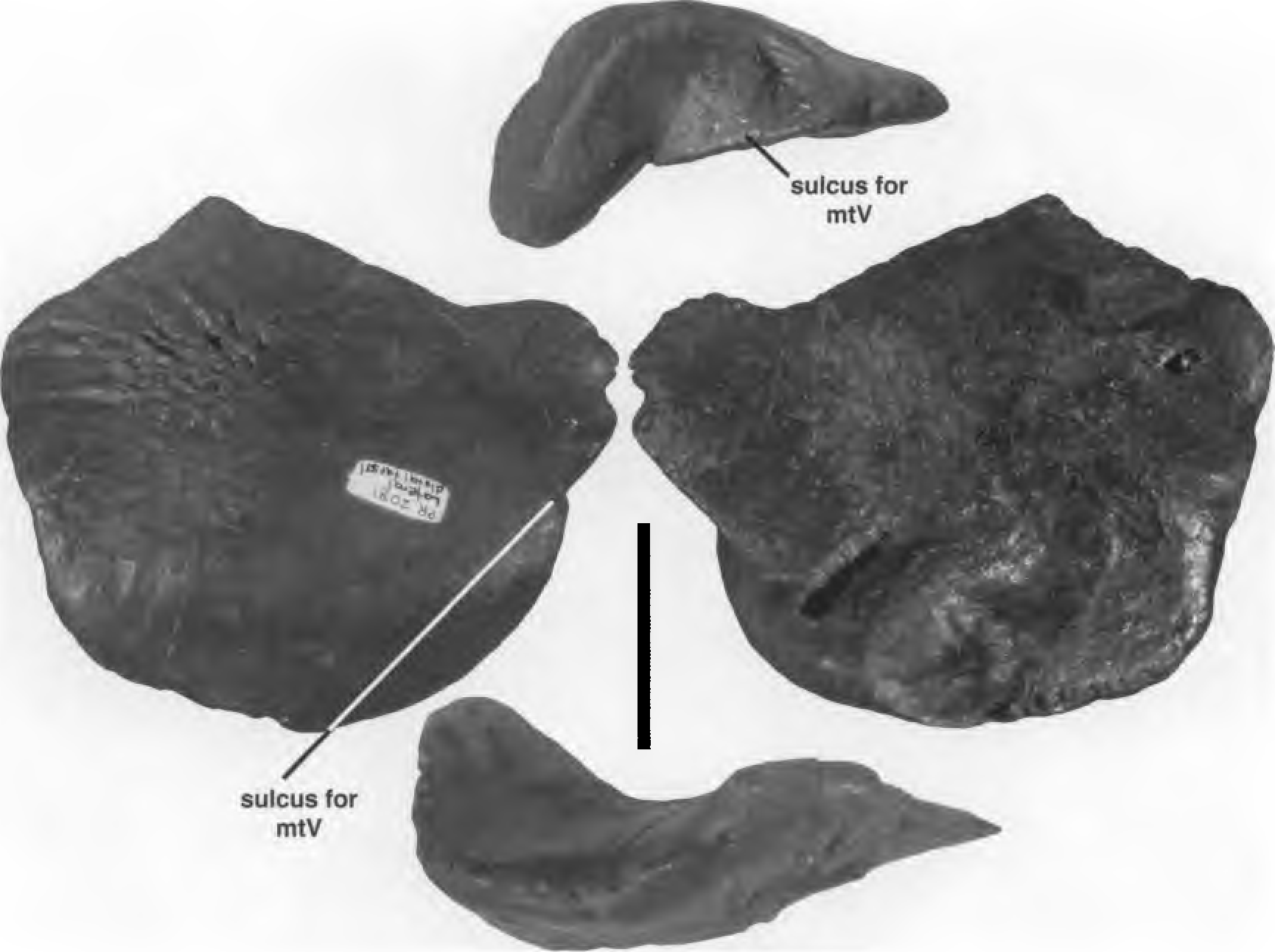
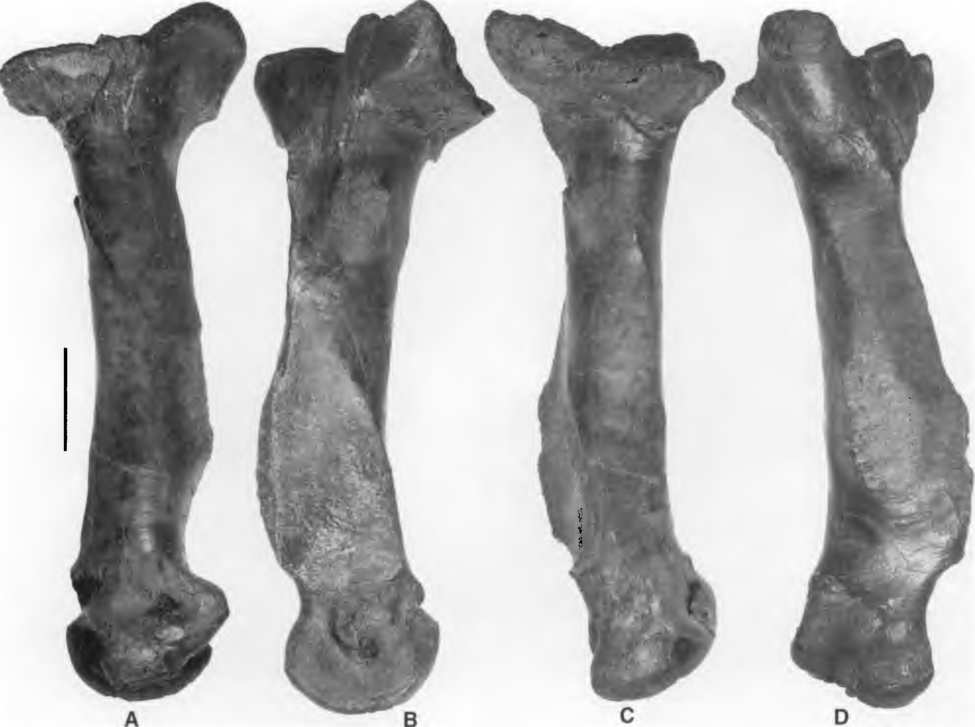
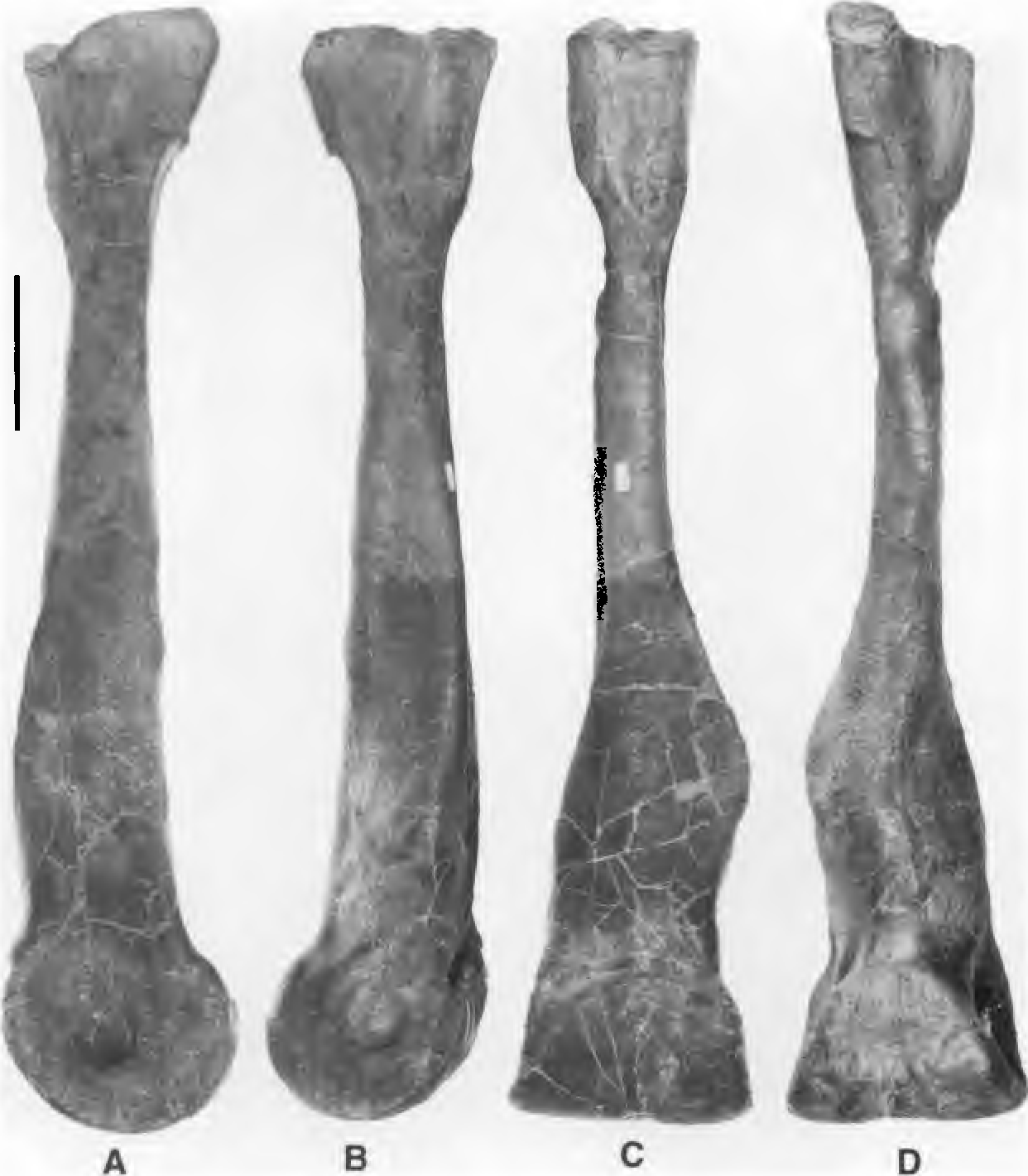

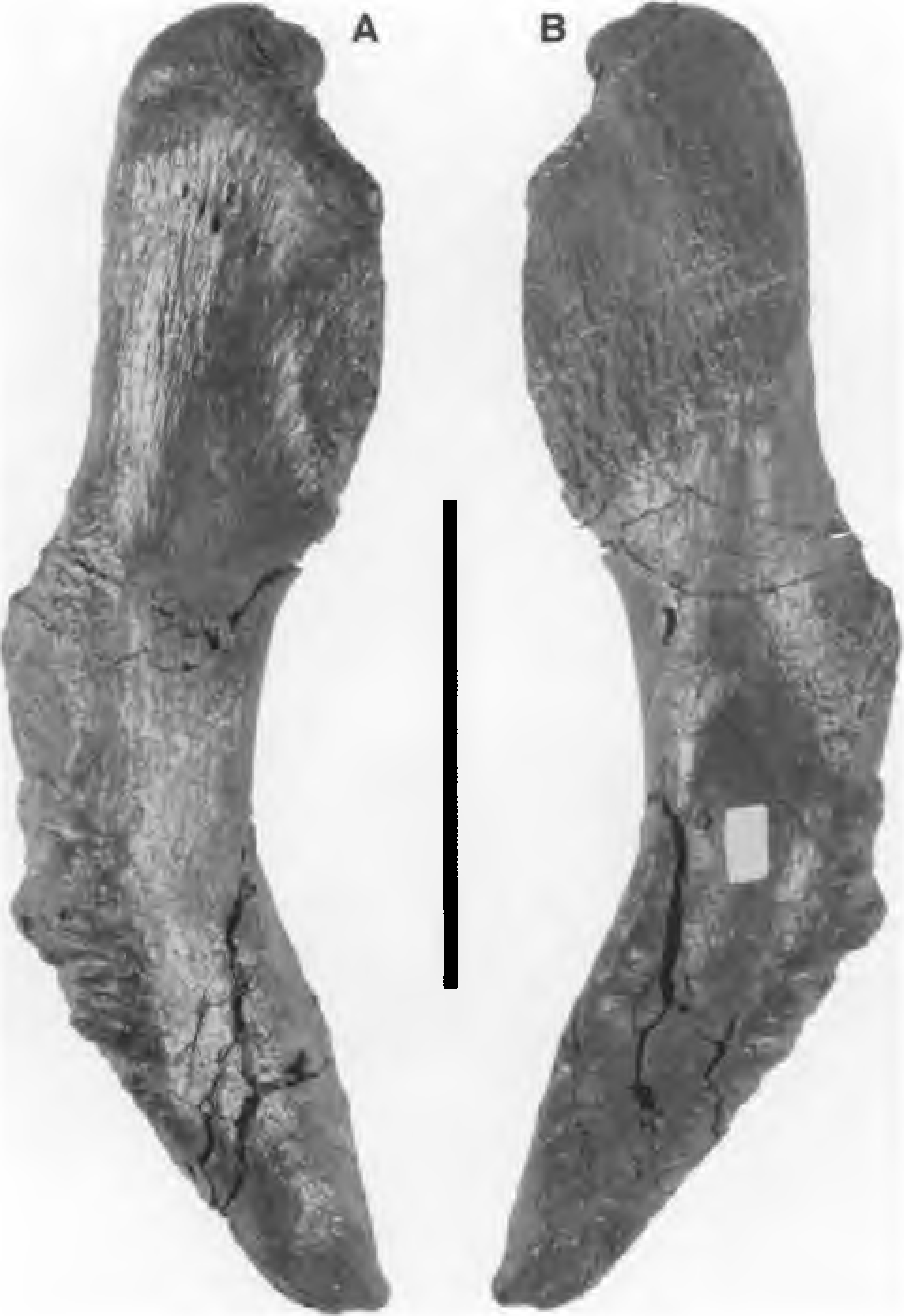
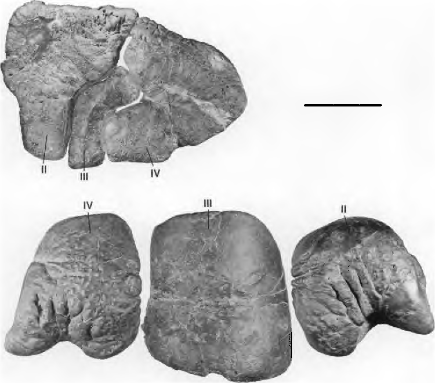
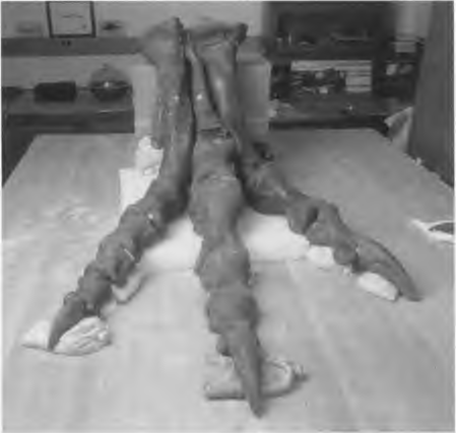
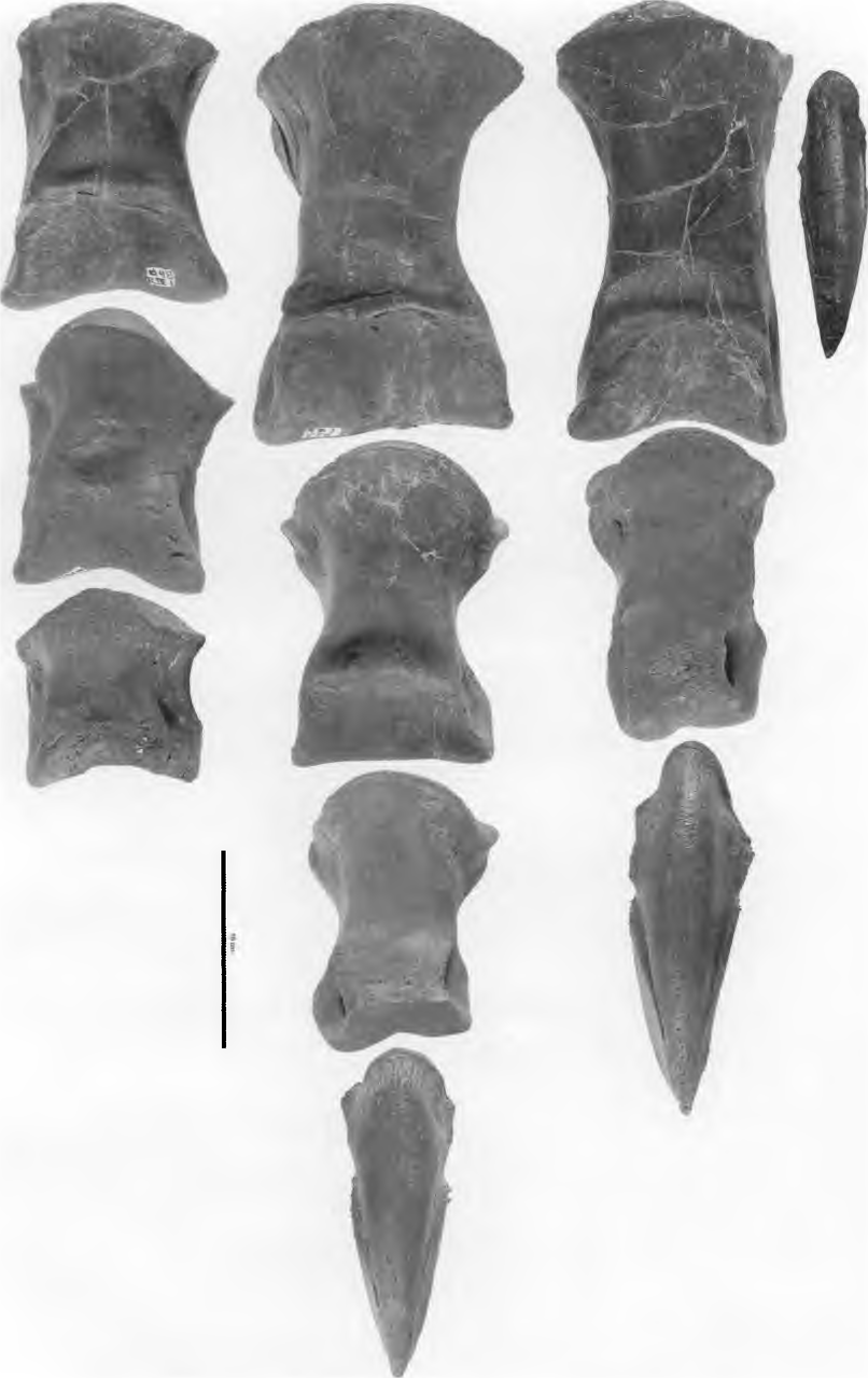
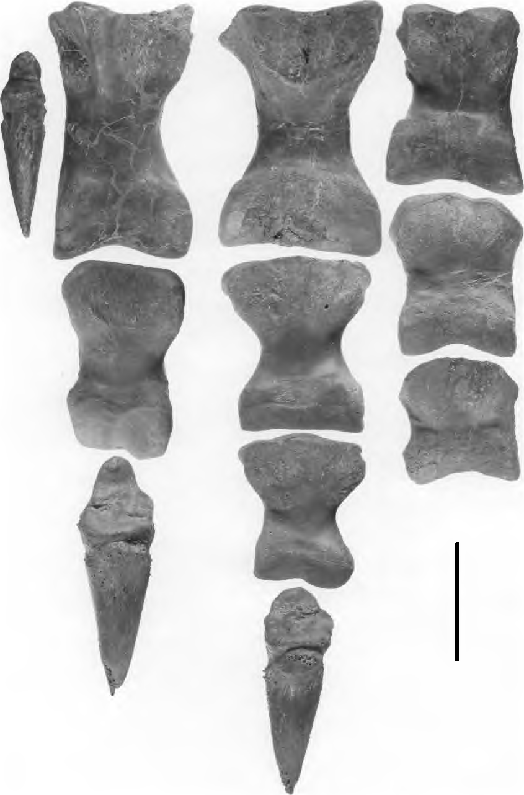
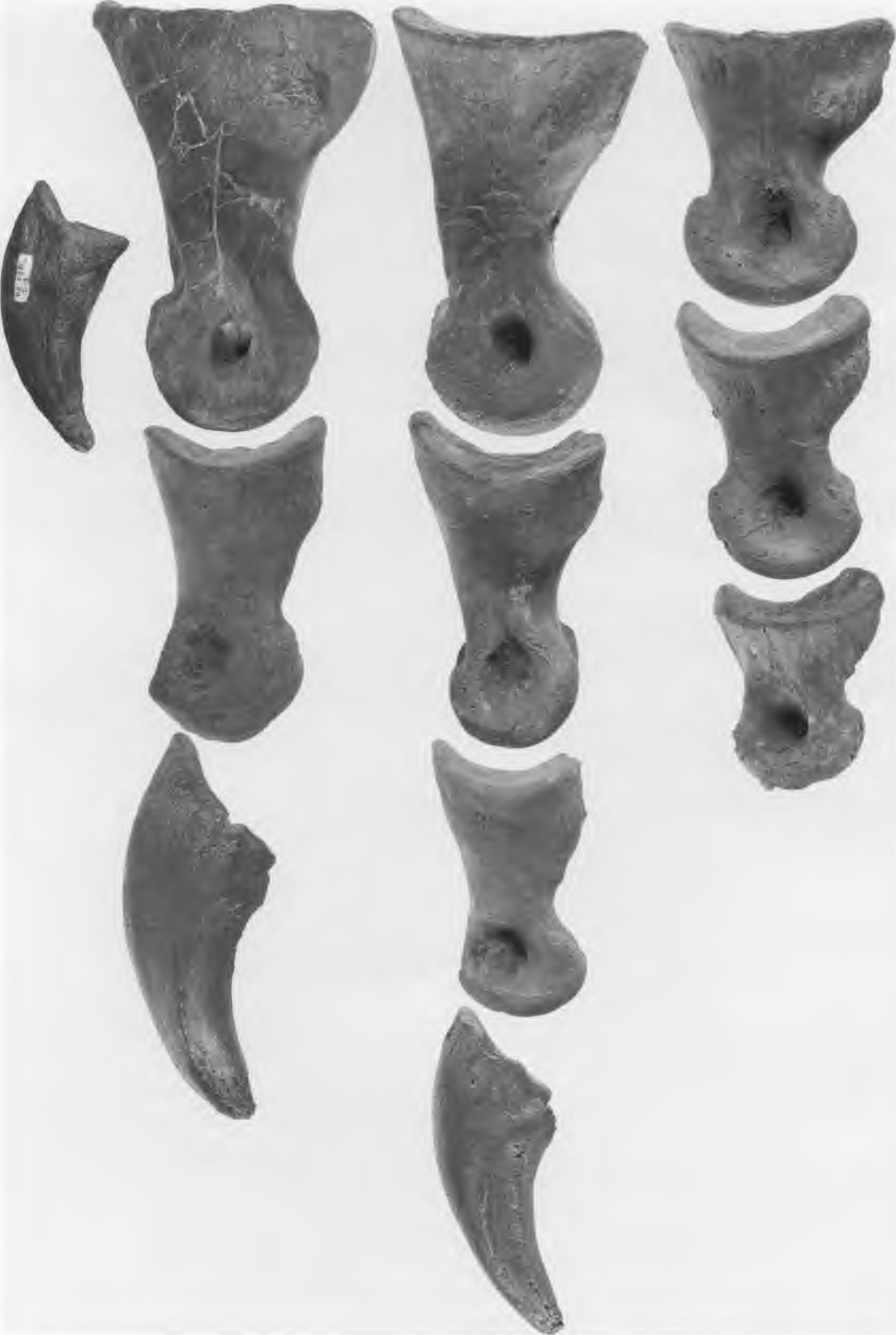
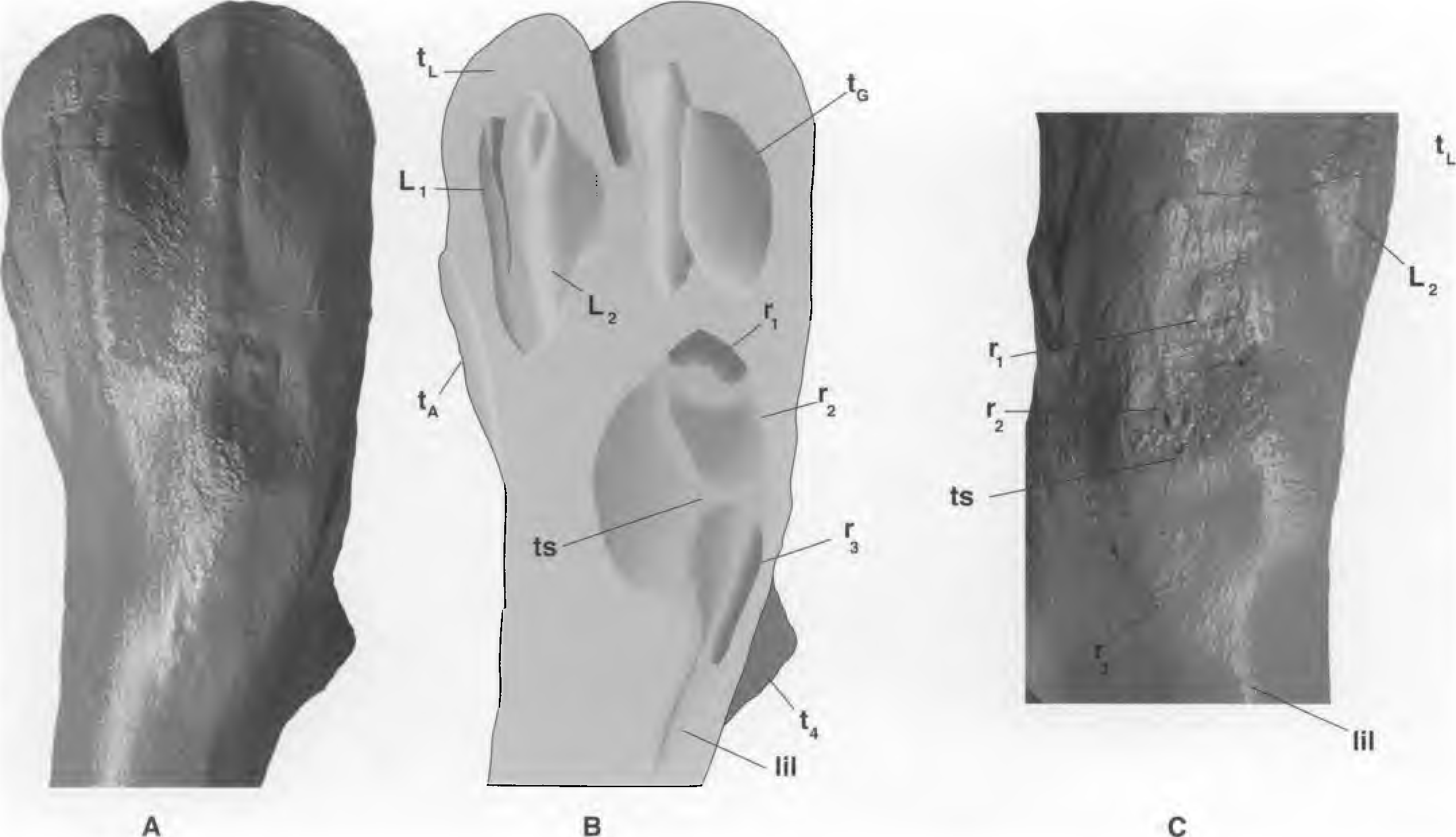
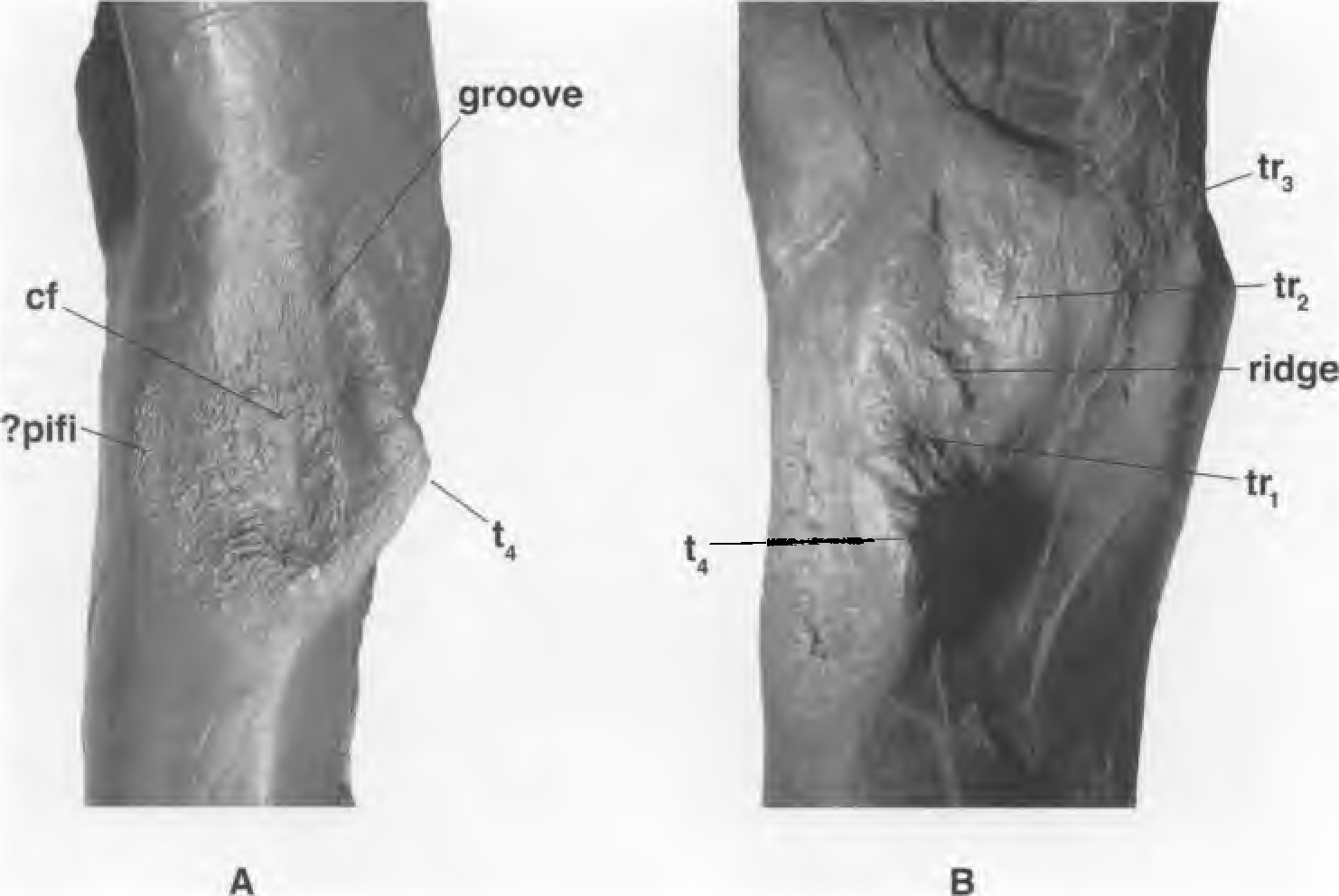

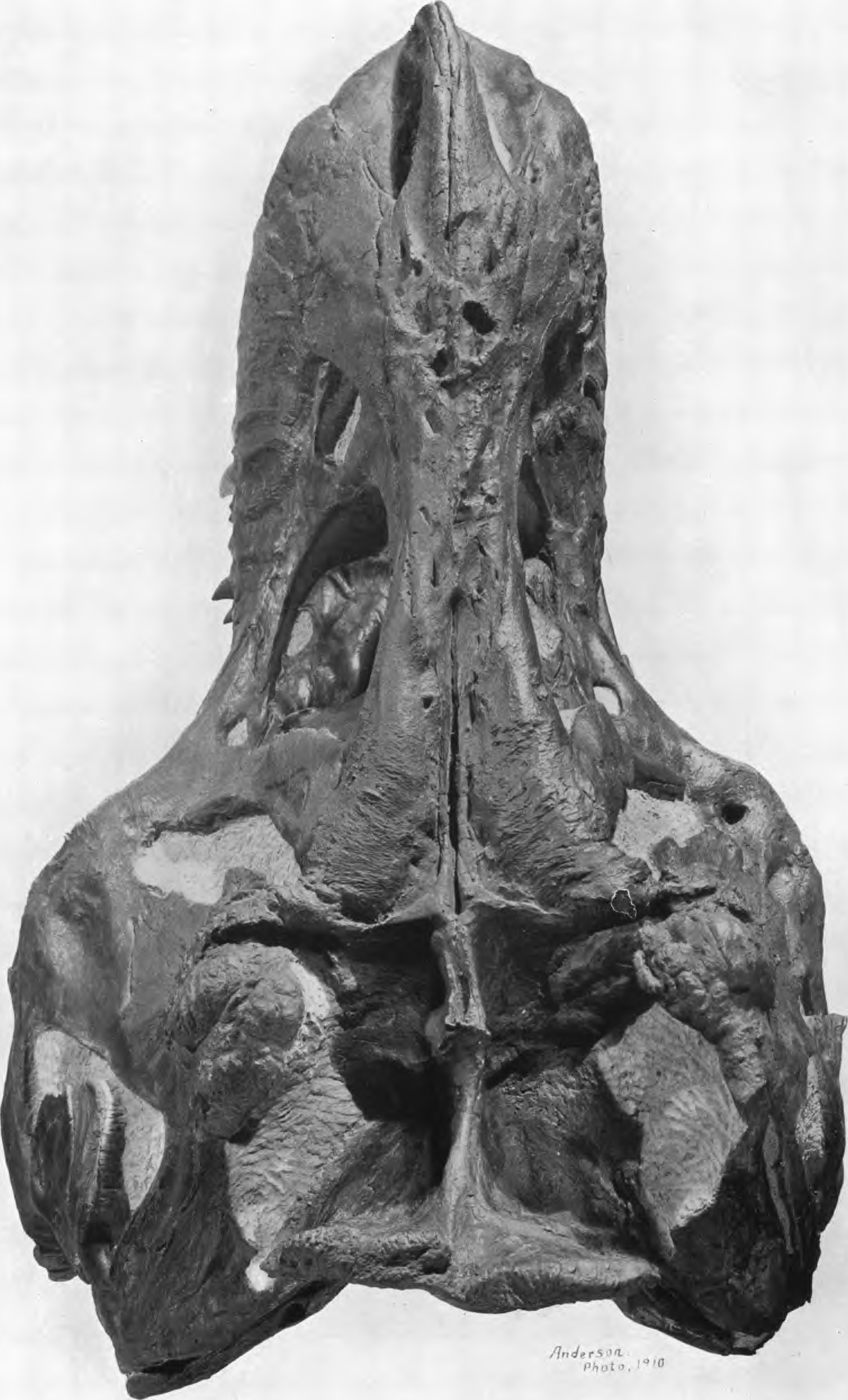
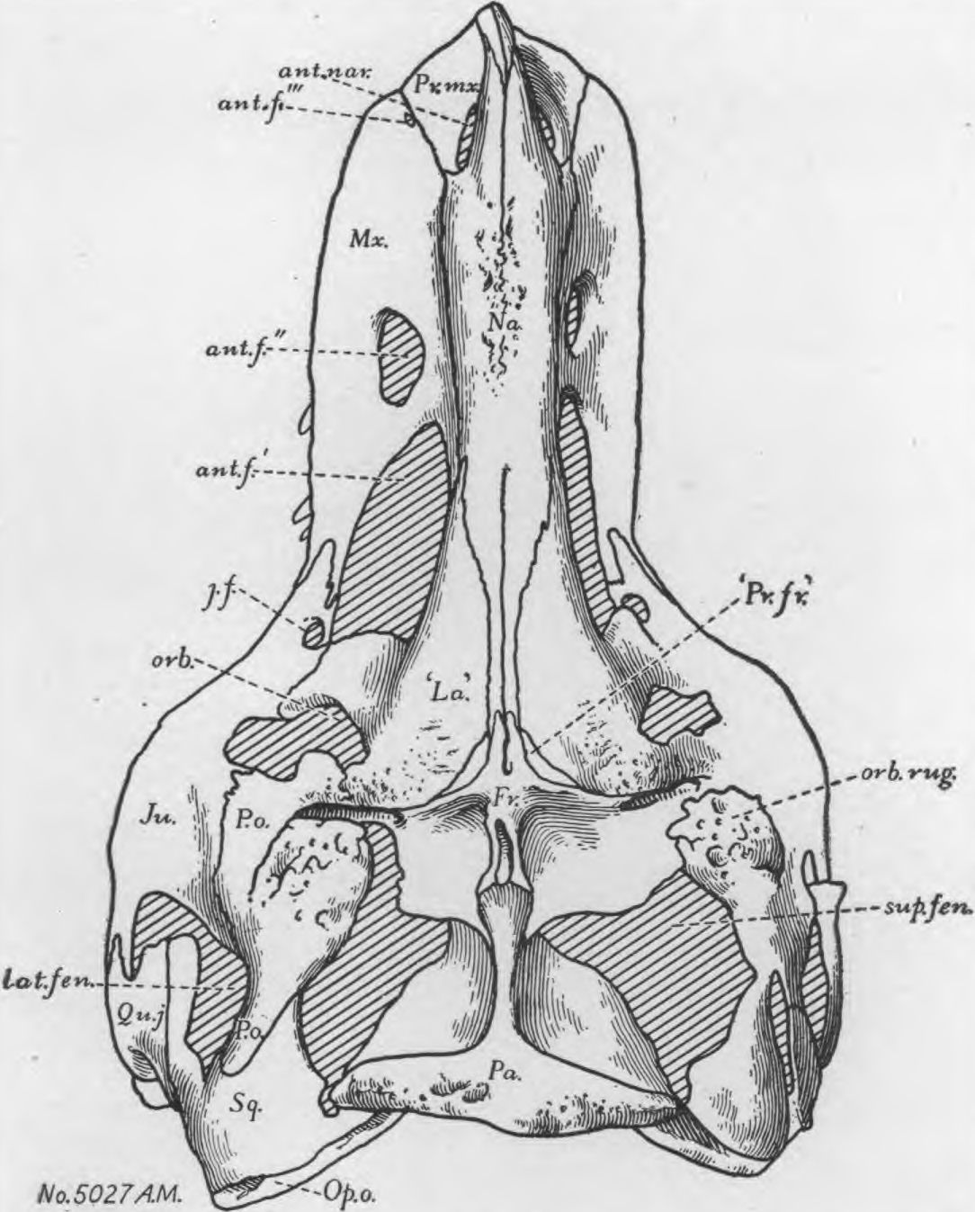
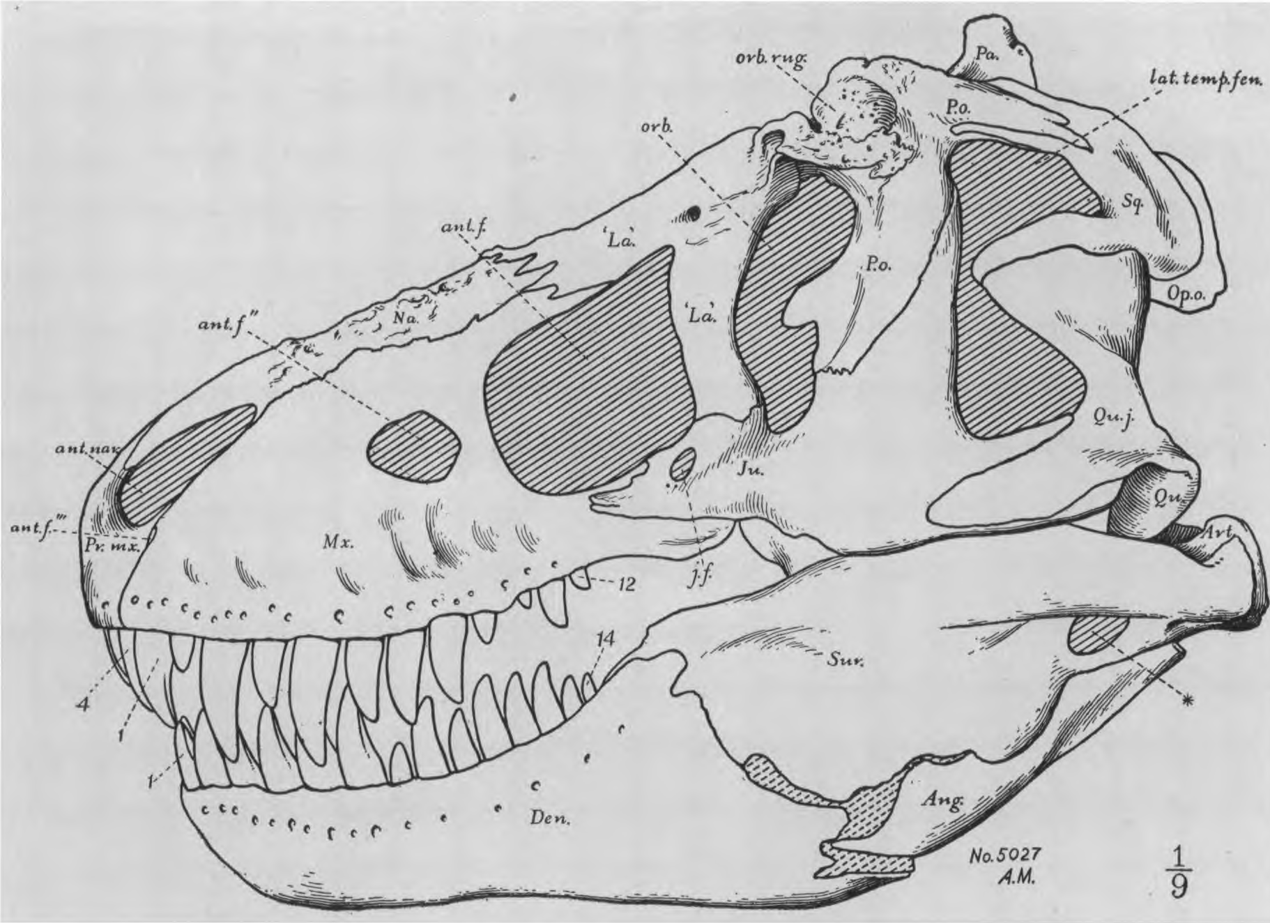
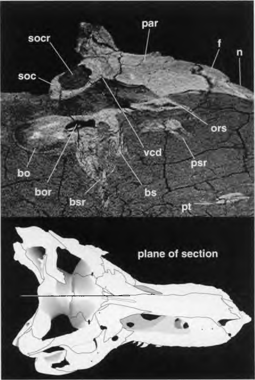
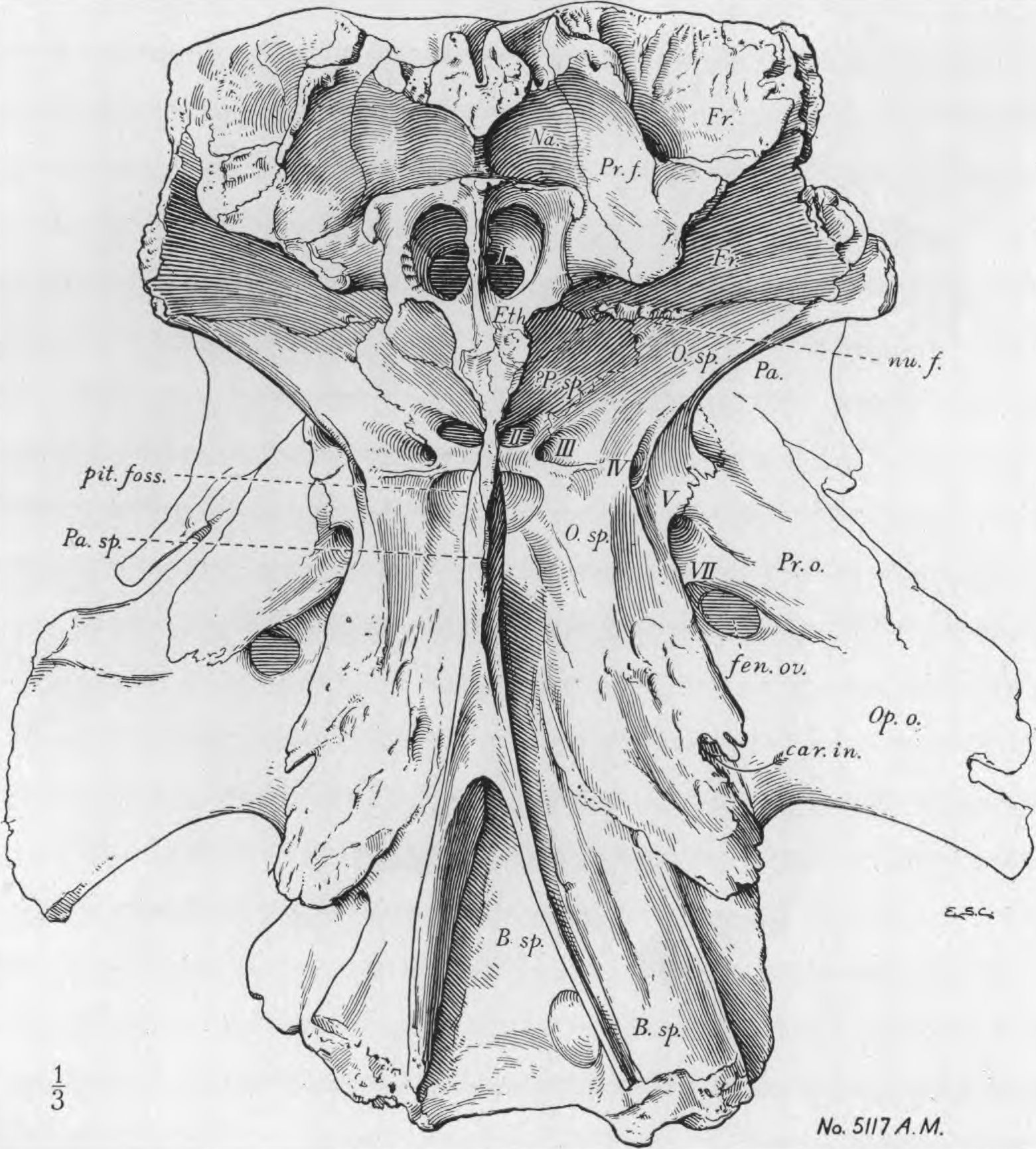
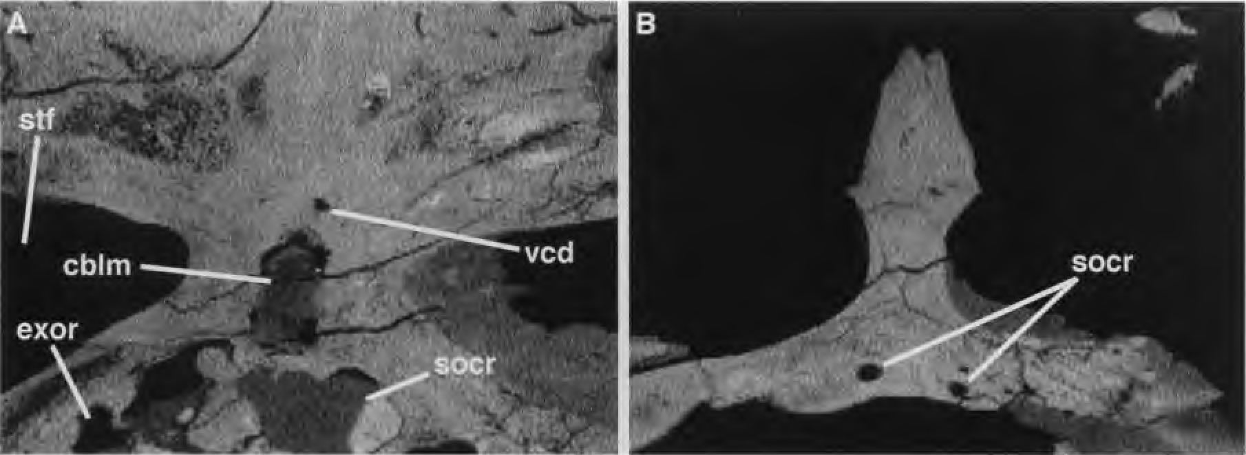

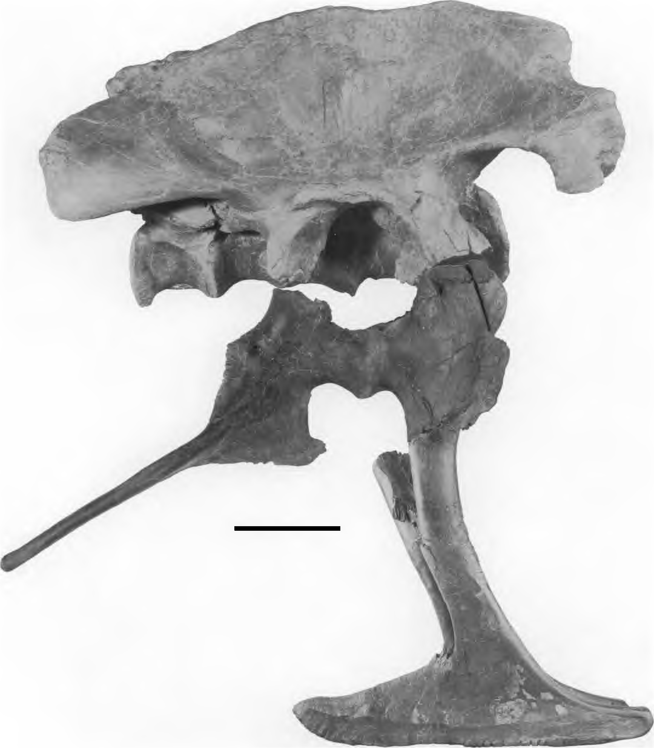
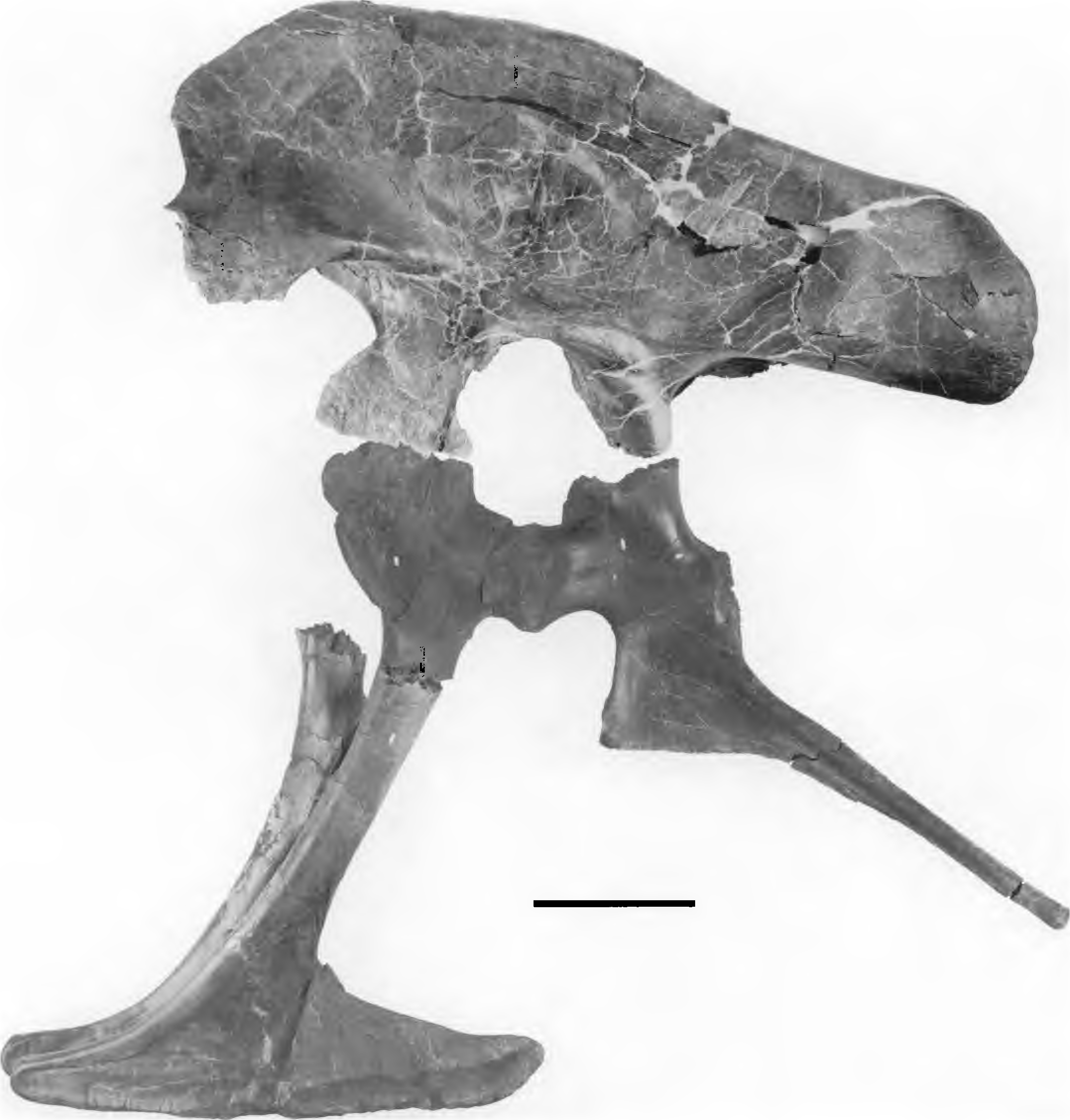
No known copyright restrictions apply. See Agosti, D., Egloff, W., 2009. Taxonomic information exchange and copyright: the Plazi approach. BMC Research Notes 2009, 2:53 for further explanation.