
Tarehylava, Stroiński, 2021
|
publication ID |
https://doi.org/ 10.37520/aemnp.2021.019 |
|
publication LSID |
lsid:zoobank.org:pub:1FA06A95-4F5D-4C7F-BAEA-42852D915D69 |
|
persistent identifier |
https://treatment.plazi.org/id/9A4E9552-9F64-FFD7-2256-F95AFE7CFF66 |
|
treatment provided by |
Marcus |
|
scientific name |
Tarehylava |
| status |
gen. nov. |
Tarehylava gen. nov.
( Figs 1–7 View Figs 1–10 , 11–63 View Figs 11–16 View Figs 17–22 View Figs 23–28 View Figs 29–34 View Figs 35–40 View Figs 41–46 View Figs 47–52 View Figs 53–63 )
Type species. Tarehylava avymaina sp. nov. (here designated).
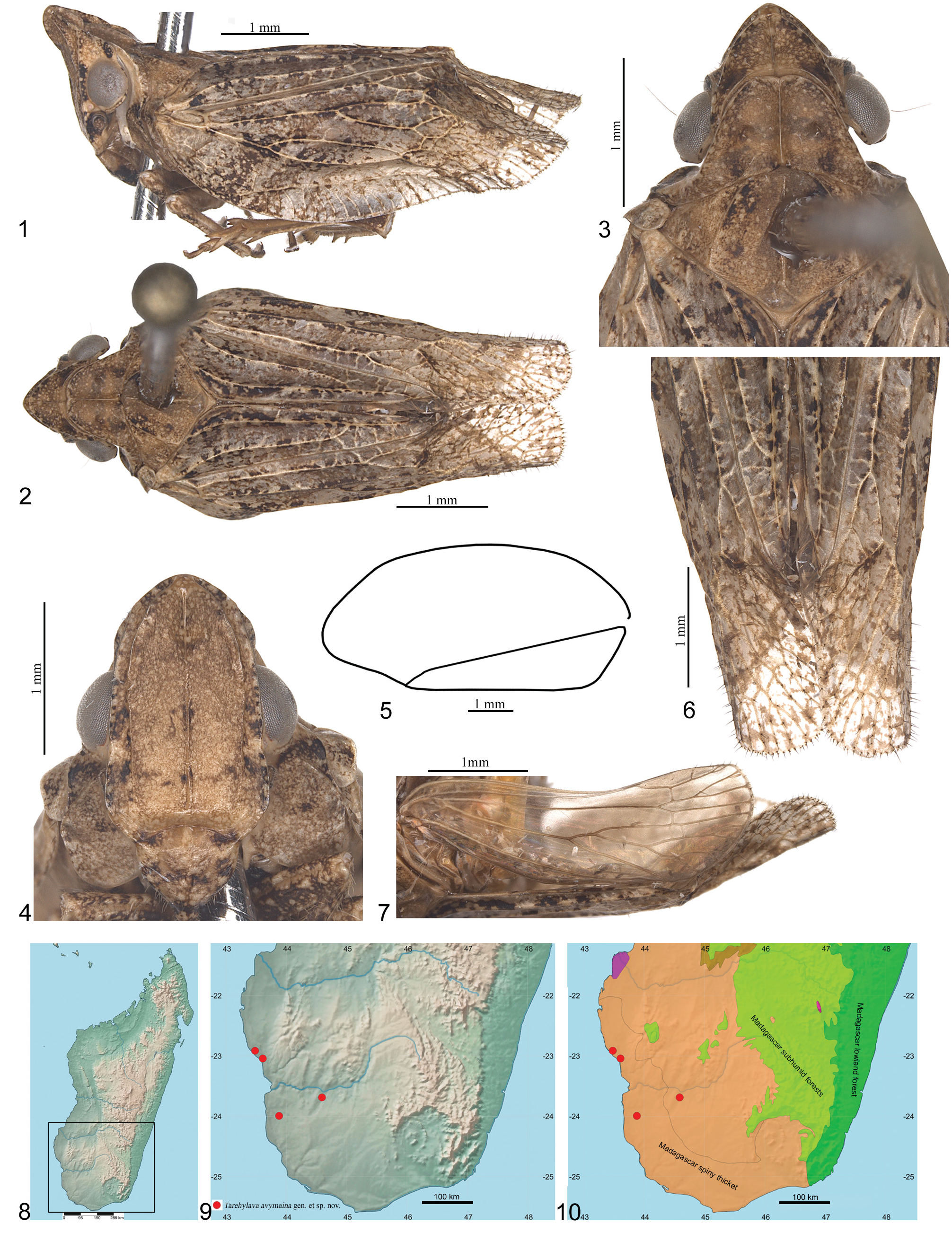
Diagnosis. Tarehylava gen. nov. differs from all other known Madagascan genera of Ricaniidae by the following characters: vertex arrowhead-shaped, frons elongate, lateral carinae of frons connected with median carina below upper margin and forming extension; lateral lobes of pronotum with transverse carina; tegmen divided in two parts, venation of posterior part swelling and flattened, with setae and sensory structures; costal margin of hind wing without precostal cell.
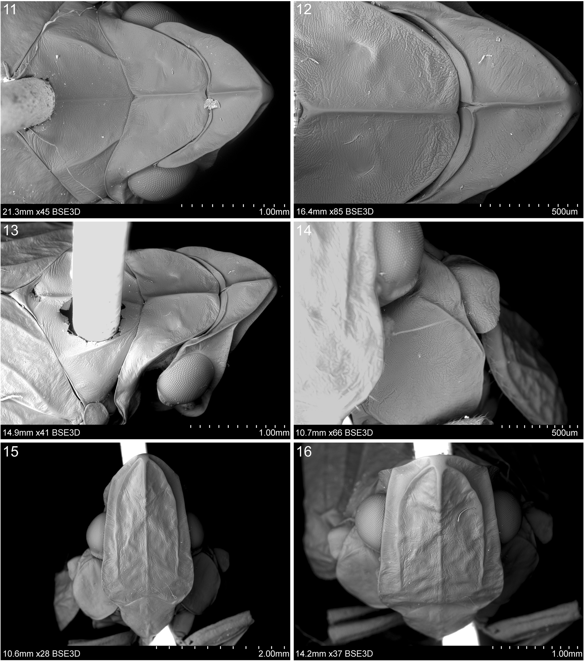
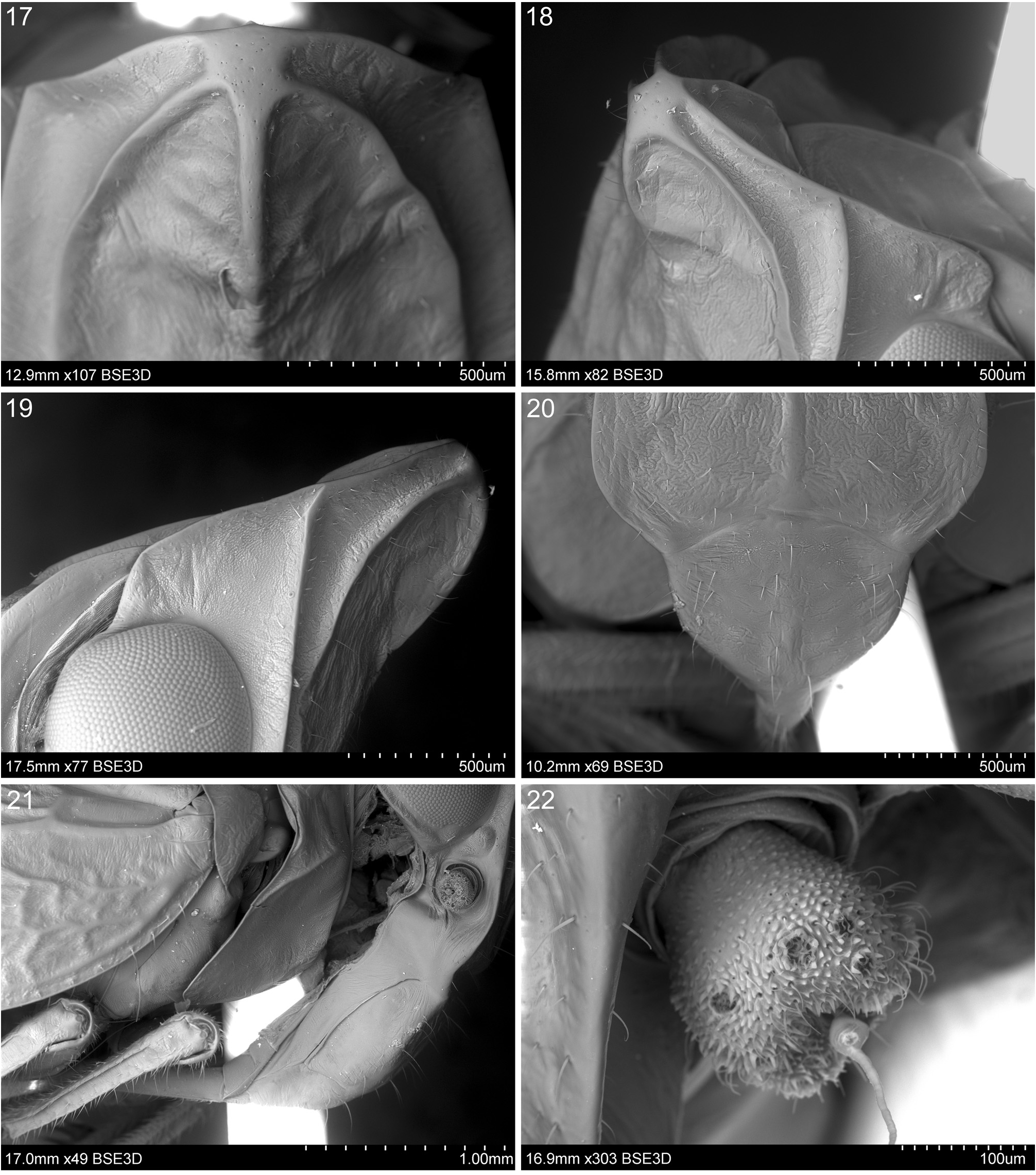
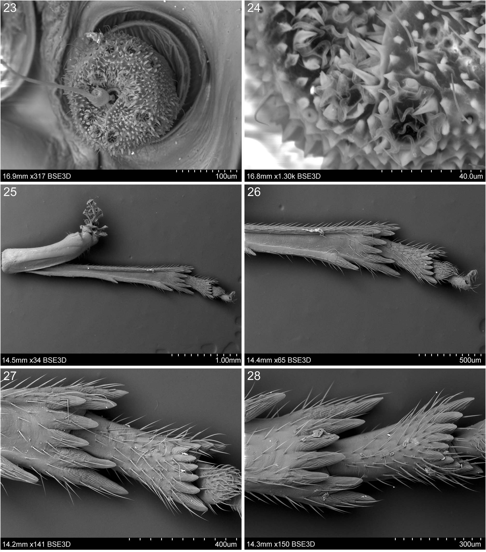
Description. Head with compound eyes (in dorsal view) slightly narrower than thorax. Vertex ( Figs 2–3 View Figs 1–10 , 11–13 View Figs 11–16 ) distinctly wider at the level of posterior angles than long at midline, arrowhead-shaped, posterior angles placed about the level of midlength of compound eyes; median portion of posterior margin placed before anterior margin of compound eyes; posterior margin strongly elevated medially. Disc of vertex with keel-shape median carina. Frons ( Figs 1, 4 View Figs 1–10 , 15–19 View Figs 11–16 View Figs 17–22 ) with all margins well carinated; frons at upper margin distinctly shorter than high at midline, widest at the lower part; lateral margins covering base of pedicel, incised at the level of ocelli. Frontal disc tricarinate, lateral carinae connected with median carina below upper margin of frons and forming extension; area between connection of frontal carinae and upper margin distinctly swollen; all carinae distinctly surpassing half of disc; median carina straight, lateral carinae in a form of horseshoe almost reaching fronto-clypeal suture, lateral carinae of same length or slightly shorter than median carina, all carinae around point of their connection distinctly elevated, keel-shaped. Disc of frons strongly wrinkled. Compound eyes with a very small callus at postero-ventral margin, elongate in dorsal view. Ocelli present. Antenna ( Figs 22–24 View Figs 17–22 View Figs 23–28 ): pedicel barrel-shaped with a slightly wider tip, with functional area (trichoid sensilla type 1 and antennal plate organs) reaching half of segment length on the dorsal and ventral surfaces, small on the lateral surface; plate organs of crenellated type, surrounded by a ring of elevated spines. Clypeus ( Figs 4 View Figs 1–10 , 15 View Figs 11–16 , 20 View Figs 17–22 ) distinctly narrower than frons, with median carina well visible in lower part. Rostrum with apical segment slightly shorter than subapical one, reaching hind coxae.
Thorax. Pronotum ( Figs 1–3 View Figs 1–10 , 11–13 View Figs 11–16 ) distinctly longer than vertex at midline; disc of pronotum with elevated median carina and two lateral impressions; lateral lobes of pronotum with fully developed postocular transverse carinae, reaching both margins; anterior margin of pronotum placed before the line of anterior margin of compound eyes. Mesonotum ( Figs 1–3 View Figs 1–10 , 11 View Figs 11–16 , 30 View Figs 29–34 ) about as long as combined length of vertex and pronotum at midline and wider in lateral angles than long at midline; disc of mesonotum tricarinate with median carina and lateral carinae present; antero-lateral carinae absent; median carina and lateral carinae separated basally, subparallel; median carina reaching scutellum, lateral carinae reaching posterior margin; lateral angles placed before midlength. Hind legs ( Figs 25–28 View Figs 23–28 ). Metatibia distinctly longer than metafemur, not widened at distal part; metatibia in basal half with two remnants of lateral spines weakly visible in some specimens, two well-developed lateral spines placed distally to each other in distal half, and apical row of well-developed teeth (2+5, 7), different in size and forming irregular line; lateral spines of equal size, larger than internal. Metatarsus with basitarsomere about as long as cumulative length of second and third tarsomeres, with asymmetrical V-shaped row of 9–10 teeth: two lateral teeth different in size, external lateral spine larger than internal lateral one which is similar in size to intermediary spines; each internal tooth bearing strong seta; mesotarsomere with pad of strong setae on ventral side.
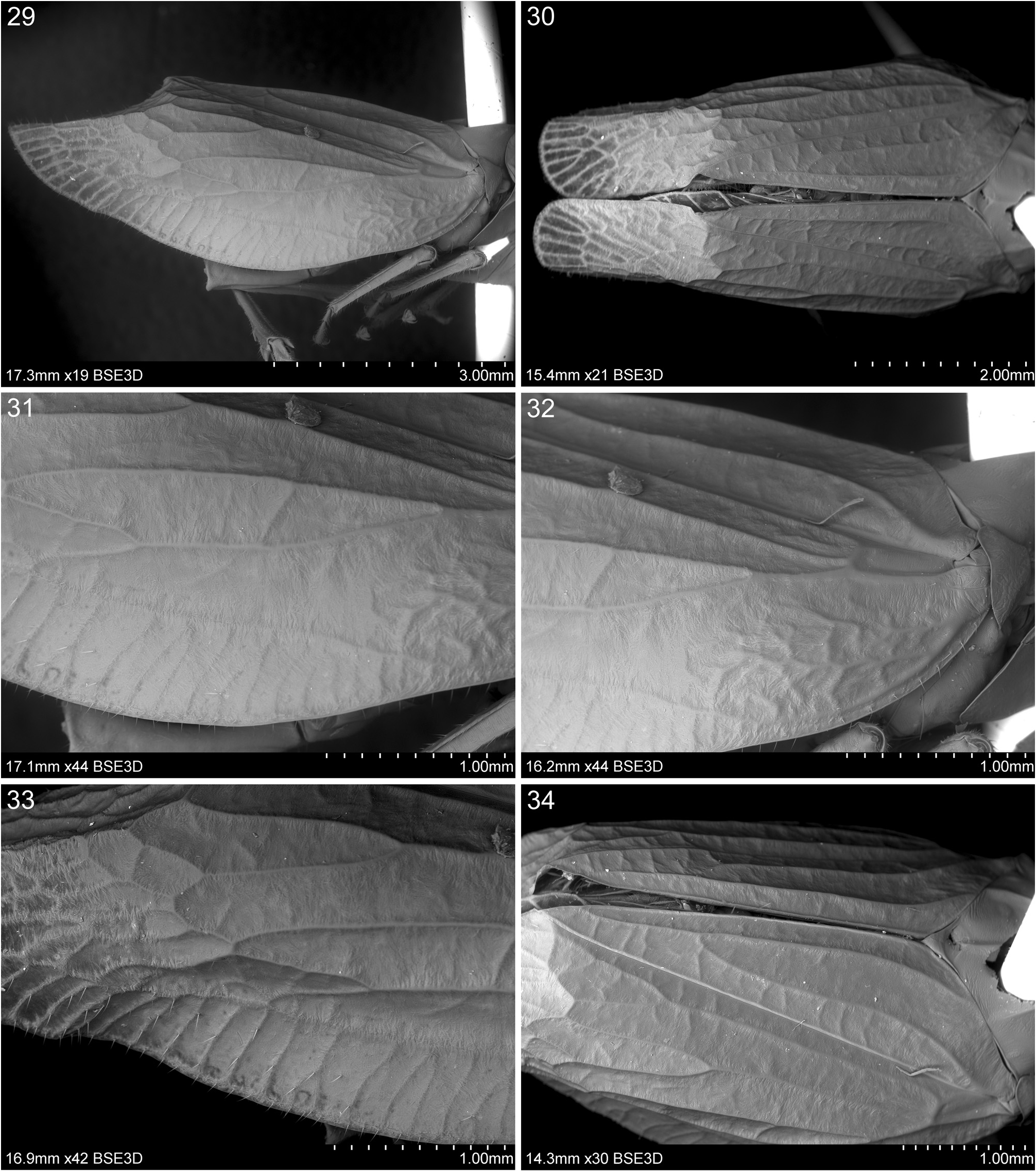
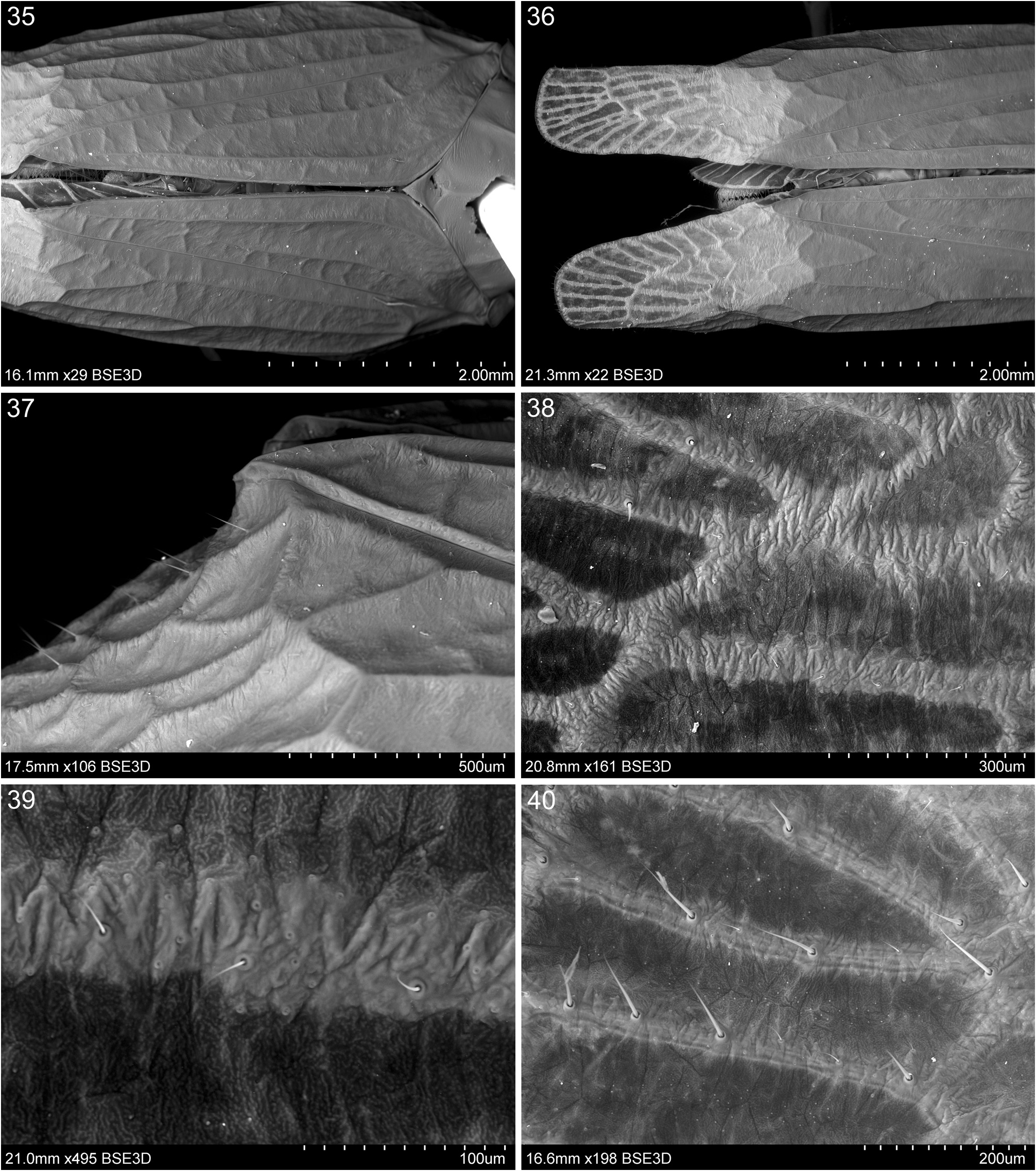
Tegmina ( Figs 1–2, 6 View Figs 1–10 , 29–40 View Figs 29–34 View Figs 35–40 ) elongate, semi-convex in basal part, partly coriaceous basally to the level of clavus apex, posterior part of tegmen from the level of clavus apex membranous, partially flexible; tegmina with distinct venation and transverse veinlets, venation of posterior part swelling and flattened, with setae and sensory structures. Costal margin arcuate, apical angle broadly rounded, posterior margin tapered with rounded tip ( Fig. 5 View Figs 1–10 ); tornus (postclaval margin) present and weakly concave. Costal area with dense transverse veinlets, ending at the level of clavus apex; very narrow basally (to 1/3 of length), distinctly wider (also wider than costal cell) more posteriorly. Costal cell with irregular net of transverse veinlets in basal part. Basal cell elongate, narrow, distinctly longer than wide (about 3 times). Longitudinal veins ScP+RA, MP and CuA leaving basal cell separated; all first forks of longitudinal veins placed distinctly before half of tegmen; veins ScRA and RP arising as long common stem from basal cell; first fork placed before first fork of MP; first fork of MP placed before or almost at claval connection (never extending beyond this level); Cu stem always longer than MP stem with first fork at the level of claval connection or more posteriorly. Nodal line absent. Cubital cell with transverse veinlets (weakly visible in some specimens). Clavus closed; CuP ending at margin, claval veins (Pcu and A1) fused distinctly after midlength of CuP; posterocubital cell (basal and posterior parts), postcubital and anal cells with transverse veinlets, transverse veinlets of postcubital and anal cells weakly visible in some specimens.
Hind wing ( Fig. 7 View Figs 1–10 ) shorter than tegmina, with posteri- or margin reaching midlength of the area between tip of clavus and posterior margin of tegmina; anterior margin without precostal cell; ScRA and MP forking distinctly after midlength of wing in distal part, first fork of MP slightly more distad than ScRA but both close each other, CuA forking approximately at wing midlength; ScRA with single terminal, RP with 2–3, MP with 3–4, CuA with 6–7 terminals; rp-m, m-cua (1–2) and icu (1–4, variable character) transverse veinlets present in distal part of wing.
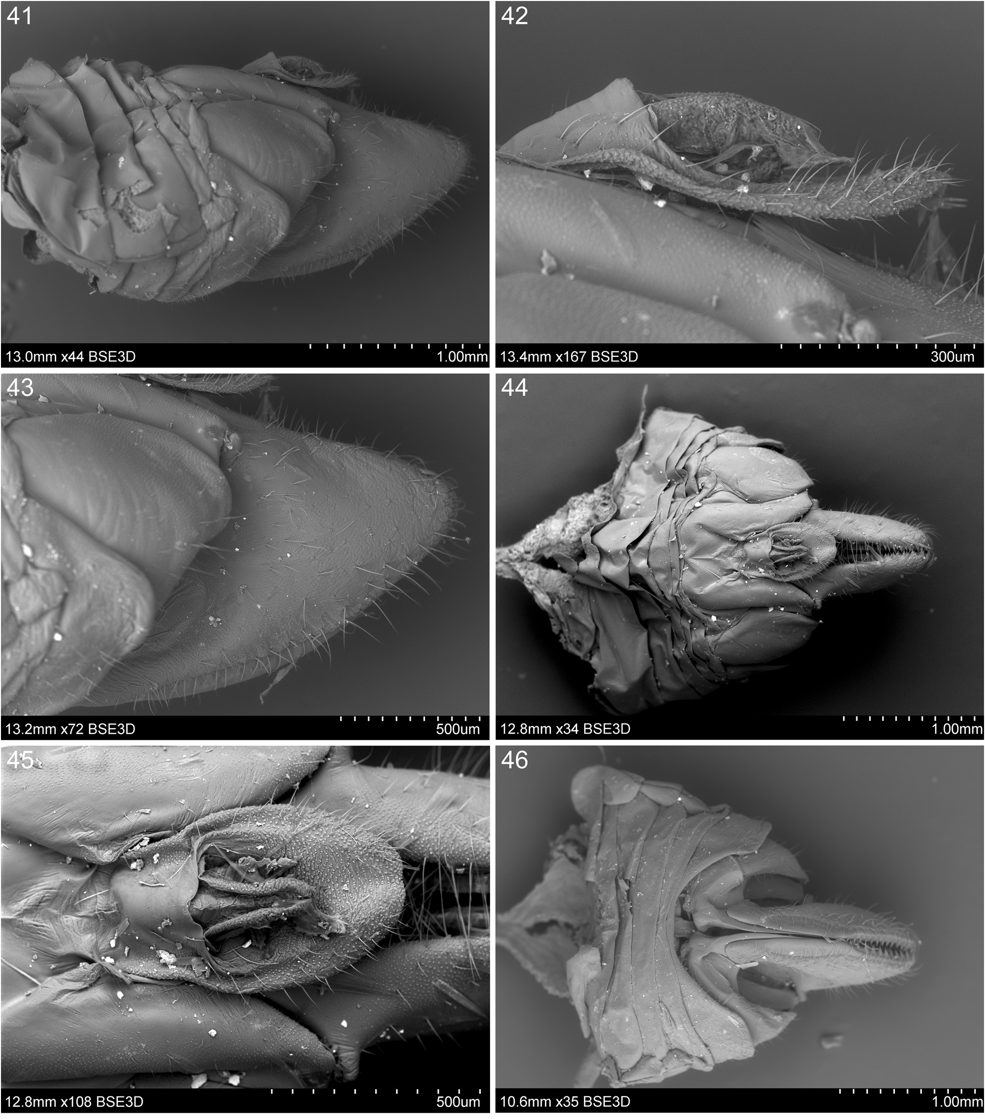
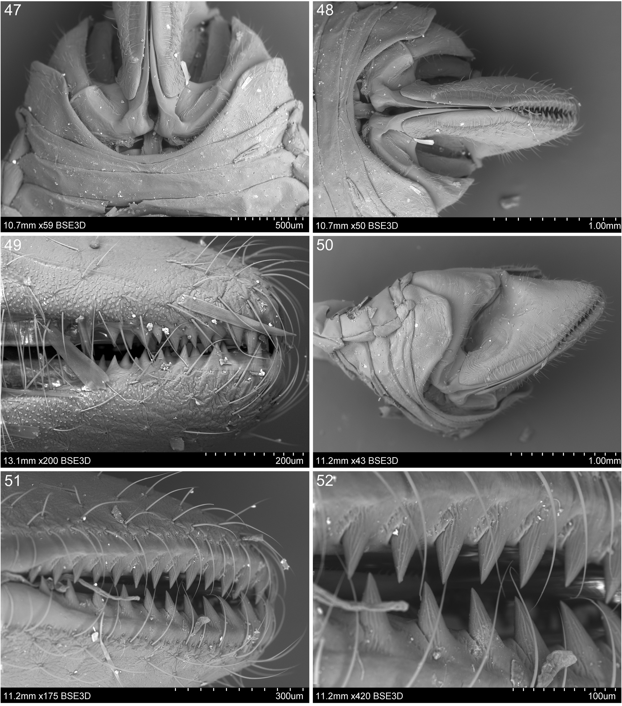
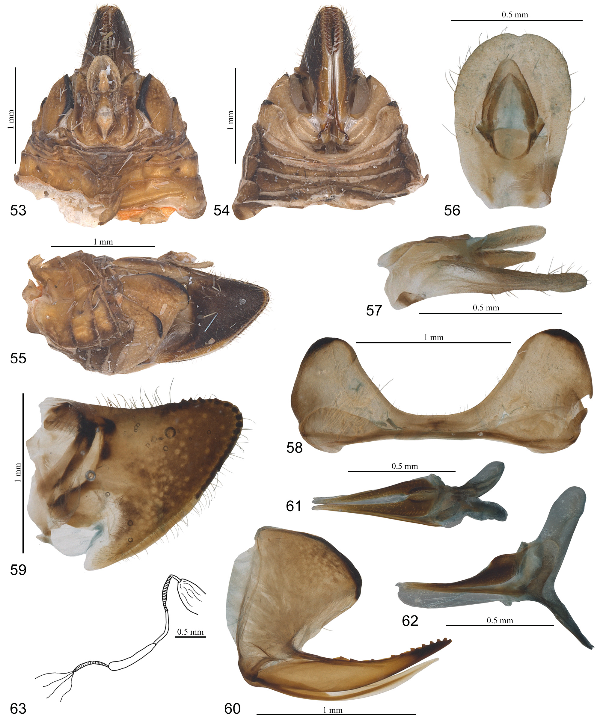
Female terminalia ( Figs 41–63 View Figs 41–46 View Figs 47–52 View Figs 53–63 ). Pregenital sternite ( Figs 46–48 View Figs 41–46 View Figs 47–52 , 54, 58 View Figs 53–63 ) with well-developed, elongate-oval and distinctly separated lateral lobes; median part of pregenital sternite narrow, anterior and posterior margins almost straight medially; posterior margin without any process. Anal tube in lateral view ( Figs 41–42 View Figs 41–46 , 55, 57 View Figs 53–63 ), elongate, not extending beyond the midlength of upper margin of gonoplac; basal part of anal tube distinctly wider than posterior one; anal opening, in dorsal and lateral views, placed before midlenght; anal tube, in dorsal view ( Figs 44–45 View Figs 41–46 , 53 View Figs 53–63 ), elongate ovoid, wider at midlenght; anal style (paraproct) and anal segment (epiproct) short, not extending beyond posterior margin of anal tube. Gonoplac ( Figs 41–43, 46 View Figs 41–46 , 49–52 View Figs 47–52 , 55, 59 View Figs 53–63 ) well developed, unilobate, laterally flattened; posterior margin of the gonoplac with single row of well-developed teeth placed alongside apical part of dorsal margin and upper half of posterior margin; membranous parts of gonoplac in two parts: first part narrow, weakly sclerotized, placed on lower part of posterior margin below teeth, second part large, fully membranous, placed ventro-basad of the gonoplac. Gonapophysis VIII ( Fig. 60 View Figs 53–63 ) elongate, sabre-like, V-shaped in cross section, with teeth at posterior part of dorsal margin; endogonocoxal process tapering apicad, slightly shorter than gonapophysis VIII, with median sclerotized belt surrounded by membranous part. Gonapophyses IX and gonospiculum bridge well developed as in Figs 61–62 View Figs 53–63 . Bursa copulatrix with two pouches connected with short ductus; first pouch elongate, with cells and sclerotized ornamentation (except dorsal part), with sclerotized plate with very large median sclerite and 5–8 small petals around; second pouch elonga- te-oval, smaller than first one, without cell but with sclerotized plates. Spermatheca ( Fig. 63 View Figs 53–63 ) well-developed; ductus receptaculi elongate and narrow, ribbed; diverticulum ductus with two parts (about the same length), distinctly longer (about twice) than ductus receptaculi, with long, narrow, smooth basal ductus, and long, smooth, wider apical ductus.
Male. Unknown.
Etymology. The generic name Tarehylava is a combination of two Malagasy words: “tarehy” (= face) and “lava” (= long), which refers to the characteristic prolongation of the frons. Gender feminine.
Distribution. South-western Madagascar: Atsimo-Andrefana region ( Figs 8–10 View Figs 1–10 ).
No known copyright restrictions apply. See Agosti, D., Egloff, W., 2009. Taxonomic information exchange and copyright: the Plazi approach. BMC Research Notes 2009, 2:53 for further explanation.
|
Kingdom |
|
|
Phylum |
|
|
Class |
|
|
Order |
|
|
Family |