
Penicillidmus, Jałoszyński, Paweł, 2014
|
publication ID |
https://doi.org/ 10.11646/zootaxa.3774.1.1 |
|
publication LSID |
lsid:zoobank.org:pub:B5A2EF46-2BF6-4ED3-A5F4-5F9951400545 |
|
DOI |
https://doi.org/10.5281/zenodo.5664982 |
|
persistent identifier |
https://treatment.plazi.org/id/9A6687E6-4F0B-3030-FF34-FE32FBE2FBB6 |
|
treatment provided by |
Plazi |
|
scientific name |
Penicillidmus |
| status |
gen. nov. |
Penicillidmus View in CoL gen. n.
Type species: Penicillidmus masseyensis sp. n.; here designated.
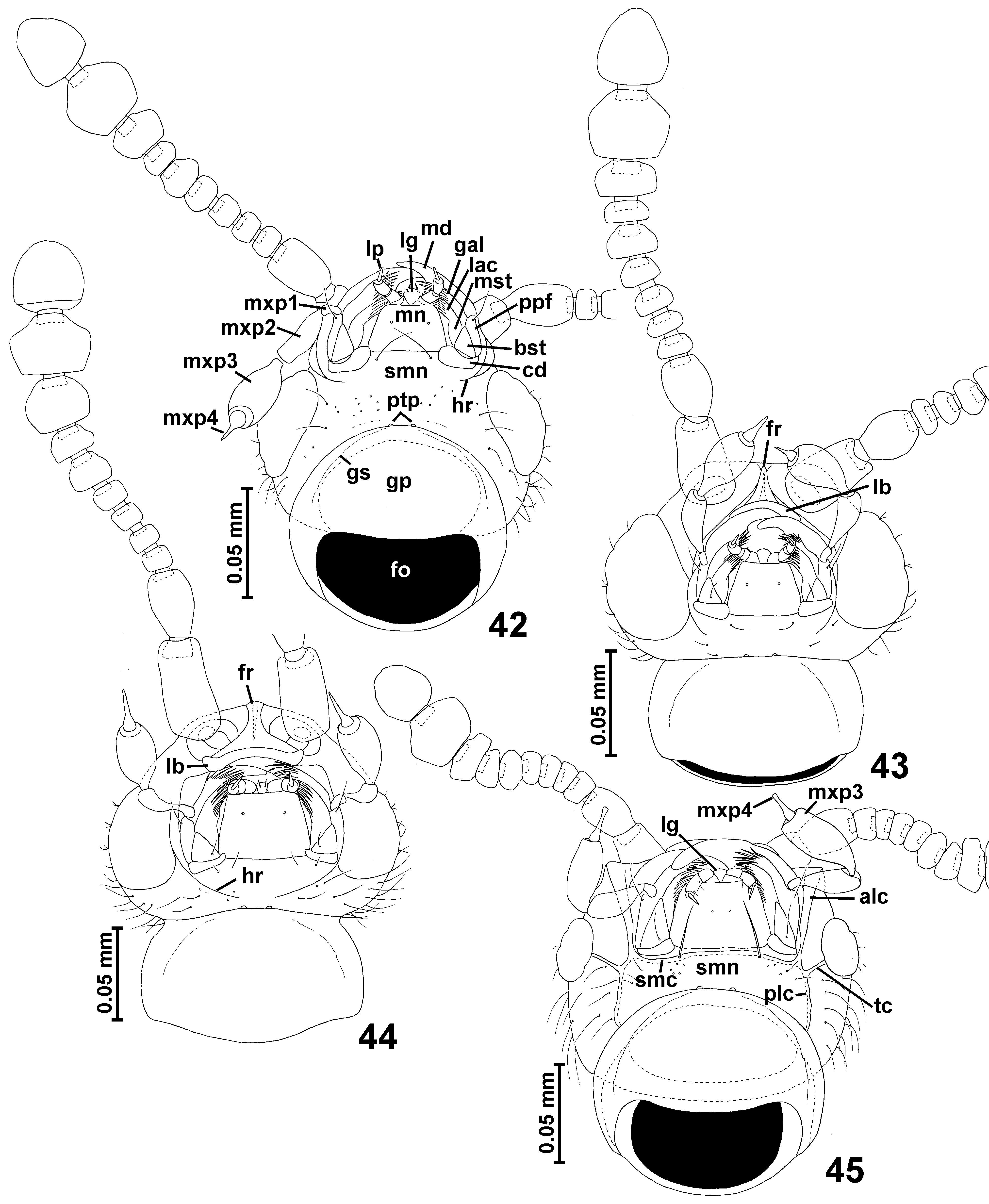
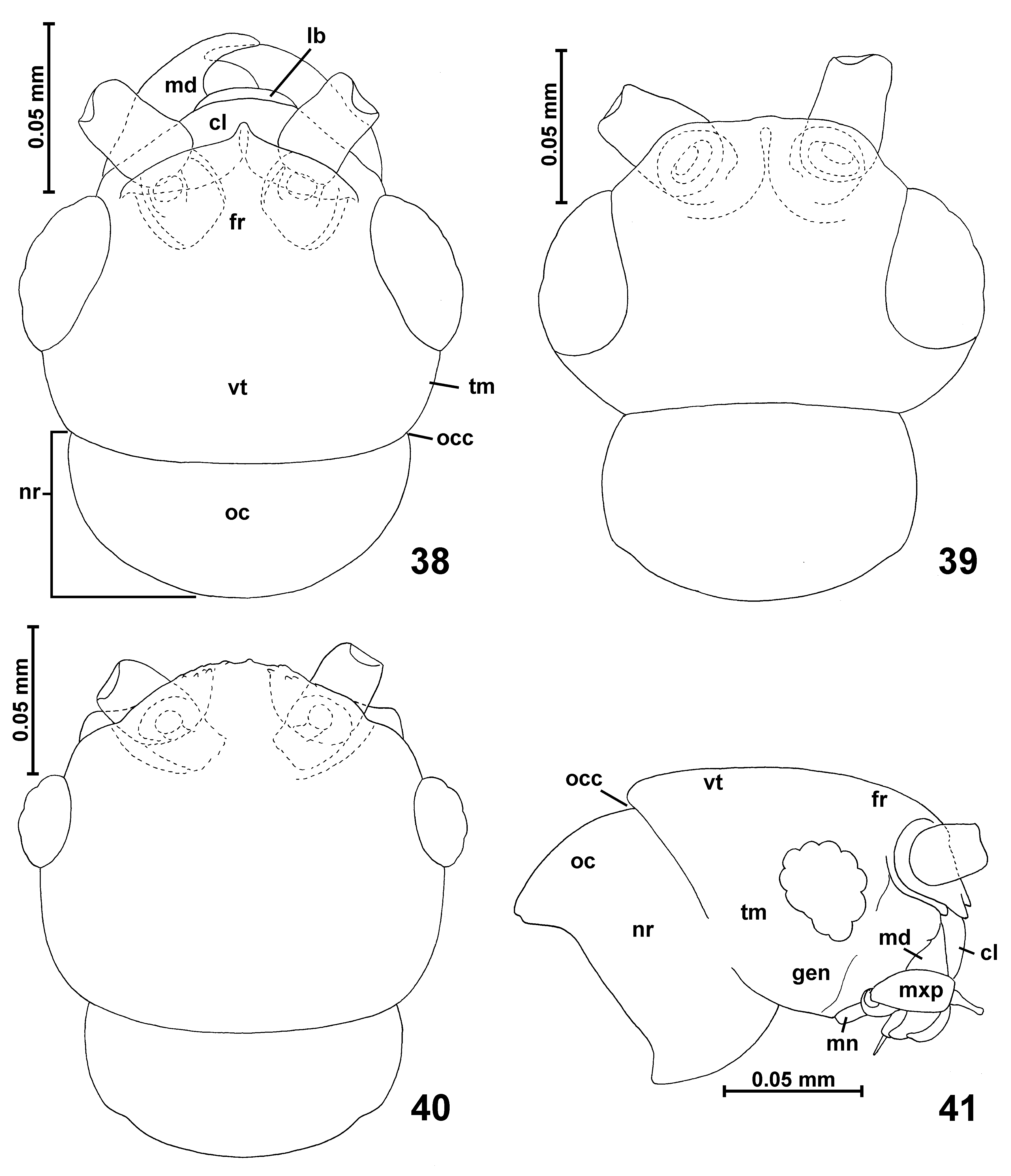
Diagnosis. Penicillidmus is defined by three characters unique among Cyrtoscydmini : i) system of carinae on ventral surface of head posterior and lateral to mouthparts composed of submental carina ( Fig. 45 View FIGURES 42 – 45 ; smc) running along anterior margin of submentum and extending laterally along cardinal insertions, longitudinal lateral carinae with posterior and anterior arms ( Fig. 45 View FIGURES 42 – 45 ; plc and alc, respectively) and transverse carinae ( Fig. 45 View FIGURES 42 – 45 ; tc) connecting longitudinal carinae with meso-ventral margins of compound eyes; ii) expanded apex of maxillary palpomere IV ( Fig. 45 View FIGURES 42 – 45 ; mxp4); and iii) postero-lateral brush of 2–4 thick bristles ( Fig. 49 View FIGURES 46 – 49 ; plb) at each side of pronotum. Additionally, Penicillidmus has the following synapomorphies shared with some other genera of Cyrtoscydmini , but only in the new genus occurring in this combination: body ( Figs. 27–28 View FIGURES 20 – 28 ) small (BL <1 mm), moderately slender and strongly convex; head short, in dorsal view subquadrate or subtrapezoidal ( Fig. 40 View FIGURES 38 – 41 ), with long tempora ( Figs. 40–41 View FIGURES 38 – 41 ; tm), weakly convex vertex (not expanded dorso-caudad) ( Figs. 40–41 View FIGURES 38 – 41 ; vt), fronto-clypeal groove absent, antennal insertions narrowly separated and located on anterior surface of head ( Fig. 41 View FIGURES 38 – 41 ); occipital constriction distinct ( Figs. 40–41 View FIGURES 38 – 41 ; occ) and the 'neck region' of the head ( Figs. 40–41 View FIGURES 38 – 41 ; nr) distinctly narrower than vertex, but only slightly broader than occipital constriction; sides of head without thick bristles, covered with regular thin setae ( Fig. 45 View FIGURES 42 – 45 ); mandibles subtriangular, without sub-apical tooth, prostheca in studied specimens not visible; posterior tentorial pits ( Fig. 45 View FIGURES 42 – 45 ) located in groove demarcating gular plate and submentum, visible in ventral view; submentum ( Fig. 45 View FIGURES 42 – 45 ; smn) short and transverse, laterally not separated from postcardinal parts of hypostomae by lateral sutures; hypostomal ridges indiscernible; maxillary palpomere III ( Fig. 45 View FIGURES 42 – 45 ; mxp3) only 3– 4 x as long as broad; prementum with short subtriangular ligula ( Fig. 45 View FIGURES 42 – 45 ; lg); prothorax with indistinct, weakly carinate prosternal intercoxal process ( Fig. 49 View FIGURES 46 – 49 ), procoxal sockets ( Fig. 49 View FIGURES 46 – 49 ) closed by broad lateral projection of coxal part of sternum; hypomera ( Fig. 49 View FIGURES 46 – 49 ) divided by complete hypomeral ridge ( Fig. 49 View FIGURES 46 – 49 ) into broad and asetose internal (adcoxal) part and external part confluent with pronotal sides, not demarcated from pronotum by lateral carinae and in anterior half covered sparsely with thick bristles ( Fig. 49 View FIGURES 46 – 49 ); basisternal part of prosternum ( Fig. 49 View FIGURES 46 – 49 ) laterally fused with internal parts of hypomera so that pronotosternal sutures are concealed under hypomera and externally not traceable (visible in transparent mounts); pronotum with four ante-basal pits ( Figs. 36–37 View FIGURES 29 – 37 ), in one species without transverse impression ( Fig. 36 View FIGURES 29 – 37 ), in the other one internal pits are connected by impression ( Fig. 37 View FIGURES 29 – 37 ); mesoventrite ( Fig. 53 View FIGURES 50 – 53 ) with asetose impressions and mesoventral intercoxal process carinate, weakly expanding ventrally (not keel-shaped) and anteriorly indistinctly fused with anterior ridge of mesoventrite; metaventral intercoxal process narrow and composed of two long and slender spines pointed at apices; mesoscutum and mesoscutellum not separated by scutoscutellar suture, forming subtriangular plate barely visible at elytral bases in intact specimens; each elytron with single large and asetose basal fovea; aedeagus ( Figs. 60–61 View FIGURES 54 – 61 ) symmetrical, drop-shaped, with thin walls of median lobe, sub-basally located foramen, paired elongated sclerites in internal armature and free, slender parameres each baring single long apical seta.
Description. Body ( Figs. 27–28 View FIGURES 20 – 28 , 36–37 View FIGURES 29 – 37 ) strongly convex, elongate and relatively slender, with moderately long appendages, BL 0.83–0.86 mm; cuticle glossy; pigmentation brown; vestiture short but distinct, lighter than cuticle.
Head ( Figs. 40–41 View FIGURES 38 – 41 , 45 View FIGURES 42 – 45 ) with anterior part (in front of occipital constriction) about as long as broad or slightly transverse, vertex together with frons slightly transverse; eyes large in males ( Figs. 27–28 View FIGURES 20 – 28 , 36–37 View FIGURES 29 – 37 ), distinctly smaller in females ( Figs. 40–41 View FIGURES 38 – 41 ); occipital constriction ( Fig. 40 View FIGURES 38 – 41 ) distinctly narrower than vertex, groove marking the constriction running nearly continuously along dorsal, lateral and ventral surface of head ( Figs. 40–41 View FIGURES 38 – 41 , 45 View FIGURES 42 – 45 ); tempora ( Fig. 40 View FIGURES 38 – 41 ) long and rounded, without bristles; vertex ( Figs. 40–41 View FIGURES 38 – 41 ; vt) broader than long, convex, not projecting dorso-caudad; frons ( Figs. 40–41 View FIGURES 38 – 41 ; fr) posteriorly confluent with vertex, transverse and subtrapezoidal with anterior margin rapidly bent anteriorly and not projecting in middle; antennal sockets located on anterior surface of head, narrowly separated; frontoclypeal groove absent; supraantennal tubercles barely marked.
Labrum transverse with rounded anterior margin. Mandibles ( Fig. 45 View FIGURES 42 – 45 ) symmetrical, subtriangular, with robust and curved apical part, in the studied specimens sub-apical tooth and prostheca not visible. Each maxilla ( Fig. 45 View FIGURES 42 – 45 ) with subtriangular basistipes, elongate galea and lacinia and moderately long maxillary palp composed of small and only slightly elongate palpomere I, strongly elongate, pedunculate palpomere II, elongate palpomere III ( Fig. 45 View FIGURES 42 – 45 ; mxp3) broadest near middle and barely noticeably narrowing distally, and narrow, strongly elongate palpomere IV ( Fig. 45 View FIGURES 42 – 45 ; mxp4) with broad and short setose basal part and rapidly narrowed, strongly elongate and slender asetose apical part with broadened apex.
Labium ( Fig. 45 View FIGURES 42 – 45 ) with short and transverse submentum ( Fig. 45 View FIGURES 42 – 45 ; smn) fused laterally with postcardinal parts of hypostomae, without lateral sutures; subtrapezoidal mentum; and short prementum with distinct subtriangular ligula ( Fig. 45 View FIGURES 42 – 45 ; lg) that separates bases of moderately long labial palps. Submental region of head forming strongly transverse, subrectangular area demarcated posteriorly by transverse groove continuous with occipital constriction, laterally by posterior arms of lateral longitudinal carinae ( Fig. 45 View FIGURES 42 – 45 ; plc) and anteriorly by transverse submental carina ( Fig. 45 View FIGURES 42 – 45 ; smc). Lateral longitudinal carinae extend anteriorly to anterior margin of head and form long anterior arms ( Fig. 45 View FIGURES 42 – 45 ; alc); additionally short transverse carina ( Fig. 45 View FIGURES 42 – 45 ; tc) connects mesal margin of eye and lateral longitudinal carina.
Gular plate ( Fig. 45 View FIGURES 42 – 45 ) large and subtrapezoidal with rounded lateral margins; gular sutures superficial; posterior tentorial pits visible in ventral view, located in transverse groove demarcating ventrally 'neck region' from submental region.
Antennae ( Figs. 27–28 View FIGURES 20 – 28 , 45 View FIGURES 42 – 45 ) short in relation to body, with distinctly delimited club composed of antennomeres IX–XI, antennomere XI much narrower than X; antennomeres moderately compact, with basal stalk of each antennomere largely or completely hidden inside preceding one.
Pronotum ( Figs. 27–28 View FIGURES 20 – 28 , 36–37 View FIGURES 29 – 37 ) in dorsal view approximately subquadrate, broadest in posterior ( Fig. 36 View FIGURES 29 – 37 ) or anterior ( Fig. 37 View FIGURES 29 – 37 ) part, with anterior margin slightly concave ( Fig. 36 View FIGURES 29 – 37 ) or convex ( Fig. 37 View FIGURES 29 – 37 ); anterior corners not marked ( Fig. 36 View FIGURES 29 – 37 ) or marked indistinctly and rounded ( Fig. 37 View FIGURES 29 – 37 ); lateral margins weakly rounded, in posterior 1/3–1/ 4 each with small protuberance bearing postero-lateral brush ( Fig. 49 View FIGURES 46 – 49 ; plb) composed of 2–4 long and thick bristles (under a stereoscopic microscope entire brush appears composed of a single thick bristle); posterior pronotal corners strongly obtuse and blunt; posterior margin slightly bisinuate. Sides of pronotum without marginal carinae or edges; base or pronotum with four small pits, internal pair not connected ( Fig. 36 View FIGURES 29 – 37 ) or connected ( Fig. 37 View FIGURES 29 – 37 ) by transverse impression. Sides of pronotum covered mostly by thin setae (similar to those on pronotal disc), only anterior part with sparse thick bristles ( Fig. 49 View FIGURES 46 – 49 ).
Prosternum ( Fig. 49 View FIGURES 46 – 49 ) with basisternal part about as long as procoxal cavities, indistinctly demarcated from procoxal cavities by diffused carina; median part of sternum with weakly developed prosternal intercoxal process visible as narrow and diffused longitudinal carina between procoxae; procoxal sockets closed by broad posterolateral lobes of coxal part of prosternum; hypomera elongate, divided into broad and setose lateral parts confluent with pronotal sides and narrow asetose internal (adcoxal) parts, adcoxal parts of hypomera anteriorly fused with prosternum, so that pronotosternal sutures are concealed under complete hypomeral ridges.
Mesonotum very small, mesoscutoscutellar suture absent so that mesoscutum and mesoscutellum form single subtriangular plate barely discernible between elytral bases in intact specimens.
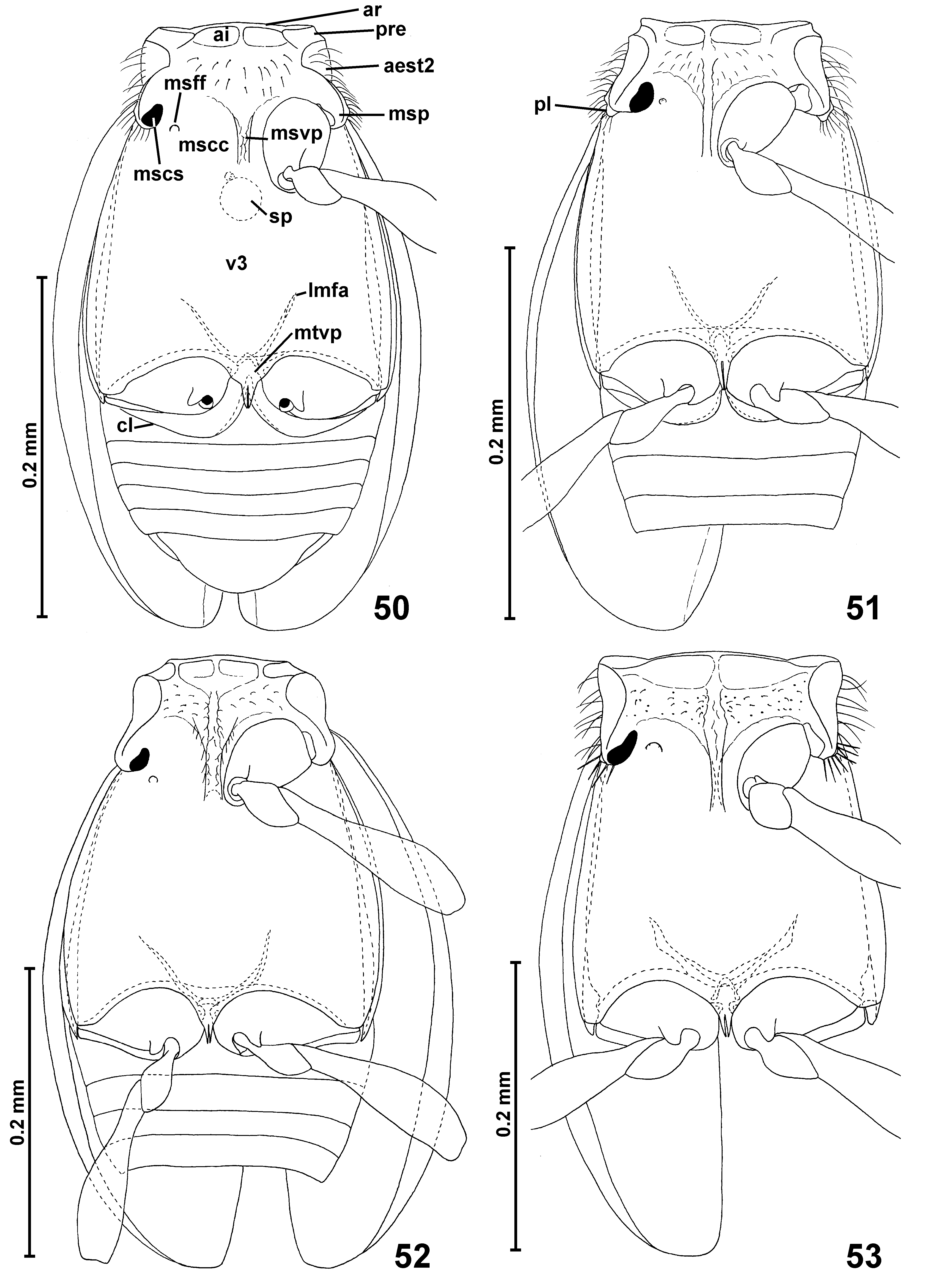
Mesoventrite ( Fig. 53 View FIGURES 50 – 53 ) with narrow anterior ridge; mesoventral intercoxal process narrow and weakly expanding ventrally, anteriorly indistinctly fused with anterior ridge; asetose lateral impressions behind anterior ridge present, area behind asetose impressions covered only with sparse and short setae; mesanepisternum with short prepectus and posterior part largely hidden in ventral view; mesepimeron not visible in ventral view; sides of mesothorax without foveae; mesocoxal projections with mesocoxal sockets located on their mesoventral surface, without projecting posterior lobes.
Metaventrite ( Fig. 53 View FIGURES 50 – 53 ) elongate, anteriorly fused with mesoventrite, posteriorly deeply bisinuate and with narrow median metaventral intercoxal process composed of two long and sharply pointed spines. Metanepisterna and metepimera narrow.
Metafurca ( Fig. 53 View FIGURES 50 – 53 ) with very short and broad stalk and divergent lateral metafurcal arms.
Elytra ( Figs. 27–28 View FIGURES 20 – 28 , 36–37 View FIGURES 29 – 37 ) oval, each with single large and asetose basal fovea located in deep and large but short basal impression; humeral calli well-marked and developed as longitudinal protuberances; elytral apices unmodified, separately rounded.
Hind wings well-developed about twice as long as elytra, with fringe of long setae along posterior margin.
Legs moderately long and slender; pro- and mesocoxae moderately elongate, metacoxae strongly transverse; all trochanters short; all femora weakly clavate; tibiae short and slightly thickening distally, without any noticeable modifications; tarsi short and stout.
Abdominal sternites unmodified, suture between VII and VIII well marked.
Aedeagus ( Figs. 60–61 View FIGURES 54 – 61 ) with symmetrical, lightly sclerotized and thin-walled median lobe, approximately drop-shaped, with internal armature composed of strongly elongate, rod-like sclerites, parameres present, not fused with median lobe, short and slender, each with one long apical seta.
Distribution and composition. Penicillidmus comprises two newly described species known from the Cape York (N Queensland) ( Fig. 63 View FIGURES 62 – 63 a–b); another undescribed species represented by a single female was found near the northern tip of the Cape York ( Fig. 63 View FIGURES 62 – 63 c).
Etymology. The name Penicillidmus is derived from the Latin penicillium (a brush, in a reference to a pair of postero-lateral pronotal penicilli), with the ending - dmus from Microscydmus . Gender masculine.
Biology and collecting methods. The only four known specimens of Penicillidmus were collected in closed or open forests by sifting leaf litter, palm frond litter and fungi, with subsequent extraction by Berlese funnels. No other details of biology are known.
Remarks. Penicillidmus is similar to Microscydmus , especially to the newly established Australian subgenus Scydmomicrus. The most important character to distinguish the new genus from Microscydmus , the system of longitudinal and transverse carinae on the ventral surface of head capsule posterad and laterad the mouthparts, is difficult to see on dry-mounted specimens and requires either SEM study or observations in transparent mounts under a compound microscope. The same methods are required to see the expanded and blunt apex of maxillary palpomere IV, another key character. However, the unique postero-lateral brushes of several thick and elongate bristles (under a stereoscopic microscope appearing as a single rod) can easily be observed at a magnification of 80x and allow for an unambiguous identification of the genus. Even if the bristles are broken off, their insertion site is located on a small lateral protuberance visible in dorsal view of the pronotum.


No known copyright restrictions apply. See Agosti, D., Egloff, W., 2009. Taxonomic information exchange and copyright: the Plazi approach. BMC Research Notes 2009, 2:53 for further explanation.
|
Kingdom |
|
|
Phylum |
|
|
Class |
|
|
Order |
|
|
Family |