
Microscydmus Saulcy & Croissandeau
|
publication ID |
https://doi.org/ 10.11646/zootaxa.3774.1.1 |
|
publication LSID |
lsid:zoobank.org:pub:B5A2EF46-2BF6-4ED3-A5F4-5F9951400545 |
|
DOI |
https://doi.org/10.5281/zenodo.5664958 |
|
persistent identifier |
https://treatment.plazi.org/id/9A6687E6-4F10-302E-FF34-FD14FDDCFE66 |
|
treatment provided by |
Plazi |
|
scientific name |
Microscydmus Saulcy & Croissandeau |
| status |
|
Microscydmus Saulcy & Croissandeau View in CoL
Microscydmus Saulcy & Croissandeau, 1893: 225 View in CoL (without species); type species: Scydmaenus nanus Schaum, 1844 (subsequent monotypy by Croissandeau, 1898: 105).
Morphology and diagnosis of Microscydmus View in CoL s. str. The nominotypical subgenus of Microscydmus View in CoL can be defined as a cyrtoscydmine member comprising beetles with typical "ant-like" appearance (i.e., with an elongate body and distinct constrictions between the head and prothorax and between prothorax and pterothorax + abdomen) and extremely small bodies (below 1 mm in length, typically 0.6-0.8 mm), sharing the following combination of characters: i) tempora, genae and sides of pronotum densely covered with straight, long and thick bristles ( Figs. 4 View FIGURES 1 – 4 , 8 View FIGURES 8 – 13 ); ii) head short, subtriangular or subtrapezoidal ( Figs. 1–2, 4 View FIGURES 1 – 4 ) with weakly convex vertex ( Fig. 2 View FIGURES 1 – 4 ; vt) and frons slightly projecting anteriorly with its narrow anterior part narrowly separating antennal insertions; iii) antennae with distinct club composed of antennomeres IX–XI; iv) submentum short and broad, without lateral sutures ( Fig. 4 View FIGURES 1 – 4 ; smn); v) posterior tentorial pits visible in ventral view and located in distinct transverse groove ( Fig. 4 View FIGURES 1 – 4 ; ptp); vi) occipital constriction only slightly narrower than vertex ( Fig. 4 View FIGURES 1 – 4 ; occ); vii) prothorax without lateral marginal carinae, with complete hypomeral ridges ( Fig. 8 View FIGURES 8 – 13 ; hyr) and basisternal part of prosternum ( Fig. 8 View FIGURES 8 – 13 ; bst) laterally fused with internal (adcoxal) part of each hypomeron; viii) pronotum with two pairs of ante-basal pits ( Fig. 9 View FIGURES 8 – 13 ; abf), internal pair connected by variously distinct transverse impression or groove ( Fig. 9 View FIGURES 8 – 13 ; abg); ix) mesoventrite with asetose impressions and setose impressions ( Figs. 10, 13 View FIGURES 8 – 13 ; ai, si) and mesoventral intercoxal process ( Figs. 10, 13 View FIGURES 8 – 13 ; msvp) carinate, moderately expanding ventrally and not connected anteriorly to anterior ridge of mesoventrite ( Figs. 10, 13 View FIGURES 8 – 13 ; ar); x) metaventral intercoxal process ( Fig. 10 View FIGURES 8 – 13 ; mtvp) with two long spines; xi) each elytron with short but distinct and deep basal impression bearing one large basal elytral fovea filled with sparse setae ( Fig. 11 View FIGURES 8 – 13 ).
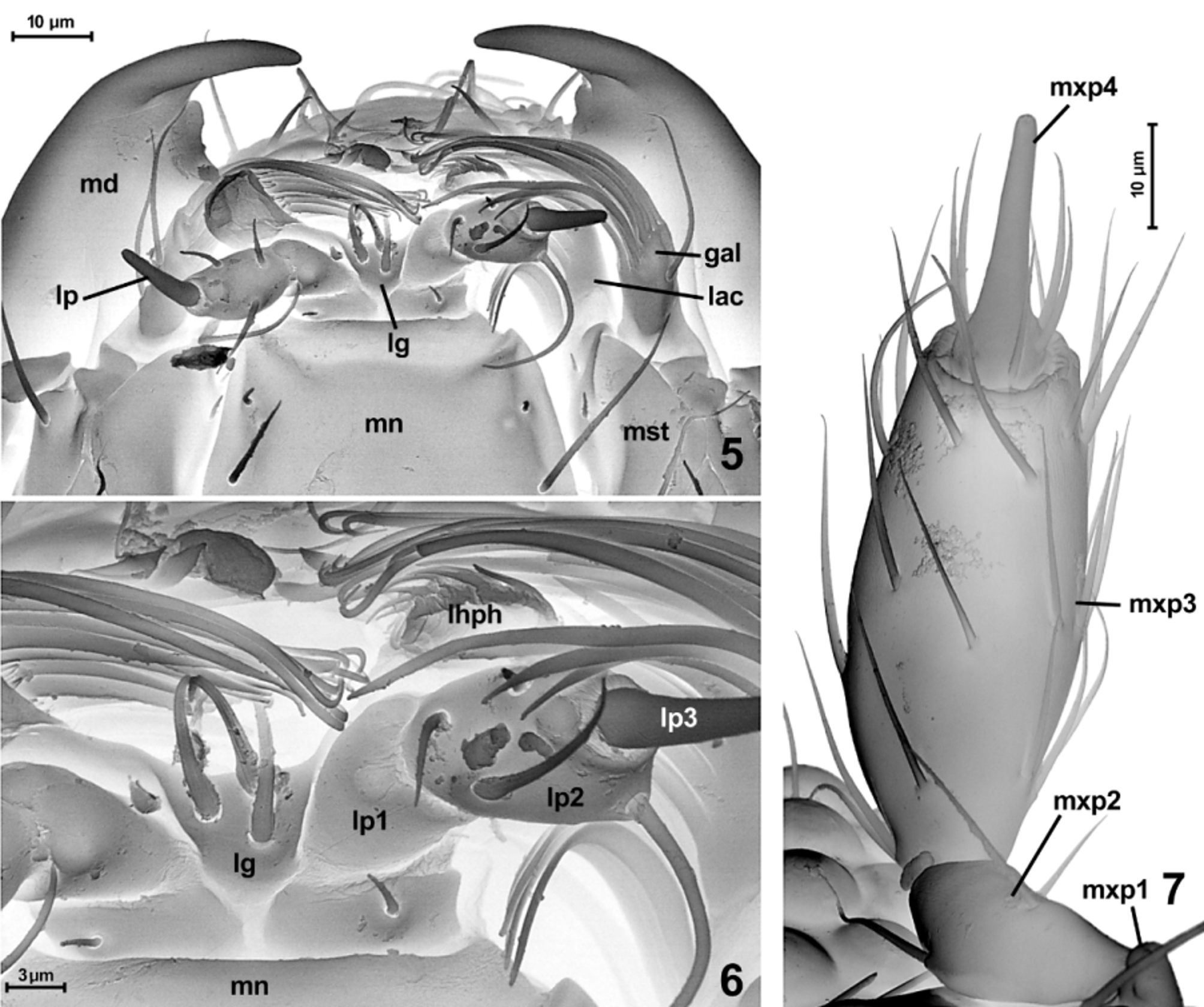
Additionally, species of Microscydmus View in CoL s. str. are characterized by distinct tempora ( Fig. 4 View FIGURES 1 – 4 ; tm); absence of fronto-clypeal groove; antennal insertions located on anterior surface of head; mandibles ( Figs. 2–5 View FIGURES 1 – 4 View FIGURES 5 – 7 ; md) subtriangular, with indistinct, small and often visible only in ventral view sub-apical tooth ( Fig. 5 View FIGURES 5 – 7 ) and large dorsomesal prostheca ( Fig. 3 View FIGURES 1 – 4 ; pst); hypostomal ridges ( Fig. 4 View FIGURES 1 – 4 ; hr) short and extending postero-mesally, not reaching anterior margin of gular plate; maxillary palpomere III ( Fig. 7 View FIGURES 5 – 7 ; mxp3) only 3– 4 x as long as broad, palpomere IV ( Fig. 7 View FIGURES 5 – 7 ; mxp4) strongly elongate and slender, with broadened short and setose basal part and distinctly demarcated, abruptly narrowing and nearly rod-like pointed distal part; prementum with short subtriangular or subtrapezoidal ligula ( Figs. 5–6 View FIGURES 5 – 7 ; lg) bearing two or three thick setae (in two specimens of M. nanus studied by SEM one has two setae, the other one, illustrated here, three setae; two setae were also found in M. minimus View in CoL ); prothorax with indistinct, weakly carinate prosternal intercoxal process ( Fig. 8 View FIGURES 8 – 13 ; psp); procoxal sockets ( Fig. 8 View FIGURES 8 – 13 ) closed by broad lateral projection of coxal part of sternum; pronotosternal sutures ( Fig. 8 View FIGURES 8 – 13 ; nss) visible only as short notches in antero-ventral part of prothorax; mesoscutum and mesoscutellum ( Fig. 12 View FIGURES 8 – 13 ; sc2+scl2) not separated by scutoscutellar suture, forming single subtriangular plate not visible between elytral bases in intact specimens; aedeagus ( Figs 17–19 View FIGURES 17 – 19 ) drop-shaped, with thin walls of median lobe, basally or sub-basally located foramen ( Fig. View FIGURES 17 – 19
17; bo) and free, slender parameres ( Fig. 17 View FIGURES 17 – 19 ; pm), typically with single robust apical seta ( Fig. 19 View FIGURES 17 – 19 ); spermatheca sclerotized and globular, usually located deeply in thorax.
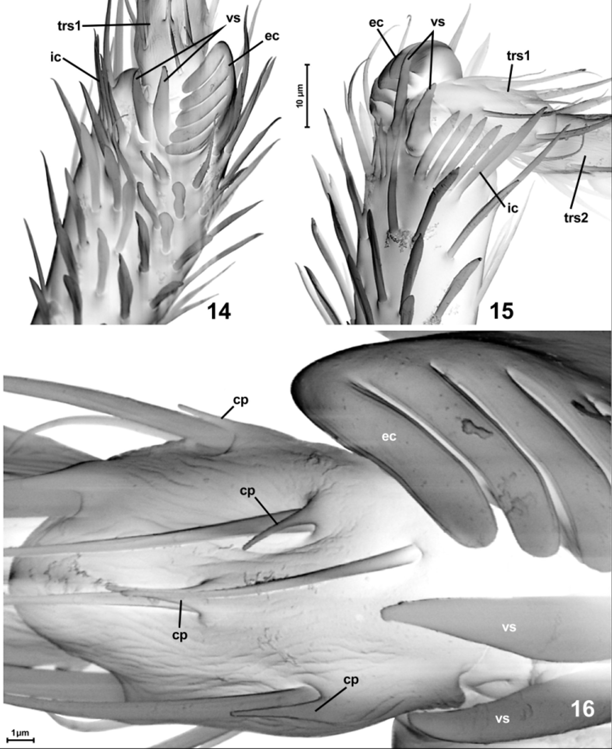
In at least some species of Microscydmus View in CoL s. str. and all in the subgenus Parastenichnus Franz, 1970 the protibiae of males have modified apices, bearing externally located combs ( Figs. 14–16 View FIGURES 14 – 16 ; ec) composed of several curved teeth fused together at base. Additionally the protibial apex can be armed with another, internally located comb ( Figs. 14–15 View FIGURES 14 – 16 ; ic) of several straight cuticular projections, and with two robust ventral spines ( Figs 14–16 View FIGURES 14 – 16 ; vs) at base of the tarsomere I. Ventral setae on tarsomeres in the type species of the genus are accompanied by cucticular projections ( Fig. 16 View FIGURES 14 – 16 ; cp).


No known copyright restrictions apply. See Agosti, D., Egloff, W., 2009. Taxonomic information exchange and copyright: the Plazi approach. BMC Research Notes 2009, 2:53 for further explanation.
|
Kingdom |
|
|
Phylum |
|
|
Class |
|
|
Order |
|
|
Family |
Microscydmus Saulcy & Croissandeau
| Jałoszyński, Paweł 2014 |
Microscydmus
| Croissandeau 1898: 105 |
| Saulcy 1893: 225 |