
Phosphaenus hemipterus ( Goeze, 1777 )
|
publication ID |
https://doi.org/ 10.11646/zootaxa.4382.3.2 |
|
publication LSID |
lsid:zoobank.org:pub:24FC59DE-4C3E-443B-B397-16E9C26DC941 |
|
DOI |
https://doi.org/10.5281/zenodo.5985356 |
|
persistent identifier |
https://treatment.plazi.org/id/9B0B7019-895D-FFBC-FF64-FC69C5A67EB8 |
|
treatment provided by |
Plazi |
|
scientific name |
Phosphaenus hemipterus ( Goeze, 1777 ) |
| status |
|
Phosphaenus hemipterus ( Goeze, 1777) View in CoL
Material examined. Lednice ( CZech Republic), three higher-instar larvae out of eleven collected in April 1987.
Diagnosis. Larvae oblong and slender; thoracic tergites subdivided with one parasagittal line of light pigmentation on each side subparallel to sagittal line; inner ventrolateral area of second antennomere with distinct longitudinal cleft; in addition to second antennomere, sensoria present also on third antennomere and distal segments of maxillary and labial palpi; mandible with short and blunt retinaculum; mandibular channel opening covered by a small hyaline appendage forming a subtriangular valve with fringing at the distal end; short fibrous setae growing from slightly sunken toroidal base covering antennae, legs and sclerites; photic organ consisting of a pair of white spots placed laterally on abdominal segment VIII.
Description of mature larva ( Figs 1–6 View FIGURES 1–9 ). Oblong and slender, cylindrical. Body length ca. 10–11 mm (from the anterior margin of protergum to the apex of caudal segment); with 3 thoracic and 10 abdominal segments. Tergites from protergum to abdominal segment VIII divided by sagittal line in dorsal view ( Fig. 1 View FIGURES 1–9 ). Thoracic tergites then subdivided with one parasagittal line of light pigmentation on each side, subparallel to sagittal line ( Fig. 1 View FIGURES 1–9 ; sg). Colouration: dorsally dark reddish-brown, ventrally pink/ochre/light brown with darker plates on laterotergites and sternum. Spiracles on laterotergites of light colouration. Paired photic organs placed laterally on venter of abdominal segment VIII.
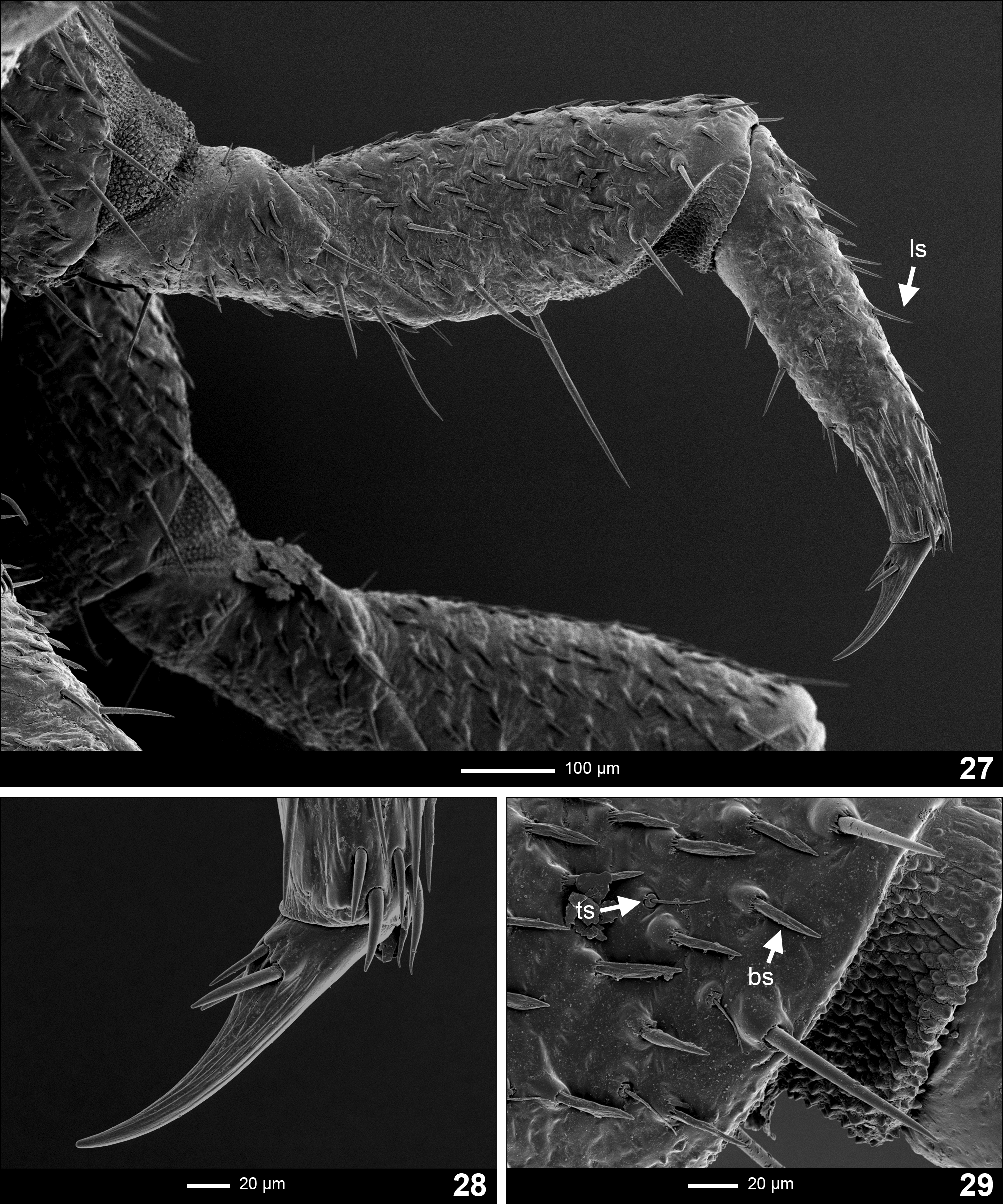
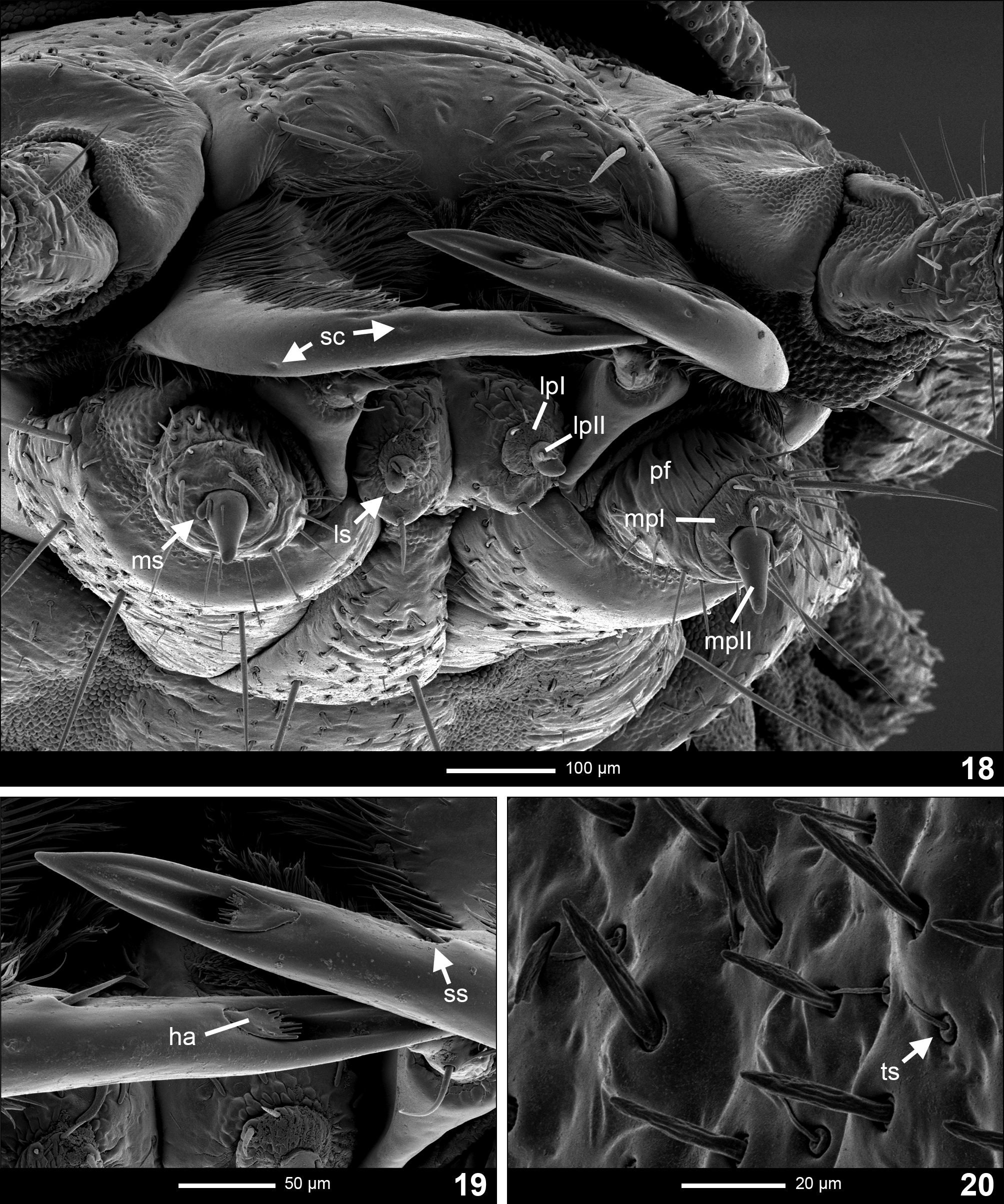
Types of general cuticular outgrowth observed. 1. Short, blunt, setae lying on surface ( Figs 15 View FIGURES 15–17 , 29 View FIGURES 27–29 ; bs); 2. stout, long setae ( Figs 15 View FIGURES 15–17 , 27 View FIGURES 27–29 ; ls); 3. flagellar setae growing from a slightly sunken toroidal socket (hereafter called toroidal setae; Figs 15 View FIGURES 15–17 , 20 View FIGURES 18–20 , 29 View FIGURES 27–29 ; ts).
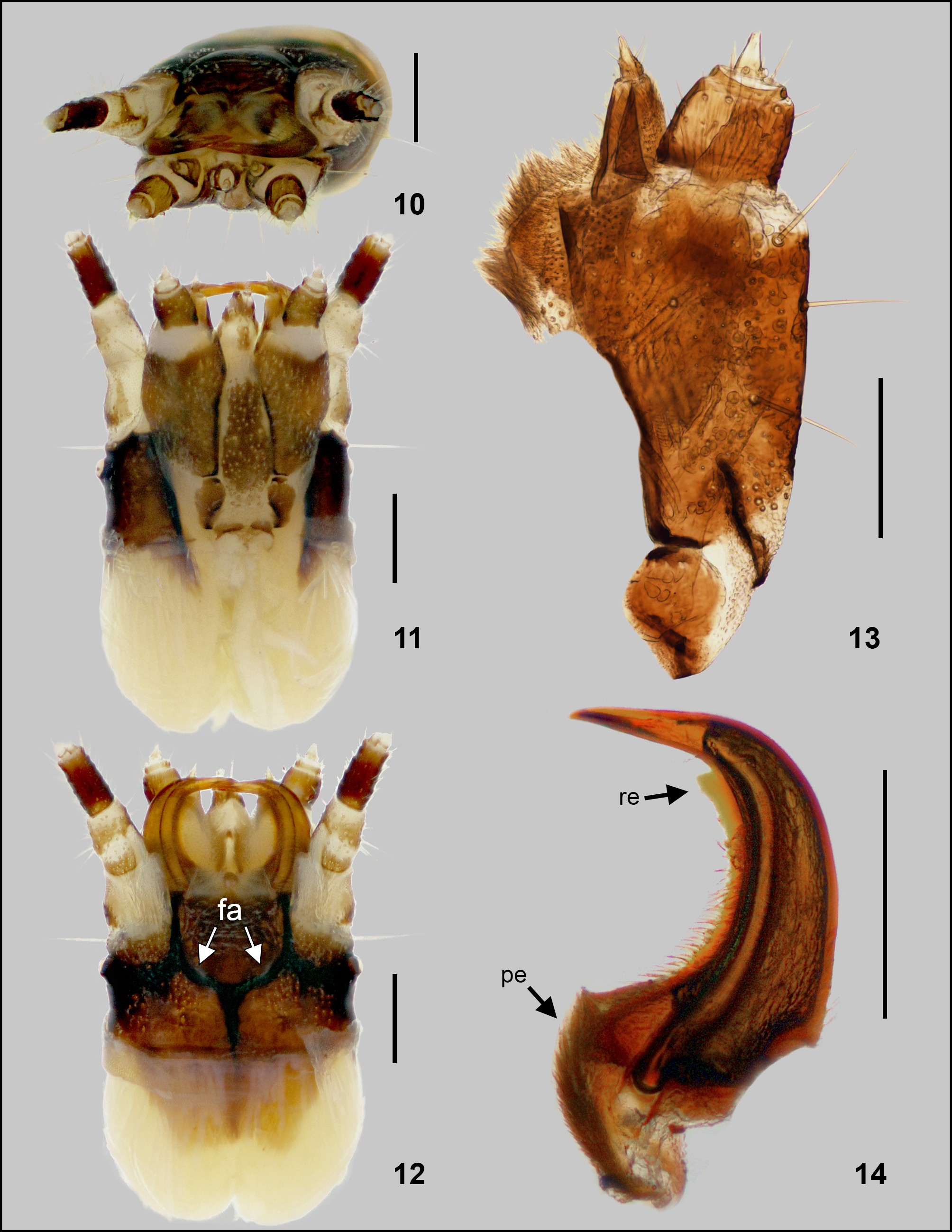
Head capsule ( Figs 10–12 View FIGURES10–14 , 15–17 View FIGURES 15–17 ). Prognathous; retractable within prothorax, extensible neck membrane covered in extremely short spines and forming a two-layered envelope around retracted head; wider than long. Epicranial plate laterally about 1/3 of the width of the head capsule, slightly concave, with one stout seta anterolateraly, close to the base of antennae. Head capsule dorsally covered with short blunt setae lying on its surface ( Fig. 15 View FIGURES 15–17 ; bs). Epicranial suture dark, Y-shaped, frontal arms U-shaped ( Fig. 12 View FIGURES10–14 ; fa). Gula not present ( Fig. 11 View FIGURES10–14 ). One stemma on each side of the head. Labrum fused with clypeus forming clypeolabrum, covering base of mandibles in dorsal view. Clypeolabrum flat in anterior view, with two setae reaching one fourth of the length of mandibles, positioned on outer lateral sides ( Fig. 15 View FIGURES 15–17 ). Epipharynx formed by two plates, and an anterior brush of long setae, which project centrally past anterior margin of the head. Hypopharynx with short setation.
Antenna ( Figs 24–26 View FIGURES 24–26 ). Trimerous, inserted on lateral distal margin of epicranial plate; partially retractable within membranous socket. Basal antennomere widest, poorly sclerotiZed, slightly bulging on the dorsal side, densely covered by three types of setae; short blunt setae lying on surface and toroidal setae mainly posterolaterally, and several stout, almost perpendicular long setae around apical region (which are longest on this antennomere in comparison with the other antennomeres) easily observable under high magnification ( Fig. 24 View FIGURES 24–26 ). Second antennomere slightly longer, narrower and laterally flattened in comparison to basal antennomere; bearing only toroidal setae and blunt setae equally and abundantly spread across the antennomere. Inner ventrolateral area of second antennomere with sensillum consisting of distinct longitudinal cleft; a sensory slot ( Fig. 24 View FIGURES 24–26 ; ses). Several sensilla coeloconica are present apically ( Fig. 26 View FIGURES 24–26 ; sc). Sensorium of second antennomere oval ( Figs 24–26 View FIGURES 24–26 ; as1), widest at the base, closely adhering to the second antennomere, slightly longer than the third antennomere, with very fine helical ridges from apex to bottom. Third antennomere ( Fig. 24 View FIGURES 24–26 ; a3) shortest, reaching about 1/2 of length of the sensorium of second antennomere and adjoining it, bearing a small sensorium, three short setae and three cuticular projections ( Fig. 25 View FIGURES 24–26 ); first longer and thick (cp1), second longer and thin (cp2) and third very short (cp3).
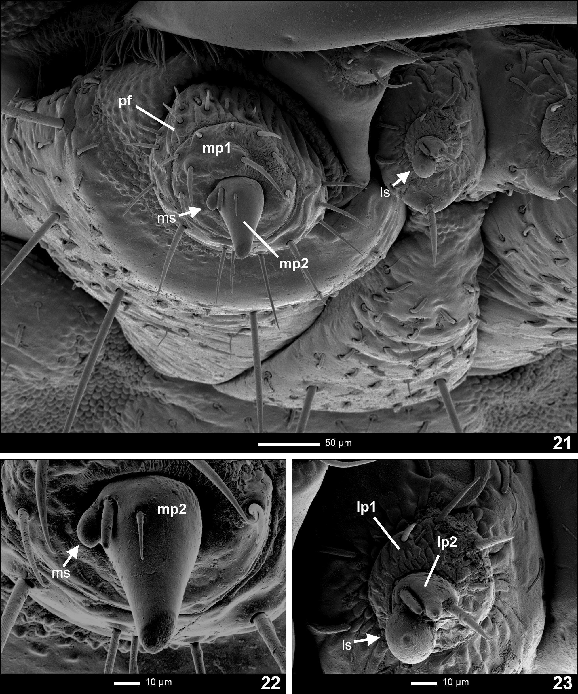
Maxilla ( Fig. 13 View FIGURES10–14 ). Consisting of five parts, attached to labium forming a maxillo-labial complex. Cardo vertical, bulbous, with wider side adjacent to stipes. Stipes elongated, subtrapeZoidal, ventrally covered with short blunt setae lying on its surface and four long and stout setae, three anteriorly and one medially. Galea bimerous, with basal part larger than distal, subtriangular in anterior view (with the tip of subtriangle aiming ventrally). Distal part conical, rotated medially with setae shorter than its body. Lacinia covered with brush of long setae on outer lateral margin. Maxillary palpifer large ( Fig. 21 View FIGURES 21–23 ; pf), subrectangular, slightly longer than wide. Maxillary palpus bimerous ( Fig. 21 View FIGURES 21–23 ; mp1, mp2), basal palpomere short and wide. Palpifer and basal palpomere covered with setae. Palpomere II ( Fig. 22 View FIGURES 21–23 ; mp2) bearing two setae, one thin and sharp placed dorsally and second slightly thicker and blunt, paired with sensorium near base on lateral surface ( Fig. 22 View FIGURES 21–23 ; ms).
Labium ( Fig. 21, 23 View FIGURES 21–23 ). Closely attached to maxilla, formed by a short prementum, mentum and mostly membranous submentum ( Fig. 11 View FIGURES10–14 ). Glossae absent. Prementum narrow, heart-shaped in ventral view; bearing three types of setae: blunt short setae lying on surface, sensory setae and a pair of long and stout setae underneath the palpi. Labial palpus bimerous ( Fig. 23 View FIGURES 21–23 ; lp1, lp2); basal palpomere wide and short, bearing several setae dorsally; second palpomere short, bearing a large sensorium ventrally ( Fig. 23 View FIGURES 21–23 ; ls), one thin seta between the apex and the sensorium, and two stout setae laterally around the apex: one sharp on the inner side and one blunt on the outer side. Mentum elongated, subtriangular, unsclerotiZed on lateral margins, ventrally bearing numerous short, blunt, setae lying on surface, numerous toroidal setae and a pair of large, stout setae posteromedially.
Mandible ( Fig. 14 View FIGURES10–14 ). Symmetrical, falcate, with an internal channel opening subapically on outer edge ( Fig. 19 View FIGURES 18–20 ). Penicillus well developed ( Fig. 14 View FIGURES10–14 ; pe). Retinaculum present, forming one thin and blunt hyaline process on apical third of mandible ( Fig. 14 View FIGURES10–14 ; re). Inner margin of mandible from retinaculum to the base covered with stout setae, lengthening towards the base of mandible ( Fig. 14 View FIGURES10–14 ). Ventrally, basal two-thirds of mandible covered with dense setation lying on surface, aimed medially. Dorsally, basal two thirds with sagittal line of dense, stout, setation lying on surface, of equal length, erect in its last third, aiming medially ( Fig. 15 View FIGURES 15–17 ). Lateral margin without setation. Sensory (hyaline) appendage on outer margin of mandible present before channel opening, forming a subtriangular valve with fringing at the distal end ( Fig. 19 View FIGURES 18–20 ; ha). A thin stout short seta present dorsally in retinaculum region on both mandibles ( Fig. 19 View FIGURES 18–20 ; ss). Several sensilla coeloconica present on the post-retinaculum apical part ( Fig. 18 View FIGURES 18–20 ; sc).
Thorax ( Figs 1–6 View FIGURES 1–9 ). Protergum wider than long, subsemicircular, wider posteriorly. Meso- and metatergum suboval, wider than long, with rounded margins. Venter of prothorax composed of subquadrate prosternum ( Fig. 2 View FIGURES 1–9 ; ps), subdivided into three well sclerotiZed areas; lateral ones narrow and transverse, extending above and to the sides of coxae fusing with episterna; medial area subrhomboid. Epimera forming thin sclerotiZed strands ( Fig. 2 View FIGURES 1–9 ; em). Lateral areas of meso- and metathorax composed of two laterotergites; anterior one sclerotiZed ( Fig. 2 View FIGURES 1–9 ; alt), bearing a well-developed bilabiate spiracle in mesothorax; posterior one membranous ( Fig. 2 View FIGURES 1–9 ; plt). Anterior ventral area of meso- and metathorax formed by mostly membranous intersternite ( Fig. 2 View FIGURES 1–9 ; is) with two darker-pigmented sagittal bands centrally, margined by paired anterior laterotergites. Posterior ventral area subdivided into triangular, wider than long basisternum and smaller sternellum. Basisternum subdivided into three darker-pigmented areas; lateral ones extending anteriorly and laterally to coxae, joining episterna; medial subrhomboid. Sternellum membranous, with sclerotiZed triangular centre touching with medial subrhomboid area of basisternum with its tip.
Legs ( Figs 27–29 View FIGURES 27–29 ). Pentamerous, all pairs similar in shape and siZe. Coxa large, stout, bearing short blunt setae lying on its surface, toroidal setae and stout long setae. Coxal-trochanteral membrane reaching more than 1/2 of coxal longitudinal length ( Fig. 2 View FIGURES 1–9 ). Trochanter smaller, subtriangular in lateral view, about the same siZe as femur, bearing short blunt setae lying on its surface, toroidal setae, and stout long setae, with long stout seta on distal venter, together with several shorter stout setae radially on distal end. Femur fusiform in lateral view, bearing short blunt setae lying on its surface, toroidal setae, and stout long setae, with one very long stout seta on the centre of ventral area ( Fig. 27 View FIGURES 27–29 ) and several shorter stout setae radially on distal end. Tibiotarsus as long as femur, narrower, tapering towards distal end, covered predominantly by stout sharp setae, lengthening dorsally. Pretarsus composed of a claw with fine ridges, ventrally bearing three short setae ( Fig. 28 View FIGURES 27–29 ).
Abdomen ( Figs 1–9 View FIGURES 1–9 ). Ten-segmented, slightly tapering towards posterior end, segments I to VIII subdivided by fine sagittal line in dorsal view ( Fig. 1 View FIGURES 1–9 ). Tergites of segments I to VII subrectangular, similar in shape and colouration, wider than long; tergite of segment VIII suboval; segment IX subrectangular, wider than long; segment X forming a narrow sclerotiZed dark ring, bearing the holdfast organ—pygopod—with several eversible processes. Segments I to VIII have single laterotergites ( Fig. 3 View FIGURES 1–9 ; lt) on each side with sclerotiZed plates bearing bilabiate spiracles; the venter of segments I to VII consists of median sternite, margined by paired narrow weakly sclerotiZed laterosternites ( Fig. 3 View FIGURES 1–9 ; ls). Sternites of segments I to VIII subrectangular, well sclerotiZed (except segment VIII) wider than long, with a pair of long stout setae on posterolateral margins. Venter and laterotergites of segment VIII membranous, bearing laterally a pair of white spots representing a functioning photic organ ( Fig. 8 View FIGURES 1–9 ; po). Sternite of segment IX sclerotiZed posteriorly, forming a thin ring supporting segment X.
Notes on life cycle and behaviour. The life cycle of Phosphaenus hemipterus lasts two or three years. Eggs are white, spherical, with a diameter of ca. 0.6 mm ( De Cock 2000). According to De Cock, late-instar female larvae tend to be larger and fatter than males and are easily recognised in the field ( De Cock 2003). Pupation takes place in April–May, the pupal stage lasts ca. 2 weeks. Mature individuals are collected occasionally and rarely from July to August ( Burakowski 2003).
Unlike most of the firefly species, whose larvae feed on snails and slugs, larvae of Phosphaenus hemipterus are obligate earthworm ( Lumbricus spp., Lumbricidae ) predators ( Majka & MacIvor 2009). Majka & MacIvor (2009) observed the larvae while feeding, using tarsal claws of the legs to anchor themselves to the body of the earthworm and their extended antennae moving over the surface of the earthworm’s body. The process of injecting a toxin into its prey in order to kill it, as observed in other species of fireflies (Schwalb 1961), has not yet been observed for this species. However, the presence of a mandibular channel suggests that a toxin is also used by this species.
Like Lampyris noctiluca , Phosphaenus hemipterus larvae also glow spontaneously by emitting bioluminescent pulses while active at night ( De Cock 2003).
Both adult sexes are flightless. Neotenic females stay in litter or lower parts of plants, are active mainly at dusk ( De Cock 2000; Burakowski 2003) and are very rarely found ( Burakowski 2003). In contrast, the males have vestigial elytra, are diurnal, and can often be found on herbaceous plants and shrubs ( Burakowski 2003). The larvae are predominantly nocturnal. Both adult sexes are feebly bioluminescent, although they appear only to glow in response to disturbance ( Majka & MacIvor 2009).


No known copyright restrictions apply. See Agosti, D., Egloff, W., 2009. Taxonomic information exchange and copyright: the Plazi approach. BMC Research Notes 2009, 2:53 for further explanation.