
Echinoderes wilberti, Anguas-Escalante & Herranz & Martínez-Arce & De Jesús-Navarrete & Sørensen, 2023
|
publication ID |
https://doi.org/ 10.1016/j.jcz.2022.12.001 |
|
persistent identifier |
https://treatment.plazi.org/id/9B181279-A269-AD20-B87E-FF2787B63B0D |
|
treatment provided by |
Felipe |
|
scientific name |
Echinoderes wilberti |
| status |
sp. nov. |
3.2. Description of Echinoderes wilberti View in CoL sp. nov
E. wilberti sp. nov.
Fig. 6–9 View Fig View Fig View Fig View Fig C-D, Tables 4–6
urn:lsid:zoobank.org:act:D6F9EC30-509A-4F5C-92DB-8BB04751B5F7 .
3.2.1. Diagnosis
Echinoderes without middorsal spines. Lateroventral acicular spines present on segments 6 to 9. Tubes present in lateroventral and subdorsal positions on segment 2, lateroventral positions on segment 5, lateral accessory positions in segment 8, and laterodorsal positions on segment 10; tubes on segment 10 show sexual dimorphism. Females with minute laterodorsal tubes on segment 10 and lateral terminal accessory spines on segment 11. Males with long laterodorsal tubes on segment 10 and three pairs of penile spines.
3.2.2. Etymology
The species is named after Wilbert Andr´es Perez Pech, who helped with the fieldwork and in the separation of kinorhynchs in the samples.
3.2.3. Material examined
Holotypic female, collected on Station T1 XC on Sept. 5, 2018, from fine sand at 1 m depth in the reef lagoon off Xcalak Quintana Roo (18 ◦ 17 ′ 14.7’’N 087 ◦ 49 ′ 56.9’’W), mounted in 100% glycerine at a glass slide with coverslip, and deposited at the Natural History Museum of Denmark under catalogue number NHMD-1176454. Paratypes include two males and seven females: one male and four females, with same collecting data and mounting as holotype, deposited under catalogue numbers NHMD-1176455 to 1176459; one male and three females, collected on station X3SF on June 6, 20222, from muddy sediment (18 ◦ 17 ′ 21.3 ′′ N 087 ◦ 49 ′ 58.0 ′′ W); specimens were used for DNA extraction and COI sequencing, and cuticles were subsequently recovered and used as hologenophores for the sequences ( NCBI Acc. No. OP 617671 to OP617674, mounted in Fluoromount G between two cover glasses attached to an H–S plastic slide, and deposited under catalogue numbers NHMD-1176460 to 1176463. GoogleMaps
Additional material includes three females and one male collected on St. T1 XC in July 11, 2021, mounted for SEM and stored in the personal reference collection of MVS .
3.2.4. Description
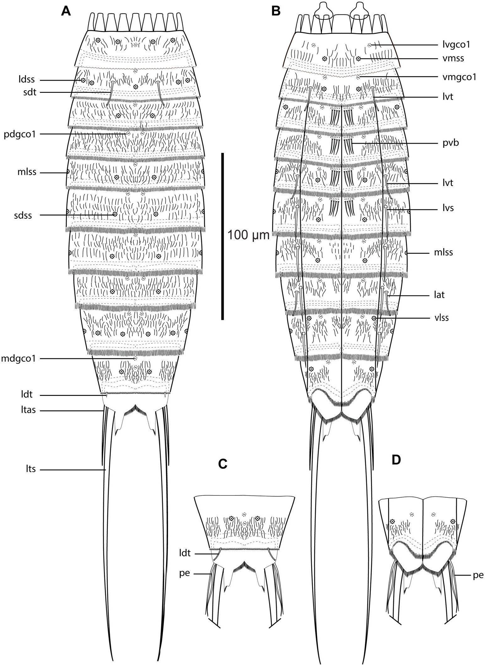
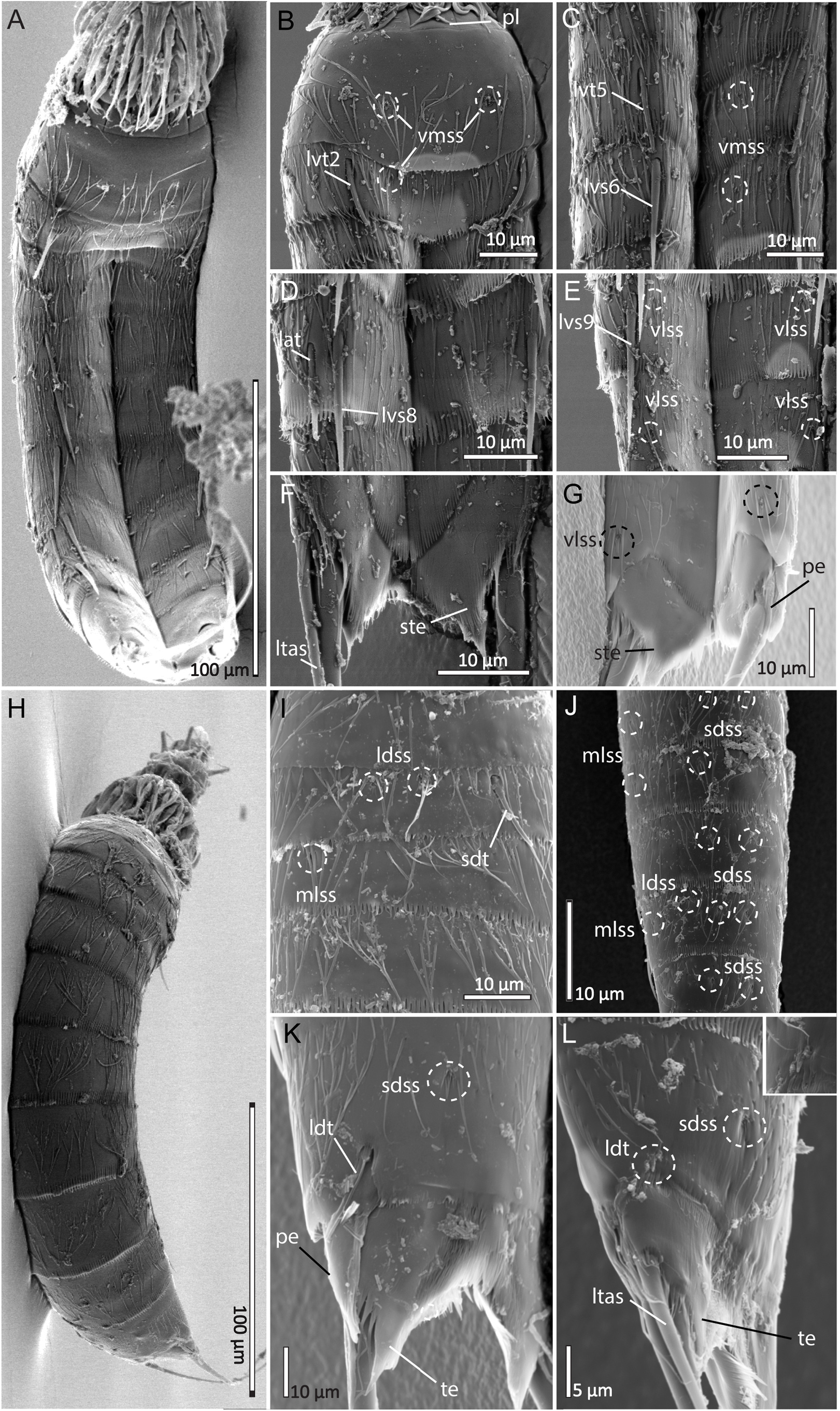
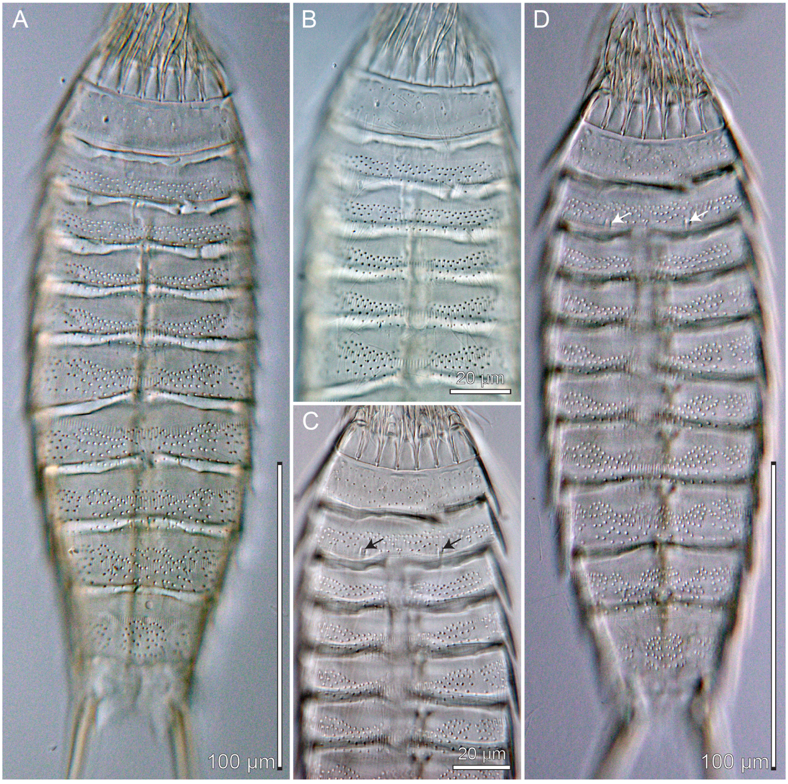
Adults with head, neck, and eleven trunk segments ( Fig. 6A–B View Fig , 7A View Fig , 8A, H View Fig ). Middorsal spines absent on all segments. For complete overview of measures and dimensions, see Table 4. Distribution of cuticular structures, i.e., sensory spots, glandular cell outlets, spines, and tubes, is summarized in Table 5, and NCBI accession numbers for COI sequences are listed in Table 6.
The head consists of a retractable mouth cone and an introvert. Detailed observations were not possible due to dirt covering.
The neck has 16 placids, measuring 12 μm in length. The midventral placid is broadest, measuring 11 μm in width at its base, whereas all others are narrower, measuring 7 μm at their bases. The trichoscalid plates are well-developed, subdorsal and laterodorsal ones narrow and elongated, and ventromedial ones hat-shaped.
Segment 1 consists of a complete cuticular ring ( Fig. 6A–B View Fig , 7B–C View Fig , 8B, I View Fig ). Sensory spots are located close to the anterior margin of the segment in subdorsal and laterodorsal positions, and in the middle of the segment in ventromedial positions; sensory spots are small and oval, with numerous micropapillae arranged radially around a central pore. Glandular cell outlets type 1 present in middorsal and lateroventral positions. Long non-bracteate cuticular hairs present, emerging through rounded perforation sites. The hair pattern on this segment consists of three transverse wavy rows, covering the dorsal, ventral, and lateral sides. The posterior segment margin is broadly convex on the ventral side, unlike dorsal and lateral sides, which are straight. Pectinate fringe of posterior segment margin crenate.
Segment 2 consists of a complete cuticular ring ( Fig. 6A–B View Fig , 7B–C View Fig , 8B, I View Fig ). Pachycyclus of the anterior segment margin is of medium thickness and uninterrupted. Tubes are present in subdorsal and lateroventral positions. Sensory spots are present in middorsal positions, as a twinpair in laterodorsal positions, and in ventromedial positions. On this and the following segments, sensory spots are more irregular in shape, and with fewer micropapillae. Glandular cell outlets type 1 present in middorsal positions and in ventromedial positions. Bracteate cuticular hairs arranged in four transverse lines around the segment, only interrupted in the area where the sensory spots and tubes are present. The posterior segment margin is straight along the dorsal and lateral sides, and ventral margin more convex. Pectinate fringe of posterior segment margin with short, pointy, uniform fringe tips along dorsal, lateral, and ventral side.
Segment 3 ( Fig. 6A–B View Fig , 7 View Fig D-E, 8I), and remaining segments, consisting of one tergal and two sternal plates. Pachycyclus of anterior segment margin of medium thickness and interrupted middorsally and at the tergosternal junctions. Sensory spots present in subdorsal and sublateral positions. Glandular cell outlets type 1 present in middorsal and ventromedial positions. Tergal cuticular bracteate hairs arranged as four wavy transversal rows with ripples progressively lower and less defined towards the central part of the plate; tergal plates with regular bracteate hairs in lateral-most parts; paraventral areas without bracteate hairs, but with one anterior, transverse row of very short stiff hairs, followed by row with four, strong bristle-like hairs. Pectinate fringe pattern of posterior segment as preceding segment but fringe tips longer and wider.
Segment 4 ( Fig. 6A–B View Fig , 7 View Fig D-E, 8I) Sensory spots absent. Glandular cell outlets type 1 present in paradorsal and ventromedial positions. Cuticular hair patterns, inclusive paraventral bristles, and pachycycli as on preceding segment. Pectinate fringe pattern of posterior segment as preceding segment but fringe tips longer and wider.
Segment 5 ( Fig. 6A–B View Fig , 7 View Fig D-E, 7C) with tubes in lateroventral positions. Sensory spots present in subdorsal, midlateral and ventromedial positions. Glandular cell outlets type 1 present in middorsal and ventromedial positions. Pachycycli and cuticular hair pattern, inclusive paraventral bristles, as on preceding segment. Pectinate fringe pattern of posterior segment as segment 4 but with fringe tips slightly longer and wider.
Segment 6 ( Fig. 6A–B View Fig , 7 View Fig F-G, 8C, J) with acicular spines in lateroventral positions. Sensory spots present in subdorsal, midlateral, and ventromedial positions. Glandular cell outlets type 1 present in paradorsal and ventromedial positions. Pachycycli and cuticular hair pattern, inclusive paraventral bristles, as on preceding segment. Pectinate fringe pattern of posterior segment with fringe tips longer and thinner than segment 5.
Segment 7 ( Fig. 6A–B View Fig , 7 View Fig F-G, 8J) with acicular spines in lateroventral positions. Sensory spots present in subdorsal, midlateral and ventromedial positions. Glandular cell outlets type 1 present in middorsal and ventromedial positions. Paraventral areas with clusters of regular cuticular hairs; otherwise cuticular hair pattern, pachycycli and pectinate fringe as on preceding segment.
Segment 8 ( Fig. 6A–B View Fig , 7 View Fig F-G, 8D, J) with acicular spine in lateroventral positions, and tubes in lateral accessory positions. Sensory spots present in subdorsal positions. Glandular cell outlets type 1 present in paradorsal and ventromedial positions. Pachycycli, cuticular hairs, and pectinate fringe as on preceding segment.
Segment 9 ( Fig. 6A–B View Fig , 7H–I View Fig , 8J View Fig ) with acicular spines in lateroventral positions. Sensory spots present in subdorsal, laterodorsal, midlateral, and ventrolateral positions; those in subdorsal positions very close to the paradorsal positions. Glandular cell outlets type 1 present in paradorsal and ventromedial positions. Small, oval sieve plate present in sublateral positions. Pachycycli, pectinate fringe of posterior segment margin and cuticular hairs as on preceding segment.
Segment 10 ( Fig. 6A–B View Fig , 7 View Fig H-J, 8G, J, K-L) with sexually dimorphic laterodorsal tubes inserted at posterior segment margin: female tubes very minute ( Fig. 8L View Fig ); male tubes long and well-developed ( Fig. 8K View Fig ). Sensory spots present in subdorsal and ventrolateral positions. Glandular cell outlets type 1 present as two longitudinally arranged middorsal ones and in ventromedial positions. The posterior segment margin of the tergal plate is straight, whereas margins of sternal plates are concave; fringe tips of pectinate fringe on the tergal plate are shorter than those on preceding segments, whereas fringe tips on sternal plates are short close to the tergosternal junction, and longer closer to the midsternal articulation. Pachycyclus as on preceding segments. Cuticular hairs arranged as on previous segments but with hairless areas next to the midsternal articulation, and with a heart-shaped hair patch present in the middle of the tergal plate.
Segment 11 with long and thin lateral terminal spines ( Fig. 6A–D View Fig , 7A View Fig , H-J, 8F-G, K-L). Sensory spots and glandular cell outlet type 1 absent. Males with three pairs of penile spines; dorsal and ventral penile spines are thin, flexible tubes, whereas the median penile spine is short, thicker, and rigid ( Fig. 6C–D View Fig , 7J View Fig , 8G, K View Fig ); females with short, thin lateral terminal accessory spines ( Fig. 6A–B View Fig , 7I View Fig , 8F, L View Fig ). Tergal extensions long and slender, laterally with a truncated part and another part extending into a thin tip; sternal extensions shorter and rounded and extending slightly into a tip, with fringe tips of pectinate fringe thinner than those on preceding segments.
3.2.5. Barcoding
Cytochrome c oxidase subunit 1 of 618 bp was partially sequenced from four specimens of E. wilberti sp. nov., and cuticles from the extracted specimens were mounted as hologenophores ( Fig. 9C–D View Fig ). The sequenced barcodes showed a similarity between 99.674% and 100% ( Table 6).

No known copyright restrictions apply. See Agosti, D., Egloff, W., 2009. Taxonomic information exchange and copyright: the Plazi approach. BMC Research Notes 2009, 2:53 for further explanation.
|
Kingdom |
|
|
Phylum |
|
|
Class |
|
|
Order |
|
|
Family |
|
|
Genus |
|
Kingdom |
|
|
Phylum |
|
|
Class |
|
|
Order |
|
|
Family |
|
|
Genus |