
Anelastes Kirby, 1819
|
publication ID |
https://doi.org/ 10.11646/zootaxa.4683.1.5 |
|
publication LSID |
lsid:zoobank.org:pub:FD42DA3D-8379-43C0-8F77-4062B878C678 |
|
DOI |
https://doi.org/10.5281/zenodo.5943513 |
|
persistent identifier |
https://treatment.plazi.org/id/9B794F14-FFAD-FB7B-58BD-5180474A88F6 |
|
treatment provided by |
Plazi |
|
scientific name |
Anelastes Kirby, 1819 |
| status |
|
Genus Anelastes Kirby, 1819 View in CoL
Type species: Anelastes drurii Kirby, 1819
= Silenus Latreille, 1834 (type species: Silenus brunneus Latreille, 1834 (= Anelastes drurii Kirby, 1819 ))
Description. Body length 3.5–13.2 mm. Body elongate, about 3.3–3.6 times as long as wide, elaterid-like, more or less convex dorsally and ventrally. Coloration reddish-yellow to dark brown; integument generally minutely granulose; vestiture uniform, short and thin.
Head transverse, strongly declined, nearly hypognathous, deeply inserted into prothorax. Posterior edge of head capsule bi-emarginate; occipital carina fine, incomplete at middle, continuing below eyes as well-defined subgenal ridges. Frons moderately convex, prominent and steeply sloping at sides to eye margins. Antennal insertions moderately widely separated, distance between inner edges of antennal insertions 0.35–0.50 times as great as distance between mandibular bases. Frontoclypeal region nearly in one plane with frons, short and wide, transversely subtrapezoidal, with poorly defined lateral carina between subantennal groove and base of mandible, anterior margin slightly convex to subtruncate. Subantennal groove very short and shallow. Anteocular and subantennal fossae superficial and nearly indistinct, preorbital furrow absent. Eyes relatively small to moderate-sized, vertically oval and slightly convex, with fine corneal facets. Gular sutures well expressed; gula slightly transverse. Cervical sclerites well developed, each divided into 2 parts.
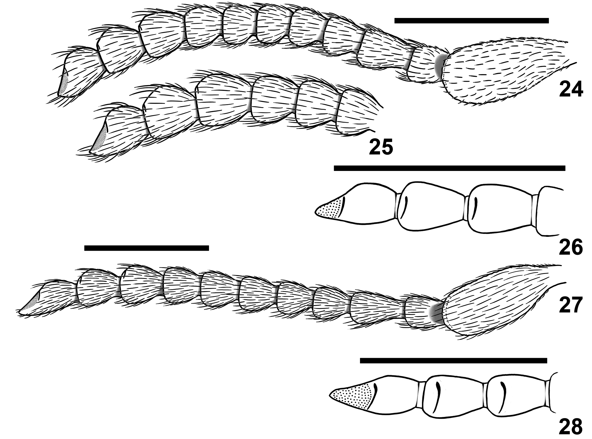
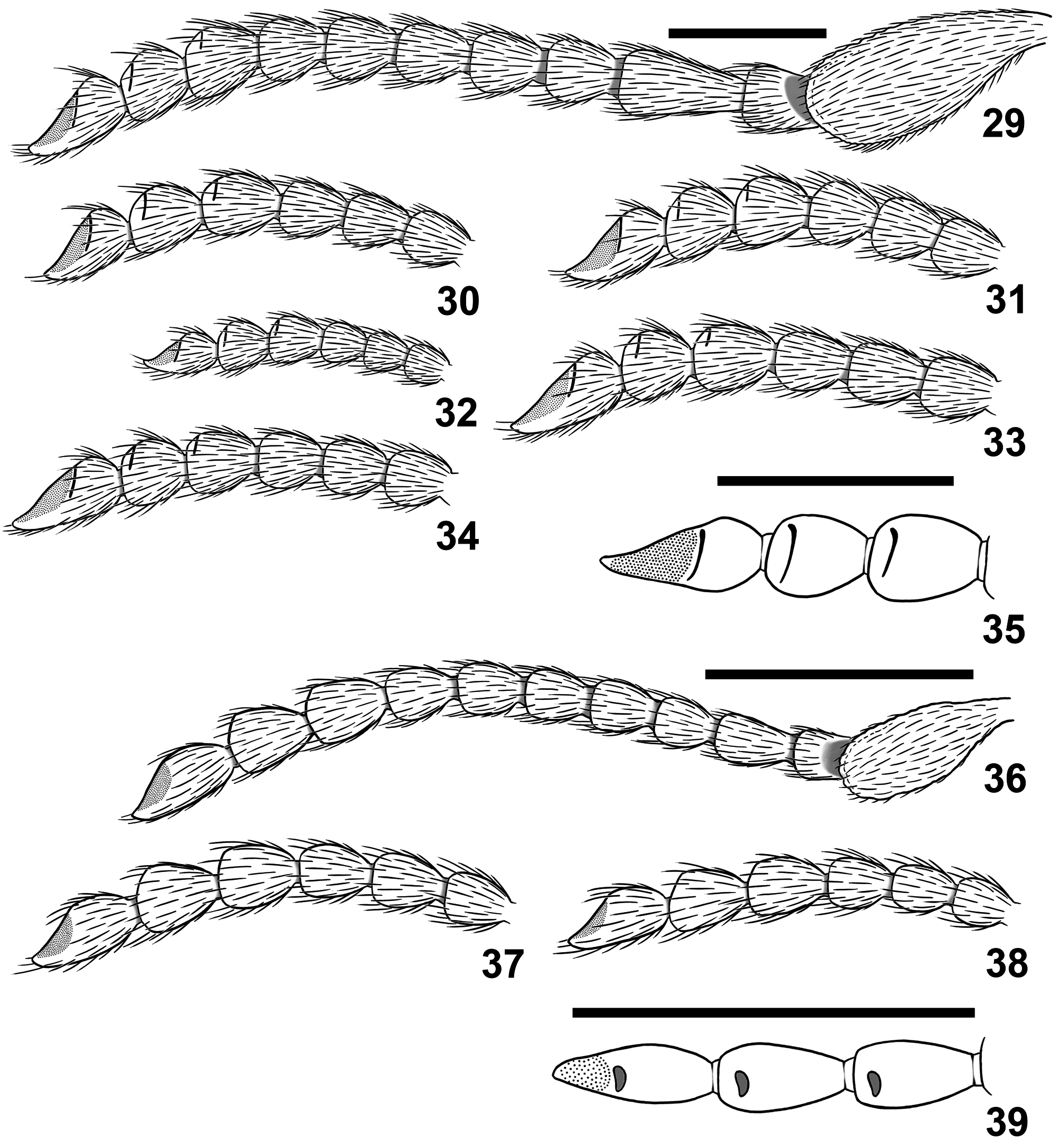
Antennae ( Figs 24–39 View FIGURES 24–28 View FIGURES 29–39 ) 11-segmented, moniliform to nearly filiform, short, not extending behind posterior angles of pronotum; scape stout and rather short, claviform, about three times as long as pedicel, without carinae on external surface; its internal surface bounded by longitudinal carinae anteriorly and posteriorly, lobed apically and densely setose; antennomere 3 more or less elongate, longer than pedicel; antennomeres 4–8 subequal and more or less thickened apically, antennomeres 9–11 slightly enlarged; antennomeres 9–10 thickened apically, each bearing sensory cavity on posterior surface before apex, sensory cavity with more or less transverse orifice; antennomere 11 with oblique surface with sensory field in apical 1/2–2/3 and sensory cavity with orifice at anterior margin of sensory field.
Labrum not exposed beneath anterior edge of epicranium, transverse, slightly sclerotized, with anterior margin fringed with long setae and notched at middle ( Fig. 19 View FIGURES 19–23 ). Mandibles stout, distinctly curved, unidentate and blunt apically, with external surfaces slightly expanded ventrally ( Figs 20, 21 View FIGURES 19–23 ). Maxillary lobes elongate and densely setose; galea subacute at apex; lacinia about twice as long as galea, subacute at apex ( Fig. 22 View FIGURES 19–23 ). Maxillary palpi elongate; terminal maxillary palpomere slightly expanded and obliquely truncate at apex ( Fig. 22 View FIGURES 19–23 ). Labium with bilobed ligula; terminal labial palpomere expanded and obliquely truncate at apex ( Fig. 23 View FIGURES 19–23 ).
Pronotum with subequal width and length to distinctly transverse, widest at about middle, sides more or less convex and with more or less expressed shallow sinuation at posterior angles; lateral carinae weakly expressed and often obliterated posteriorly, dorsally scarcely visible; anterior angles not expressed; posterior angles more or less produced and rounded or obliquely subtruncate at apex; posterior edge more or less bisinuate with subtruncate prescutellar lobe; interlocking device well-developed. Disc more or less convex, with median groove posteriorly. Hypomeron subtriangular, with shallow and not delimited anteriorly profemoral groove; pronotosternal sutures complete, open and divergent anteriorly. Prosternum about five times as long as procoxal cavity, moderately convex, produced anteriorly into short, slightly deflexed ventrally and subtruncate at apex chin-piece. Prosternal process at base about as wide as coxal cavity, between procoxae raised along their margins and impressed medially, abruptly declined just behind procoxae, posteriorly forming short spine (apical part of prosternal process fitting to cavity of mesoventrite) with small tooth on upper surface ( Figs 40, 42 View FIGURES 40–43 ).
Scutellum elongate, subtrapezoidal, abruptly elevated at base, basal edge slightly convex, lateral edges near straight, apex rounded.
Elytra elongate, subparallel-sided, completely covering pygidium and conjointly subacute at apices; anterior edge with strong transverse carina extending from sides of scutellum to humerus and concealing deep cavity receiving pronotal process when prothorax and elytra interlocked; disc with nine impressed punctate striae; epipleura not delimited by carina, wide at base and gradually narrowing up to level of metacoxae.
Mesoventrite almost as long as wide, separated by incomplete sutures from mesanepisterna, in lateral view gently obliquely raised before mesocoxal cavities; anterior edge at middle not raised, with weak notch, continued posteriorly as wide, nearly horizontal, laterally delimited by ridges, and medially grooved slide, leading into a moderately large and deep mesoventral cavity, extending well beyond anterior edges of mesocoxal cavities ( Fig. 44 View FIGURES 44–46 ). Mesocoxae globular and separated by about half of longest mesocoxal diameter. Mesanepisternum fused to mesepimeron, pleural suture slightly discernible. Metaventrite about as long as wide, moderately convex, with discrimen about two-thirds as long as ventrite; postmesocoxal lines weakly developed, bordering edge of mesocoxal cavities. Visible portion of metepisternum wide, distinctly broadened posteriorly, about four times as long as wide ( Fig. 45 View FIGURES 44–46 ). Metepimeron completely concealed by elytra. Metacoxae slightly oblique; metacoxal plates nearly cariniform laterally; mesal third abruptly angularly expanded, with posterior margin strongly emarginate at coxal-trochanter joint ( Fig. 46 View FIGURES 44–46 ).
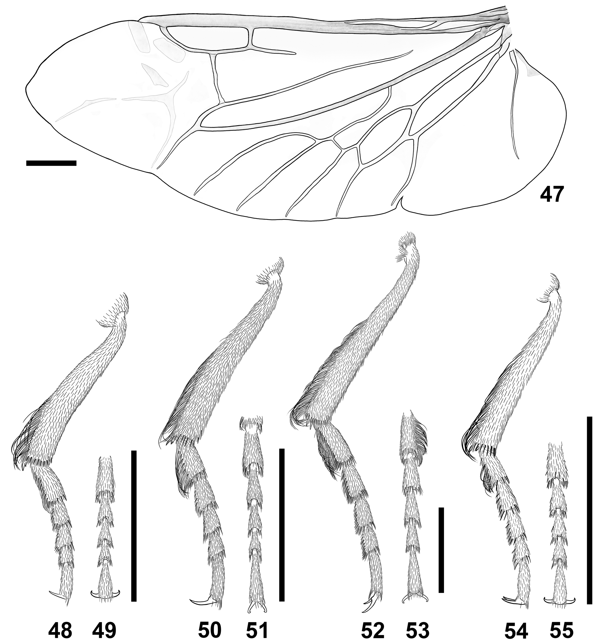
Hindwing ( Fig. 47 View FIGURES 47–55 ) transparent, 2.65 times as long as wide; apical region 0.25 times total wing length, with vague anterior sclerotizations and posterior linear sclerites forming X-shaped mark. Radial cell 3.7 times as long as wide with right posterobasal angle; cross-vein r3 slightly oblique; cross-vein r4 arising near middle of radial cell, long and slightly sinuate. Basal portion of RP long, extending to basal third of wing, slightly sinuate; radio-median loop relatively narrow; medial spur slightly curved, reaching wing margin. Medial field with five free veins, all reaching wing margin; MP 3+4 with oblique basal cross-vein, CuA1 joining it before MP 3 -MP 4 fork; wedge cell slightly longer than medial spur, three times as long as wide, with apex strongly oblique, CuA 1+2 arising in apical third of cell; AA 3 meeting CuP in basal half of wedge cell.Anal notch shallow; AP nearly straight, not reaching wing margin.
Legs stout; fore and mid legs with femur subequal in length to tibia, hind leg with tibia distinctly longer than or subequal to femur. All tibiae with two apical spurs; slightly compressed laterally, elliptic in cross section; meso- and metatibiae covered with short and fine setae; setation of pro- and metatibiae showing sexual dimorphism (see below). Tarsi distinctly compressed laterally; tarsomeres 1–4 gradually decreasing in length, widened apically and excavate at apex, beneath clothed with tiny spines and median stripe with minute setae; setation of metatarsomere 1 showing sexual dimorphism (see below); tarsomere 5 about as long as two previous tarsomeres combined. All tarsal claws simple, evenly curved.
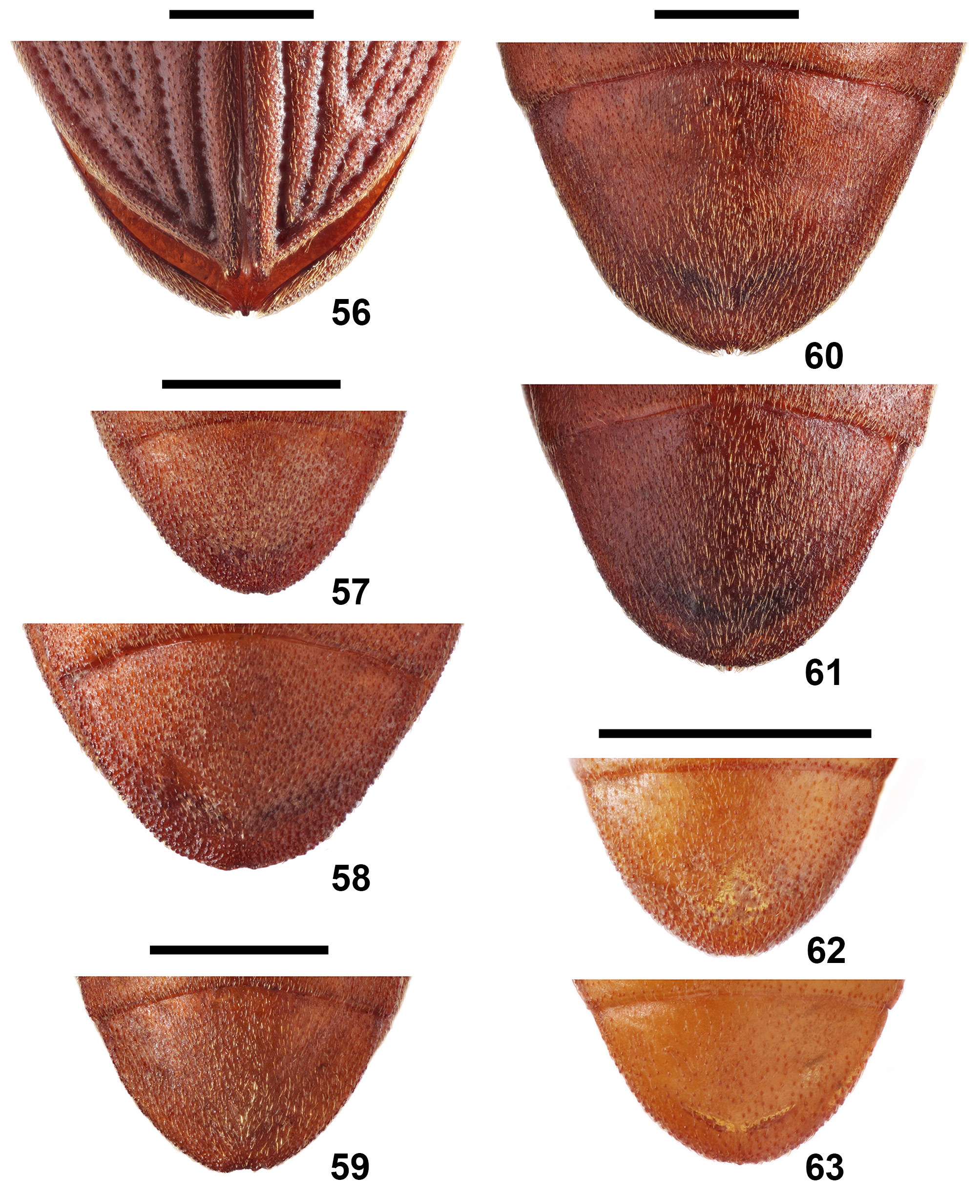
Abdomen with five connate ventrites; ventrites 1–4 of about subequal in length, ventrite 5 somewhat longer than two preceding ones combined; apex of ventrite 5 more or less rounded and often with small median emargination; posterior stripe along edge of ventrite 5 inflected dorsally and interrupted in middle and with more or less prominent central projection ( Figs 56–63 View FIGURES 56–63 ). Abdominal tergites lightly sclerotized.
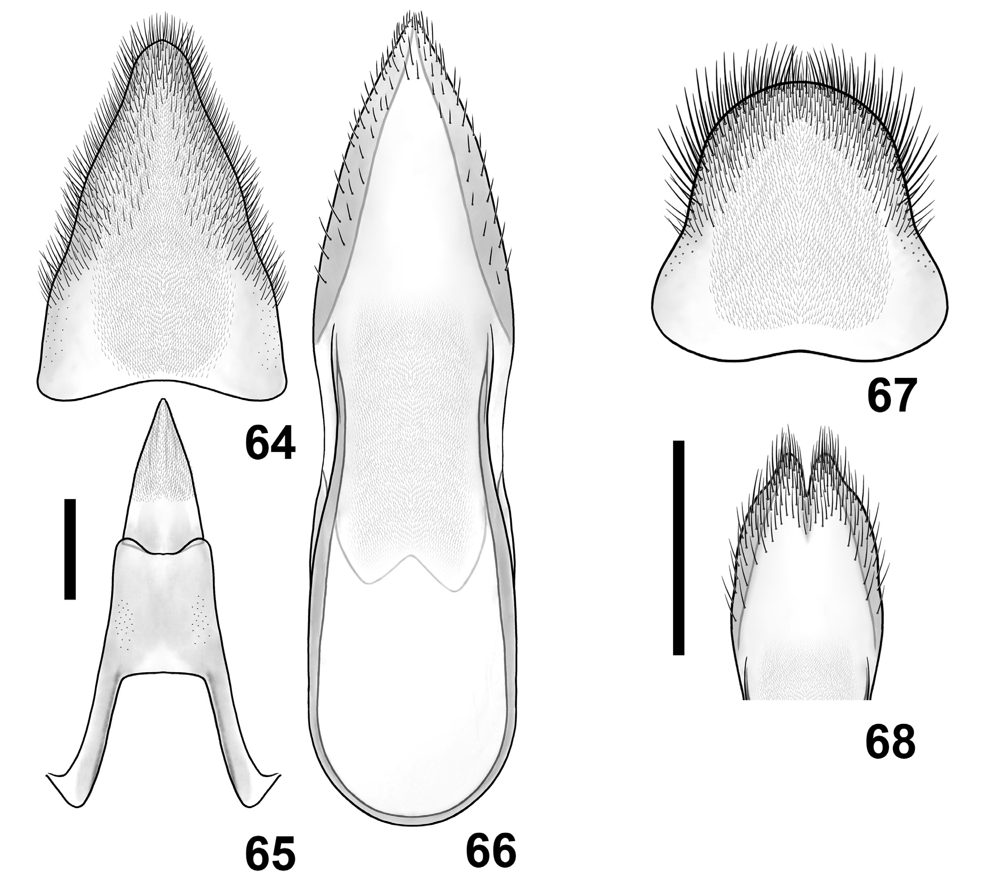
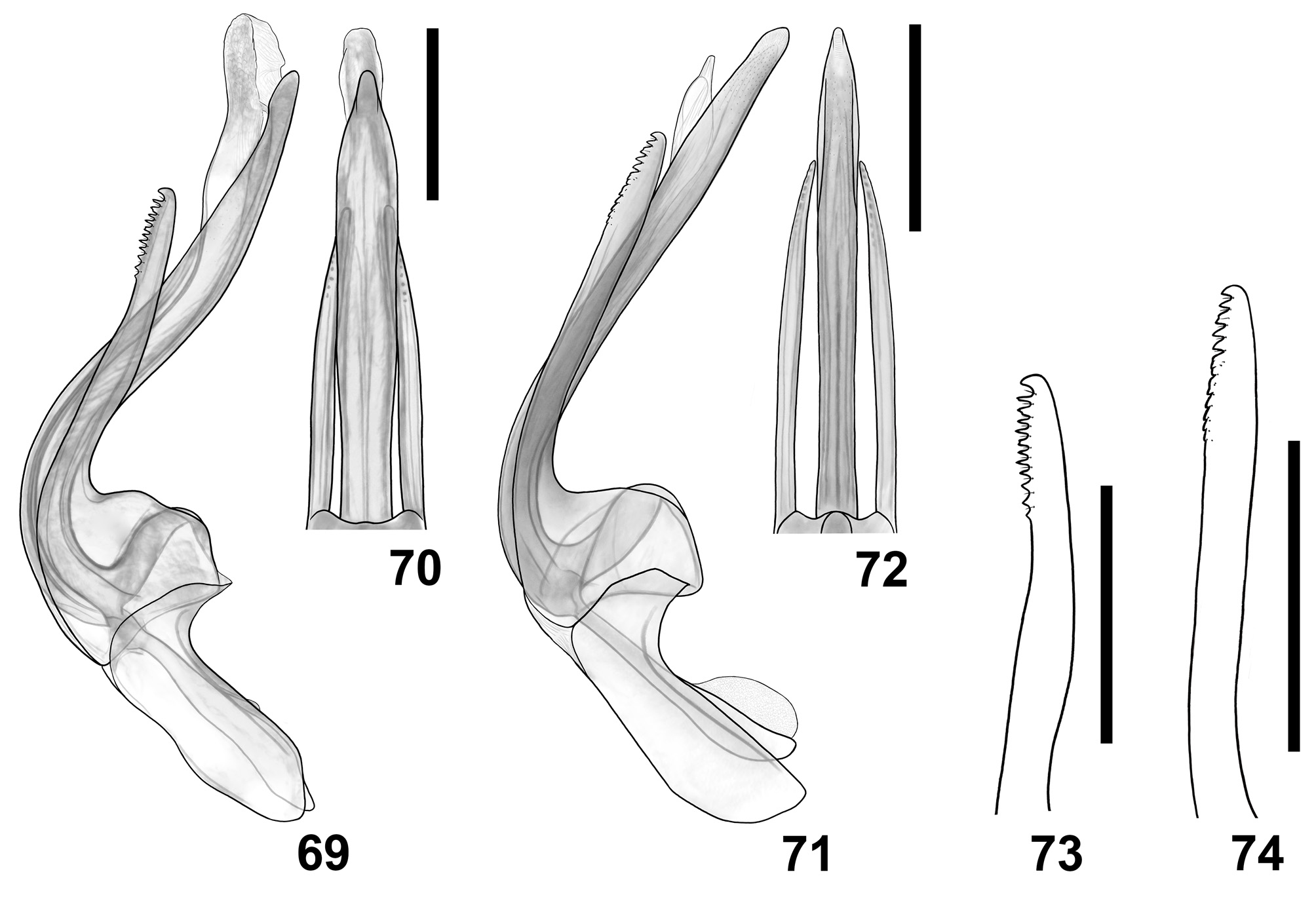
Male. Protibia with fringe of golden setae along inner margin; protarsomere 1 not modified. Metatibiae bearing a fringe of long golden setae along inner margin in distal half, metatarsomere 1 fringed beneath with long golden setae. Tergite VII (pygidium) ( Figs 64, 67 View FIGURES 64–68 ) semielliptical to subtriangular and rounded at apex. Sternite 9 narrowly rounded or notched at apex; laterobasal apophyses narrow, fused together at base and forming elongate lobe rounded at base; tergite IX with pair of laterobasal apophyses and shallowly emarginate at apex; tergite X (proctiger) distinctly separated from IX, elongately subtriangular. Aedeagus ( Figs 69–92 View FIGURES 69–74 View FIGURES 75–85 View FIGURES 86–92 ) more or less compressed laterally, resting into abdomen on its right side. Phallobase subtrapezoidal, dorsally open, symmetrical and emarginated at base. Parameres shortly fused dorsally at base and separated ventrally, with bases strongly expanded dorsally, and then curved; each paramere with apex more or less curved ventrally, ventral margin before apex serrate. Penis elongate, extending well beyond apices of parameres, anteriorly with paired ventral apophyses and dorsal process fused with expanded part of parameres. Flagellum elongate, tubular.
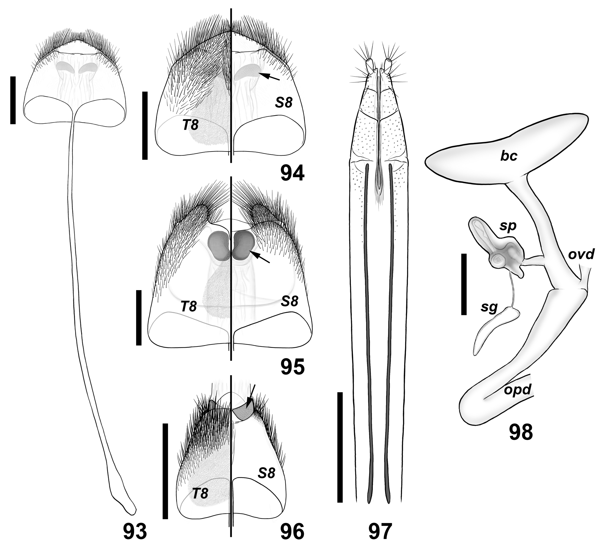
Female. All tibiae with uniform and simple vestiture; metatarsomere 1 clothed beneath as following tarsomeres. Tergite VIII ( Figs 94–96 View FIGURES 93–98 ) rounded, subtruncate or excised at apex. Sternite 8 ( Figs 93–96 View FIGURES 93–98 ) with spiculum ventrale long and narrow, fused with its base. Membranous tube, connecting abdominal segment 8 with ovipositor, flanked at base by pair of more or less developed sclerites, retractable inside abdominal segment VIII in rest. Ovipositor ( Fig. 97 View FIGURES 93–98 ) elongate and slender, weakly sclerotized; paraprocts about four times as long as coxites, with longitudinal baculi; coxite moderately short, narrowing apically, divided into three part; styli well developed, terminal. Female genital tract ( Fig. 98 View FIGURES 93–98 ) with large, bilobed anteriorly bursa copulatrix; spermatheca soft, attached through a short duct to tubular basal part of bursa copulatrix; spermathecal gland narrow, attached by duct to base of spermatheca.


No known copyright restrictions apply. See Agosti, D., Egloff, W., 2009. Taxonomic information exchange and copyright: the Plazi approach. BMC Research Notes 2009, 2:53 for further explanation.