
Andrena (Truncandrena) petrae Wood, 2022
|
publication ID |
https://doi.org/ 10.11646/zootaxa.5185.1.1 |
|
publication LSID |
lsid:zoobank.org:pub:D34A7F04-8EAD-4441-A859-CFD79F7740D2 |
|
DOI |
https://doi.org/10.5281/zenodo.7074107 |
|
persistent identifier |
https://treatment.plazi.org/id/9C1E87C4-C72C-FF85-FF0B-F919FB17B1B8 |
|
treatment provided by |
Plazi |
|
scientific name |
Andrena (Truncandrena) petrae Wood |
| status |
sp. nov. |
Andrena (Truncandrena) petrae Wood sp. nov.
( Figs. 182–190 View FIGURES 179–187 View FIGURES 188–194 )
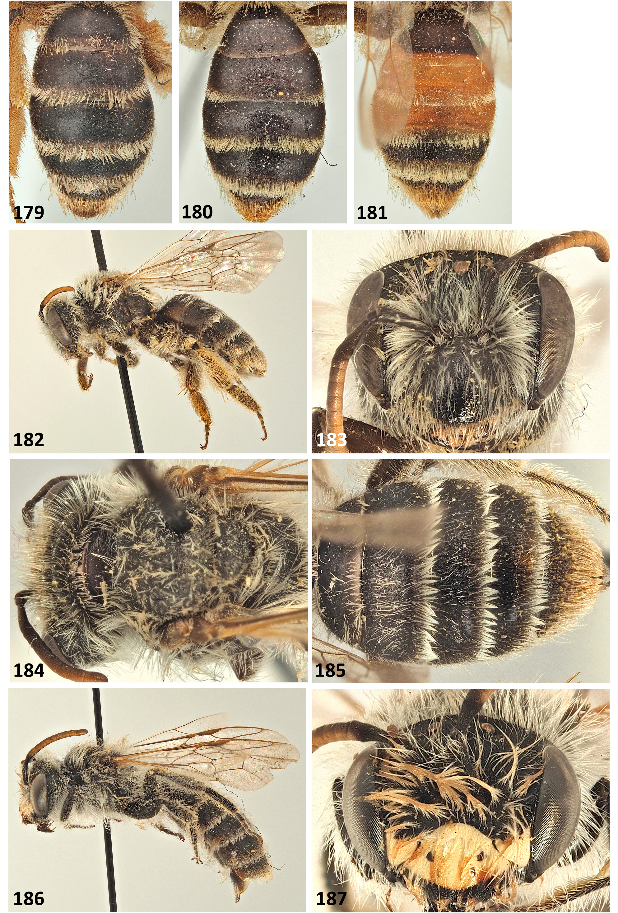
Female ( Fig. 182 View FIGURES 179–187 ).
Body length: 10–12 mm.
Colour. Body uniformly dark grey ( Fig. 182 View FIGURES 179–187 ). Flagellum dark basally, becoming orange from apex of segment 2 onwards, particularly ventrally ( Figs. 182–183 View FIGURES 179–187 ). Legs dark, apical tarsal segments slightly lightened brownish. Wings hyaline, stigma dark orange centrally, dark brown laterally, venation dark orange ( Fig. 182 View FIGURES 179–187 ). Tergal margins apically lightened brown-whitish hyaline ( Fig. 185 View FIGURES 179–187 ).
Pubescence. Face, gena, vertex, scape with long white hairs, becoming light brownish on vertex, longest on gena equaling length of scape. Clypeus apically, mandibles and labrum with faintly golden hairs ( Figs. 183–184 View FIGURES 179–187 ). Fovea with short brownish hairs. Mesonotum and scutellum with short white hairs, becoming longer on mesepisternum, not exceeding length of scape ( Figs. 182–184 View FIGURES 179–187 ). Propodeal corbicula weakly complete, composed of white plumose hairs; surface of propodeal corbicula almost hairless, with few simple white hairs laterally. Leg hair white to faintly golden apically, scopa white ( Fig. 182 View FIGURES 179–187 ). Flocculus weakly complete, composed of white plumose hairs. Tergal discs with short to long white hairs, marginal zones 1–4 with complete white hairbands, on 2–4 thick and obscuring underlying surface. Prepygidial fimbria golden centrally, white laterally; pygidial fimbria golden ( Fig. 185 View FIGURES 179–187 ).
Head ( Figs. 183–184 View FIGURES 179–187 ). 1.3 times broader than long. Labral process trapezoidal, slightly broader than long, apical margin straight. Clypeus weakly domed, basally shagreened, in apical half almost without shagreenation, shining. In apical 2/3 clypeus densely punctured with exception of impunctate longitudinal central line, punctures large and separated by <0.5–1 puncture diameters ( Fig. 183 View FIGURES 179–187 ). Paraocular area with inconspicuous punctures; frons with longitudinal striations, underlying surface very weakly shining. Flagellomere 1 exceeds 2+3, slightly shorter than 2+3+4. Facial fovea dorsally occupying half distance between lateral ocellus and compound eye, in length slightly exceeding level of antennal insertions, not narrowed below ( Fig. 183 View FIGURES 179–187 ). Fovea dorsally separated from lateral ocellus by 1.5 diameters of lateral ocellus. Ocelloccipital distance equal to 1.5 diameter of lateral ocellus. Genal area slightly exceeding width of compound eye.
Mesosoma ( Fig. 184 View FIGURES 179–187 ). Pronotum without elevated dorsolateral angle or lateral carina. Mesonotum and scutellum shagreened, very weakly shining, majority of surface with irregular extremely shallow and inconspicuous punctures, indicated by slightly raised margins, punctures separated by <0.5–1 puncture diameters. Mesepisternum with fine granular microreticulation, with faint raised reticulation above. Anterolateral face of propodeum with only fine granular microreticulation, posterolateral part of propodeum with fine granular microreticulation and faint raised reticulation above; propodeal triangle narrow, indicated by absence of raised reticulation. Inner side of hind femur rounded, not carinate. Tarsal claws with small inner tooth. Recurrent vein 1 reaching submarginal cell 2 clearly beyond its middle. Submarginal crossvein 1 meets marginal cell 5 vein widths from stigma. Nervulus interstitial ( Fig. 182 View FIGURES 179–187 ).
Metasoma ( Fig. 185 View FIGURES 179–187 ). Terga with fine microreticulation, weakly shining, strongest on tergum 1, becoming finer and weaker on following terga. Tergal discs with fine, sparse punctures, punctures separated by 3–4 puncture diameters. Marginal zones weakly depressed, occupying 0.2–0.3 of tergal disc, with denser punctures, separated by 2–3 puncture diameters. Pygidial plate rounded triangular, flat, internal surface dull.
Male ( Fig. 186 View FIGURES 179–187 ).
Body length: 10–10.5 mm.
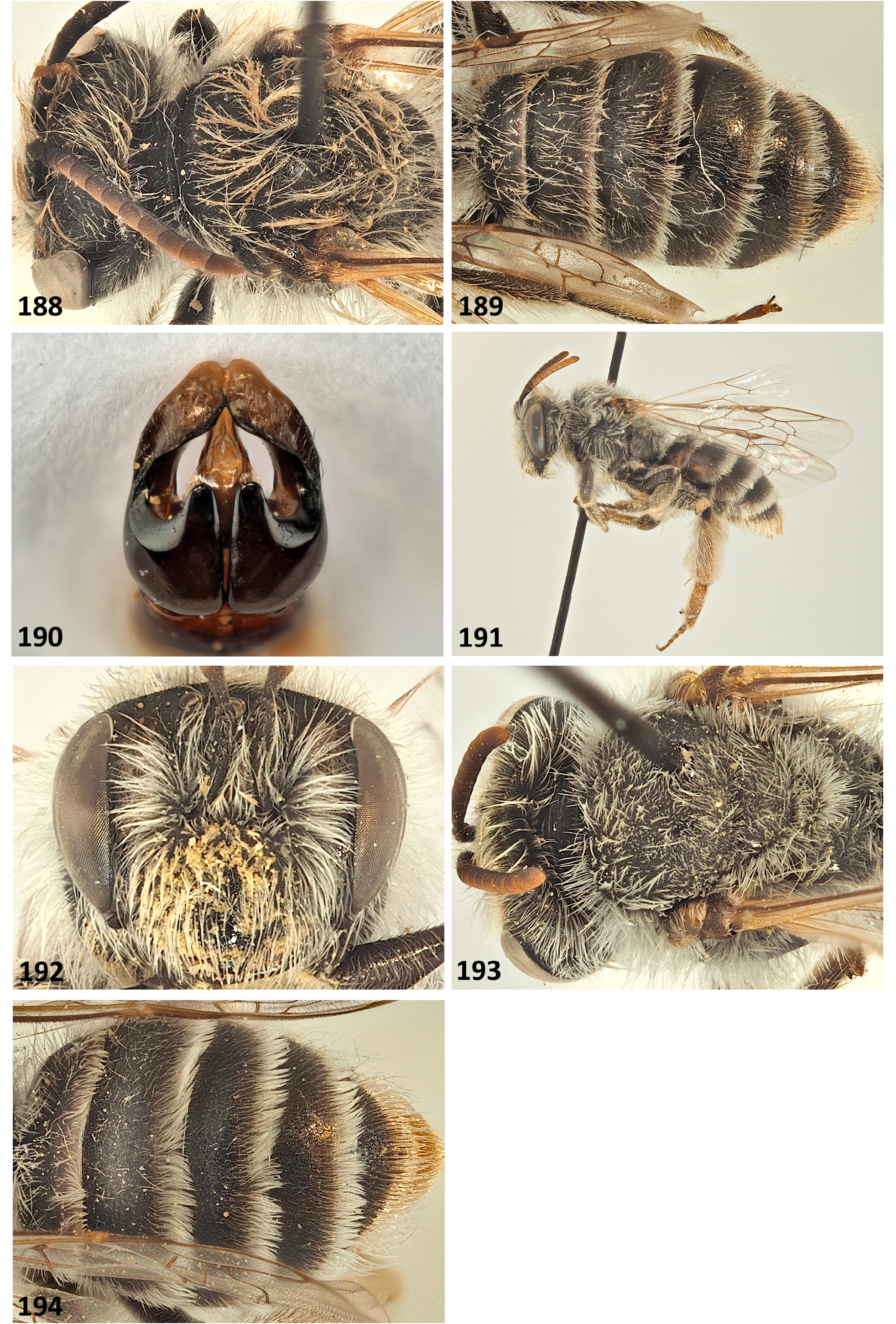
Colour. Body uniformly dark grey ( Fig. 186 View FIGURES 179–187 ). Clypeus and lower paraocular areas yellow, clypeus entirely yellow with exception of two lateral black maculae ( Fig. 187 View FIGURES 179–187 ). Flagellum dark basally, strongly orange below from segment 2–11. Legs dark, apical tarsal segments slightly lightened brownish. Wings hyaline, stigma dark orange centrally, dark brown laterally, venation dark orange ( Fig. 186 View FIGURES 179–187 ). Tergal margins apically lightened brown-whitish hyaline ( Fig. 189 View FIGURES 188–194 ).
Pubescence. Face, gena, vertex and scape with long white hairs, on gena longest hairs equaling length of scape ( Figs. 186–187 View FIGURES 179–187 ). Mesosoma all over with long white hairs, becoming brownish above, majority equaling or exceeding length of scape ( Figs. 186 View FIGURES 179–187 , 188 View FIGURES 188–194 ). Leg hair white ( Figs. 186 View FIGURES 179–187 ). Tergal hairs as in female, though hair bands less dense ( Fig. 189 View FIGURES 188–194 ).
Head ( Figs. 187–188 View FIGURES 179–187 View FIGURES 188–194 ). 1.3 times broader than long. Labral process short, slightly broader than long, apical margin emarginate. Clypeus weakly domed, finely shagreened, weakly shining. Clypeus punctate with exception of ill-defined longitudinal impunctate midline, punctures separated by 0.5–1 puncture diameters ( Fig. 187 View FIGURES 179–187 ). Paraocular area with inconspicuous punctures; frons with longitudinal striations, underlying surface very weakly shining. Flagellomere 1 as long as 2+3. Ocelloccipital distance equal to 1.5 diameter of lateral ocellus ( Fig. 188 View FIGURES 188–194 ). Genal area equaling width of compound eye.
Mesosoma ( Fig. 188 View FIGURES 188–194 ). Pronotum without elevated dorsolateral angle or lateral carina. Mesonotum and scutellum with fine granular shagreen, dull to weakly shining centrally, essentially impunctate. Mesosoma structure otherwise as in female. Tarsal claws with strong inner tooth. Recurrent vein 1 reaching submarginal cell 2 at its middle. Submarginal crossvein 1 meets marginal cell 5 vein widths from stigma. Nervulus interstitial ( Fig. 186 View FIGURES 179–187 ).
Metasoma ( Fig. 189 View FIGURES 188–194 ). As in female.
Genitalia and hidden sterna ( Fig. 190 View FIGURES 188–194 ). Gonocoxites with pronounced rounded dorsal lobes, gonostyli broadened apically, with strong raised internal margin, apically with slightly narrowed and rounded points. Penis valves centrally broad, with hyaline lateral extensions, narrowed apically ( Fig. 190 View FIGURES 188–194 ). Sternum 8 narrowed centrally, thus narrowly triangular, apically truncate. Ventral surface covered with plumose white hairs projecting laterally.
Diagnosis. Andrena petrae is superficially similar to the Andrena doursana species group and related species (in the Levant these include A. dorchini sp. nov., A. mizorhina Warncke stat. nov., A. mucronata Morawitz and A. ulula Warncke ) but can quickly be separated by the punctation of the mesonotum which is subtle (dense and essentially contiguous in the doursana group). It is very similar to A. syriensis Wood (female described below); females are almost identical and very difficult to separate, and more specimens are required in order to understand intra- and interspecific differences. The female of A. petrae can potentially be separated by a greater ocelloccipital distance (1.5 times diameter of lateral ocellus versus 1 times diameter of lateral ocellus in A. syriensis ), the slightly sparser mesonotal punctures, the position of the first recurrent vein, and the dark hind tarsi and basitarsi.
In the male sex, A. petrae is also most similar to A. syriensis , both possessing yellow facial markings extending onto the lower paraocular areas. However, the two taxa can be clearly separated by the construction of the genital capsule, specifically by the gonocoxal teeth which are pronounced and apically rounded, not strongly diverging apically (teeth weakly formed, very strongly diverging apically in A. syriensis ), and by the lateral extensions of the penis valves which are hyaline but relatively short, thus forming lateral wings (elongate, finger-like, clearly extending laterally away from the penis valves in A. syriensis , compare illustrations in Wood 2021a).
Distribution. Southern Jordan and south-central Turkey. Likely present also in Syria.
Flight period: April.
Flower records: None.
Holotype: JORDAN: N of Petra , SE Shobak [Shawbak], 1.iv.2013, M. Snižek, ♂ ( OLML).
Paratypes: JORDAN: same as holotype (1♂); TURKEY: Şanlıurfa [ Harran / Urfa], 19.iv.1976, K. Warncke (1♀, 1♂) ( OLML, SMNHTAU) .
Etymology. Named after the ancient city of Petra in Jordan, the name of which means ‘rock’ in Ancient Greek. The species epithet is an adjective.
Remarks. There is some variation across this range, most obviously in the males where Jordanian specimens have two lateral black maculae on the clypeus, but these are absent in the sole Turkish specimen. However, the genital capsule is the same across this distance, so this is considered to be simple variation.
| OLML |
Oberösterreichisches Landesmuseum |
No known copyright restrictions apply. See Agosti, D., Egloff, W., 2009. Taxonomic information exchange and copyright: the Plazi approach. BMC Research Notes 2009, 2:53 for further explanation.