
Pimelodella yaharo, Conde-Saldaña & Albornoz-Garzón & García-Melo & Dergam & Villa-Navarro, 2019
|
publication ID |
https://doi.org/ 10.11646/zootaxa.4668.4.8 |
|
publication LSID |
lsid:zoobank.org:pub:3A0501FD-2C81-4F26-935F-D33306B64E44 |
|
persistent identifier |
https://treatment.plazi.org/id/9C71A71E-026C-FFAA-53D2-F948FC8B35F3 |
|
treatment provided by |
Plazi |
|
scientific name |
Pimelodella yaharo |
| status |
sp. nov. |
Pimelodella yaharo , new species
( Figures 1–2a View FIGURE 1 View FIGURE 2 ; Table 1 View TABLE 1 )
urn:lsid:zoobank.org:act:EB2DEC53-73D1-47DD-83D4-A9EC1E87B53F
Pimelodella chagresi View in CoL [non Steindachner, 1876].— Villa-Navarro et al. 2014:334 [list of species]
Holotype. CZUT-IC 10922 , 74.7 mm SL, Colombia, Departamento La Guajira, Dibulla, Río Jeréz , 11º12’26.1”N, 73º15’16”W, 69 masl, F. A. Villa-Navarro, 10 September 2012. GoogleMaps
Paratypes. All from Colombia, Departamento La Guajira, Dibulla : CZUT-IC 10942 (1, 69.9 mm SL), same locality as holotype, F. A. Villa-Navarro, 19 September 2011 GoogleMaps ; CZUT-IC 12602 (2, 72.4–82.0 mm SL), Río Cañas , 11°12’41.78”N, 73°24’10.60”W, 49 masl, J. G. Albornoz-Garzón, 22 September 2014 GoogleMaps ; CZUT-IC 15262 (2, 1 C&S, 66.9–76.9 mm SL); IAvH-P 22004 (2, 1 C&S, 68.7–72.7 mm SL), Río Cañas , 11°12’42.80”N, 73°24’11.44” W, 49 masl, C. Conde-Saldaña & J. G. Albornoz-Garzón, 12 January 2015 GoogleMaps .
Diagnosis. Pimelodella yaharo can be diagnosed from all other trans-Andean species of Pimelodella except P. chagresi , P. grisea and P. reyesi , by having greater maximum depth of dentations in posterior margin of pectoralfin spine (1.40–1.68 times in the width of the spine at its base vs. 1.69–3.27 times in P. elongata , 2.22–6.22 times in P. eutaenia , 1.71–2.90 times in P. floridablancaensis , 2.54–4.13 times in P. macrocephala , 2.03–3.27 times in P. modesta , 1.73–2.67 times in P. odynea , and 1.73–2.28 times in P. yuncensis ). Pimelodella yaharo differs from P. chagresi and P. reyesi by having shorter dorsal-fin spine length (8.3–13.7% SL vs. 14.1–16.9% in P. chagresi and 14.6–19.5% in P. reyesi ) and by having a shorter pectoral-fin spine length (12.9–14.5% SL vs. 15.1–17.5% in P. chagresi and 15.3–19.7% in P. reyesi ). Moreover, P. yaharo differs from P. reyesi by its shorter maxillary barbels (53.0–68.3% SL vs. 70.9–89.7%) and from P. chagresi by its shorter dorsal-fin base (12.3–14.9% SL vs. 14.9–16.4%). The new species differs from P. grisea by having a narrower bony interorbital width (17.6–21.6% HL vs. 22.0–26.4%), a conspicuous paired dorsal dark brown stripe (vs. dorsal surface of body without a conspicuous dark brown stripe) and by having a wide dark brown midlateral stripe (vs. narrow dark brown midlateral stripe).
Pimelodella yaharo further differs from remaining trans-Andean species by the following combination of characters: shorter distal portion without dentations of the posterior margin of pectoral-fin spine (21.6–29.8% in pectoral-fin spine length vs. 41.0–64.0% in P. eutaenia , 32.2–52.0% in P. floridablancaensis , 38.8–49.4% in P. macrocephala and 39.9–53.1% in P. odynea ); shorter adipose-fin base (22.8–26.4% SL vs. 30.0–36.1% in P. elongata , 27.5–32.9% in P. odynea and 30.1–36.5% in P. yuncensis ); shorter bony interorbital width (17.6–21.6% HL vs. 26.8–32.1% in P. macrocephala , and 23.2–27.4% in P. modesta ); deeper caudal-peduncle (9.2–10.5% SL vs. 6.4–8.7% in P. elongata and 7.1–8.8% in P. odynea ); longer inner mental-barbels (14.4–20% SL vs. 8.4–12.6% in P. modesta ); shorter head (22.2–23.7% SL vs. 24.9–27.4% in P. macrocephala ); wider body (17.6–21.1% SL vs. 15.3–16.5% in P. elongata and 15.0–17.5% in P. odynea ) 40 vertebrae (vs. 41–43 in P. elongata , 42 in P. eutaenia , 41–43 in P. floridablancaensis , 41–43 in P. odynea and 37–38 in P. yuncensis ) and by having a wide dark brown midlateral stripe (vs. narrow to no conspicuous stripe in P. yuncensis ).
Description. Morphometric data given in Table 1 View TABLE 1 . Body slightly deeper than wide, depth at dorsal-fin origin 4.66–5.11 times in standard length. Body elongated, somewhat triangular in cross-section at dorsal-fin origin, progressively compressed to caudal-fin ( Fig. 1 View FIGURE 1 ). Dorsal profile slightly convex from snout tip to dorsal-fin origin; slightly concave just posterior to dorsal-fin base to adipose-fin origin. Profile convex along adipose-fin base, straight and ascending along caudal peduncle. Ventral profile of body slightly convex from snout to margin of branchiostegal membrane, convex between branchiostegal membrane and pelvic-fin origin, straight or slightly concave from this point to anal-fin origin, straight along anal-fin base and concave from base of last anal-fin ray to caudal fin.
Head conical and depressed, dorsally covered by thin skin. Snout short and slightly convex. Mouth subterminal, premaxillary arranged in two rectangular patches, tooth patch not exposed. Premaxilla and dentary with conical, small and narrow teeth. Eye elliptical, its greatest diameter along horizontal axis, placed dorsolaterally. Eye with well-defined orbital rim. Bony interorbital distance narrower than eye diameter.Anterior naris tubular. Posterior naris bordered anteromedially by low fleshy margin. Barbels thin and slightly depressed. Maxillary barbel, adpressed parallel to main body axis, reaching between verticals through pelvic-fin base terminus and first anal-fin ray base. Outer mental barbel adpressed reaching between verticals through posterior end of pectoral-fin base and dorsal-fin spine insertion. Inner mental barbel reaching between verticals through pectoral-fin origin and pectoral-fin base terminus. Branchiostegal membrane free, supported by six rays and joined to isthmus only at anteriormost point. Gill rakers on first gill arch 9–10 (2). Supraoccipital process triangular, reaching anterior nuchal plate. Anterior nuchal plate with a short anterior process fitting into bifid supraoccipital process posterior tip.
Posterior process of cleithrum triangular, narrow, its dorsal border concave and its ventral border straight or slightly convex. Axillary pore present at pectoral-fin base. Urogenital papilla triangular and tubular, located posterior to anus, separated by a gap.
Dorsal fin lepidotrichia I,6* (8); spinelet narrow and its distal tip sharp. Dorsal-fin spine strong, straight, sharp, and shorter than first branched ray; margins almost smooth, distal third of anterior and posterior margins with minute serrae. Distal margin of dorsal-fin convex. Posteriormost dorsal-fin pterygiophore located anterior to neural spine of vertebrae 11. Distance between terminus of dorsal-fin base and adipose-fin origin longer than dorsal-fin base.
Pectoral-fin rays I,7 (3) or I,8* (5), pectoral fin triangular with slightly convex to straight distal border. First pectoral-fin ray curved with proximal portion rigid, forming spine ( Fig. 2a View FIGURE 2 ), and short distal tip soft. Anterior margin of pectoral-fin spine with minute dentations, except for a smooth, distalmost margin; posterior margin with 8–12 (2) very strong recurved dentations, distalmost portion without dentations comprises 21.6–29.8% of pectoral-fin spine length. Maximum depth of dentations 1.40–1.68 times in the width of spine at its base.
Pelvic-fin rays i,5* (8), with distal border convex. Pelvic-fin origin posterior or slightly posterior to vertical through base of last dorsal-fin ray. First unbranched ray shortest, second, third and fourth branched rays longer.
Anal-fin rays v,7 (1), 8* (4), 9 (2) or 10 (1); distal margin of anal fin convex. Unbranched rays distinctly shorter than first, second and third branched rays. Anal-fin origin posterior to vertical through adipose-fin origin. Tip of anteriormost anal-fin pterygiophore inserted posterior to hemal spine of vertebra 20 (2). Tip of posteriormost analfin pterygiophore inserted ahead of hemal spine of vertebra 28 (2).
Adipose fin relatively short (22.8–26.4% in SL); dorsal-fin to adipose fin distance 0.72–0.92 times in dorsal-fin base. Adipose-fin emerging gradually, its maximum height at second third, posterior lobe free. Adipose-fin origin anterior to anal-fin origin.
Caudal fin deeply forked, upper lobe pointed and slightly longer than lower lobe; lower lobe rounded. Upper lobe with 14–16 (2) procurrent fin-rays, 1 unbranched ray, and 6 (1) or 7 (7)* branched rays. Lower lobe with 17–18 (2) procurrent fin-rays, 1 unbranched ray and 8 (8)* branched rays. Middle caudal-fin rays not articulated directly to caudal plate. Caudal skeleton, parhypural; 1+2, 3+4, 5. Total vertebrae 40 (2). Ribs 9 (2).
Lateral line canal complete, extending to basal portion of interadial membrane of middle caudal-fin rays. Head laterosensory canals with simple (unbranched) tubes usually ending in a single pore. Supraorbital pore S1 medially adjacent to anterior naris. S2+ I2 between anterior and posterior nares, slightly closer to posterior naris. S3 just at posterior rim of posterior naris and S4 located slightly anterior to anterior eye margin. Branches S6, S7 and S8 contained in the frontal. Epiphyseal branches (S6) fused to each other, bearing a single symphyseal pore (S6+S6). S8 (parietal branch) posteromedial to eye. Infraorbital pore I1 laterally adjacent to maxillary barbel base, just between anterior naris and barbel base. I3 posterior to maxillary barbel base, at vertical through posterior naris. I4 posterior to anterior eye margin. I5 at vertical through posterior eye margin. I6 posterior to eye. Preoperculomandibular canal with 11 lateral-line branches and pores, along dentary, subpreopercle and preopercle. Posteriormost preopercular pore (PO1+PM11) at the end of membranous branch. Pterotic branch 2 (PO2) located dorsal to dorsoposterior corner of opercular margin. Postotic sensory branch 3 (PO3) dorsal to lateral line canal.
Coloration in alcohol. Body light brown fading to a pale abdominal region ( Fig. 1 View FIGURE 1 ). Dorsal region of head with heavy concentration of dark brown chromatophores on cephalic region. Ventral region of head pale. Pseudotympa- num area slightly darker than surrounding areas. A dark brown midlateral stripe well defined, extending from snout to centralmost caudal-fin rays. Dorsal surface of body with paired conspicuous dark brown stripe, extending from posterior margin of head to caudal-fin insertion, getting diffuse posterior to adipose-fin base. Dorsal surface of maxillary barbels brown and ventral surface yellowish. Mental barbels yellow. Fins yellowish with dark chromatophores disperse. Interradial membrane of dorsal fin with stripes of conspicuous dark chromatophores along anterior edge of rays ( Fig. 1 View FIGURE 1 ).
Distribution and habitat. Pimelodella yaharo is only known from Río Jerez and Río Cañas. Both are coastal drainages of the Sierra Nevada de Santa Marta (SNSM) which drains directly into the Caribbean Sea ( Fig. 3 View FIGURE 3 ). The localities in these rivers (Río Jerez: 69 masl, pH 7.89, conductivity 75 µS/cm; Río Cañas: 49 masl, pH 7.90, conductivity 40 µS/cm) are associated with Dry Tropical Forest areas, have clear waters, moderate to strong current and substrates with sand, pebbles and rocks ( Fig. 4 View FIGURE 4 ). Pimelodella yaharo is generally found syntopically with Astyanax sp., Creagrutus affinis , Gephyrocharax sp., Hemibrycon sp., Roeboides dayi , Saccoderma hastata , Rhamdia guatemalensis , Trichomycterus sp., Hypostomus sp., Eigenmannia sp., Eleotris pisonis , Awaous banana , Sicydium salvini , Synbranchus marmoratus , Andinoacara latifrons , Caquetaia kraussii , Poecilia caucana , Agonostomus monticola , and Gobiesox sp.
Etymology. Yaharo is a noun in apposition and refers to the pre-conquest name of the region where today is the municipality of Dibulla, where the Río Jerez and Río Cañas flow.

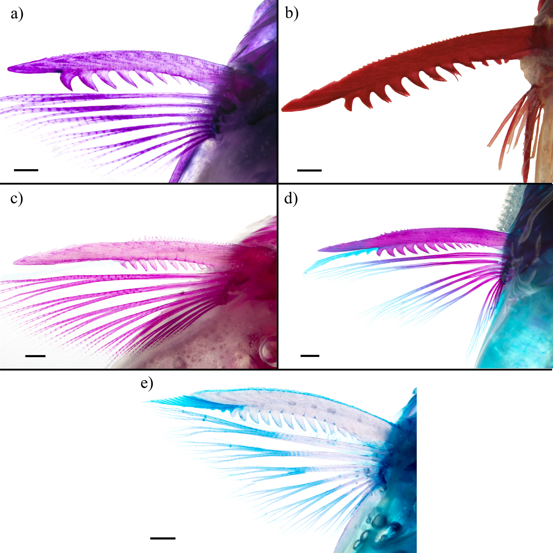


TABLE 1. Morphometric data for Pimelodella yaharo based on holotype and paratypes (n= 7). Min= minimum; Max= maximum; SD= standard deviation.
| Holotype | Range | Mean | SD | |
|---|---|---|---|---|
| Standard length (mm) | 74.7 | 66.9–82.0 | 73.0 | |
| Percent of SL | ||||
| Body depth | 21.5 | 19.6–21.5 | 20.4 | 0.7 |
| Body width | 17.7 | 17.6–21.1 | 18.4 | 1.2 |
| Head length | 22.2 | 22.2–23.7 | 22.8 | 0.5 |
| Maxillary-barbel length | 63.8 | 53.0–68.3 | 60.1 | 5.2 |
| Outer mental-barbel length | 25.8 | 17.8–33.8 | 26.7 | 4.7 |
| Inner mental-barbel length | 18.1 | 14.4–20.0 | 17.5 | 1.6 |
| Posterior process of cleithrum length | 10.2 | 9.6–10.9 | 10.3 | 0.5 |
| Predorsal length | 34.4 | 34.2–36.5 | 35.2 | 0.8 |
| Dorsal-fin base | 14.7 | 12.3–14.9 | 14.0 | 1.0 |
| Dorsal-fin spine length | 12.8 | 8.3–13.7 | 12.0 | 2.2 |
| Dorsal fin to adipose fin | 17.2 | 16.3–18.8 | 17.5 | 0.9 |
| Preadipose length | 63.6 | 63.4–67.0 | 64.8 | 1.2 |
| Adipose-fin base | 25.6 | 22.8–26.4 | 25.1 | 1.3 |
| Adipose-fin depth | 5.1 | 3.4–5.1 | 4.4 | 0.7 |
| Preanal length | 65.6 | 65.6–71.0 | 67.7 | 1.8 |
| Anal-fin base | 12.3 | 11.8–14.7 | 13.1 | 1.0 |
| Prepectoral length | 22.5 | 21.7–27.0 | 23.3 | 1.8 |
| Pectoral-fin spine length | 13.4 | 12.9–14.5 | 13.9 | 0.6 |
| Pectoral-fin spine length without posterior dentations | 3.5 | 3.1–4.2 | 3.5 | 0.4 |
| Pectoral-fin spine width | 1.5 | 1.5–2.0 | 1.7 | 0.1 |
| Maximum depth of pectoral-fin posterior dentations | 0.9 | 0.9–1.4 | 1.1 | 0.1 |
| Prepelvic length | 46.6 | 44.8–52.5 | 48.9 | 2.4 |
| Caudal-peduncle length | 21.4 | 18.8–21.4 | 20.1 | 0.9 |
| Caudal-peduncle depth | 10.3 | 9.2–10.5 | 9.9 | 0.5 |
| Dorsal caudal-fin lobe length | 27.7 | 23.9–31.7 | 27.0 | 2.4 |
| Ventral caudal-fin lobe length | 24.3 | 21.2–25.9 | 24.5 | 1.5 |
| Percent of HL | ||||
| Head depth | 71.5 | 65.5–71.5 | 68.1 | 2.2 |
| Head width | 78.1 | 72.2–83.1 | 76.8 | 3.7 |
| Fleshy interorbital width | 29.9 | 28.6–32.7 | 30.9 | 1.6 |
| Bony interorbital width | 19.7 | 17.6–21.6 | 19.6 | 1.5 |
| Eye diameter | 23.4 | 22.7–26.5 | 25.0 | 1.5 |
| Snout length | 39.5 | 39.5–45.1 | 41.3 | 1.9 |
| Anterior-posterior nares distance | 15.3 | 14.3–19.7 | 17.3 | 1.9 |
| Anterior internarial width | 21.7 | 14.8–21.7 | 17.5 | 2.2 |
| Posterior internarial width | 22.7 | 17.2–22.7 | 20.0 | 1.7 |
| Mouth gape | 36.6 | 29.7–36.6 | 33.0 | 2.3 |
No known copyright restrictions apply. See Agosti, D., Egloff, W., 2009. Taxonomic information exchange and copyright: the Plazi approach. BMC Research Notes 2009, 2:53 for further explanation.
|
Kingdom |
|
|
Phylum |
|
|
Class |
|
|
Order |
|
|
Family |
|
|
Genus |