
Paraustrosimulium colboi ( Davies & Györkös), 1988
|
publication ID |
https://doi.org/ 10.11646/zootaxa.4337.4.1 |
|
publication LSID |
lsid:zoobank.org:pub:BA7E7DE5-25C2-41BA-8642-9B429FDC5294 |
|
DOI |
https://doi.org/10.5281/zenodo.6052668 |
|
persistent identifier |
https://treatment.plazi.org/id/9D4187CA-FFF0-FFE6-ADF6-520AFAACB1A5 |
|
treatment provided by |
Plazi |
|
scientific name |
Paraustrosimulium colboi ( Davies & Györkös), 1988 |
| status |
|
Paraustrosimulium colboi ( Davies & Györkös), 1988
( Figs. 1–42 View FIGURES 1 – 6 View FIGURES 7 – 11 View FIGURES 12 – 17 View FIGURES 18 – 23 View FIGURES 24 – 25 View FIGURES 30 – 31 View FIGURES 35 – 40 View FIGURES 41, 42 )
Austrosimulium colboi Davies & Györkös, 1988: 111 View in CoL . Original designation. Provisional placement to genus. Austrosimulium (Austrosimulium) colboi View in CoL . Crosskey 1989: 221.
Austrosimulium colboi View in CoL . Crosskey & Howard 1997: 26. Unplaced to subgenus. Austrosimulium (Austrosimulium) colboi View in CoL . Bugledich 1999: 330.
“ Austrosimulium colboi View in CoL ”. Moulton 2003: 47.
Austrosimulium colboi View in CoL . Crosskey & Howard 2004: 18. Adler & Crosskey 2009: 19; 2017: 30. Unplaced to subgenus.? Austrosimulium colboi View in CoL . Craig et al. 2012: 53.
Austrosimulium colboi View in CoL . Hernández-Triana et al. 2017: 350.
Paraustrosimulium colboi . This work, new combination.
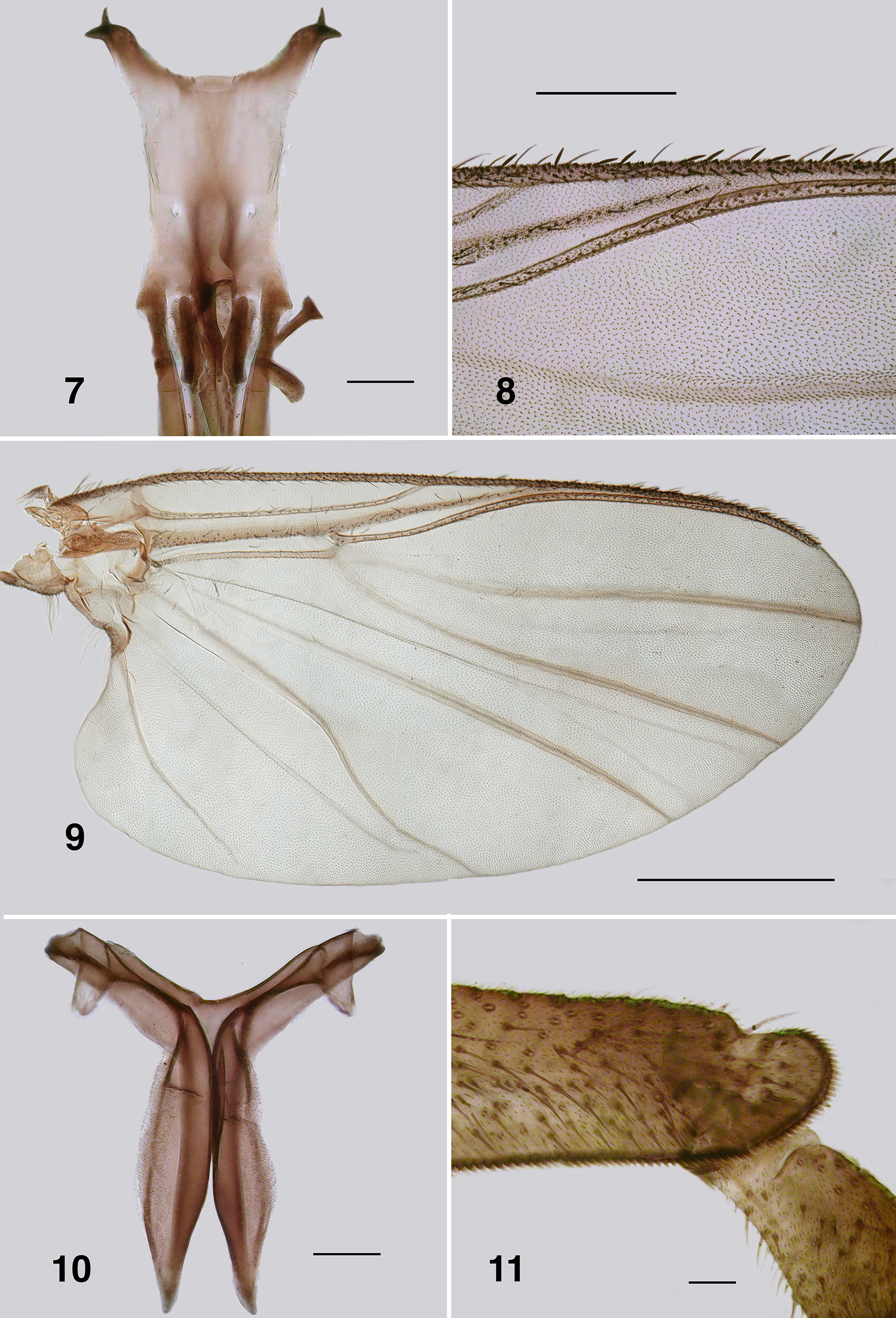
Redescription. Adult female (based on numbers of reared specimen). Body ( Fig. 1 View FIGURES 1 – 6 ): general body colour in alcohol evenly blackish brown; total length 1.9–2.3 mm. Head ( Fig. 2 View FIGURES 1 – 6 ): width 0.57–0.60 mm; depth 0.4 mm; postocciput black, hirsute; frons black; frons–head ratio (narrowest width of frons: greatest width of head) 1.0:6.3. Eyes: slightly bicolourous, interocular distance 0.09 mm; ommatidia 0.015 mm in diameter; 35–39 rows up and across at mid–eye. Clypeus: black; 0.17mm wide; vestiture of sparse black hairs. Antenna ( Fig. 3 View FIGURES 1 – 6 ): total length 0.41–0.46 mm; pedicel small, scape enlarged, both blackish brown, remainder brown; 8 flagellomeres, basal ones wider than long, distally ones more quadratic, overall tapered, apical flagellomere distinctly so. Mouthparts: substantial, ca.
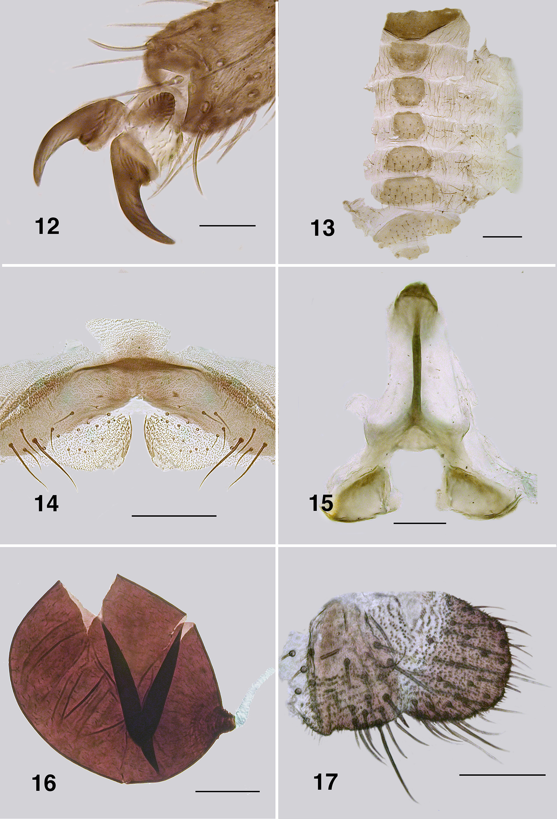
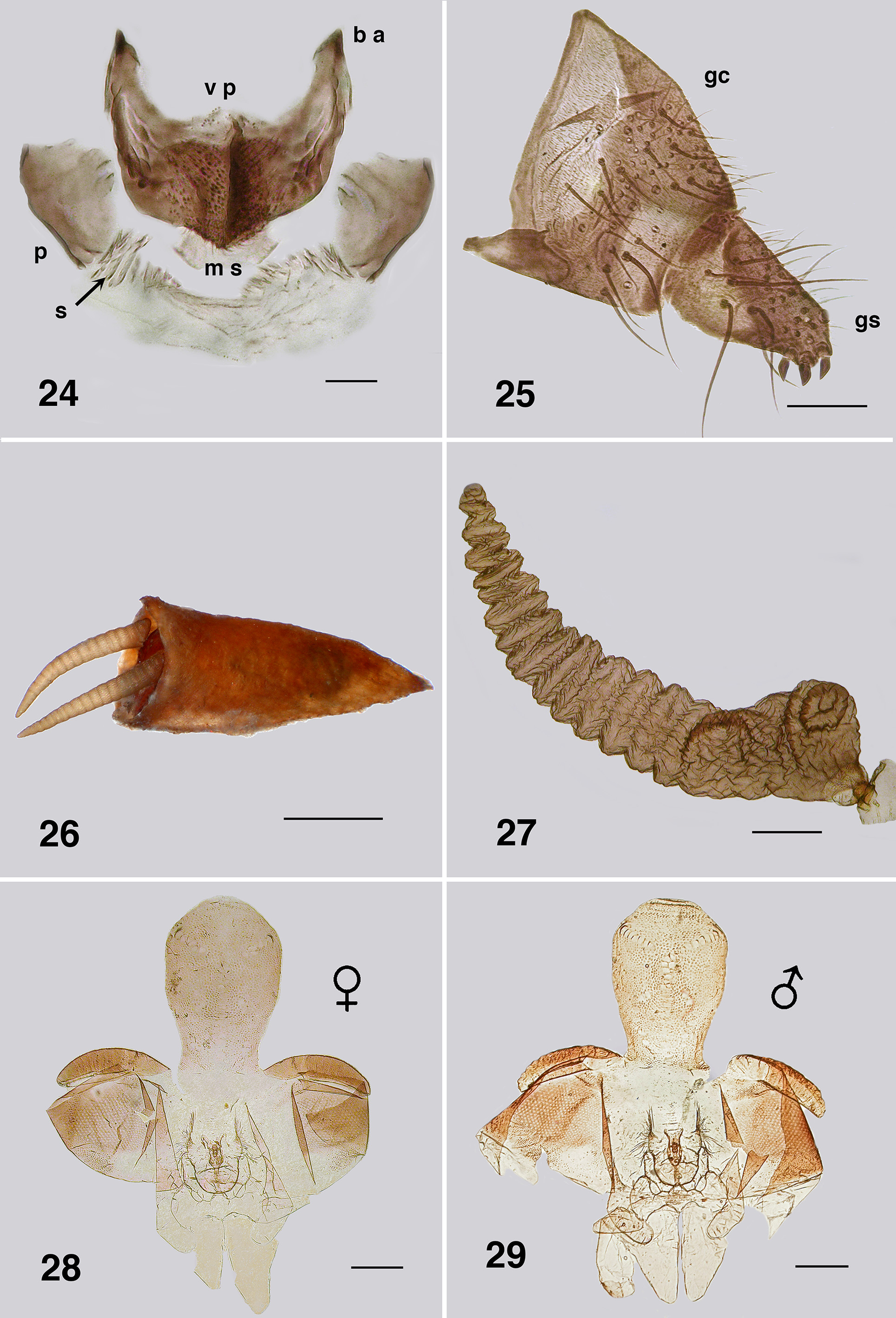
0.7 length of head depth; maxillary palpus ( Fig. 4 View FIGURES 1 – 6 ), total length 0.45 mm, articles overall brownish black, 3rd article darker; proportional length of 3rd, 4th and 5th articles 1.0:0.8:1.5; sensory vesicle ovoid, 0.5x width of 3rd article, opening 0.3x width of vesicle; mandible ( Fig. 5 View FIGURES 1 – 6 ), broadly triangular apically, sharply pointed with 48–50 inner teeth and 9–11 finely pointed outer teeth; lacinia ( Fig. 6 View FIGURES 1 – 6 ) with 13–15 inner teeth and 22–26 outer teeth; cibarial cornuae ( Fig. 7 View FIGURES 7 – 11 ) (partly reconstructed) lacking apical fluting or sculpture, sharply terminated, central depression broad. Cervical sclerites markedly developed ( Fig. 1 View FIGURES 1 – 6 ). Thorax: moderately domed; length 0.80–0.96 mm; width 0.60–0.63 mm; scutum evenly blackish brown, vestiture of evenly distributed recumbent silver hairs; postpronotal lobe with longer hairs; antepronotal lobe with longer hairs; scutellum and postnotum concolourous with scutum; scutellar depression and scutellum with long substantial black hairs; pleuron and anepisternal membrane blackish brown, bare. Haltere: dark. Wing ( Figs. 8, 9 View FIGURES 7 – 11 ): length 2.1–2.4 mm; width 0.9–1.2 mm, veins lightly pigmented, costa not extended to wing apex with spiniform setae distally, absent from other veins; radial veins closely applied to costa; basal cell absent; a:b ratio 1.0:2.8. Metathoracic furcasternum ( Fig. 10 View FIGURES 7 – 11 ): dorsal arm with distinct projection. Legs ( Fig. 1 View FIGURES 1 – 6 ): overall blackish yellow; hind basitarsus about 5.5x as long as its greatest breadth, ventral row of stout spines absent, calcipala as long as wide 0.75x width of basitarsus, pedisulcus present, but not markedly developed ( Fig. 11 View FIGURES 7 – 11 ); tarsal claw ( Fig. 12 View FIGURES 12 – 17 ) with moderately developed basal heel and markedly small tooth. Abdomen ( Fig. 13 View FIGURES 12 – 17 ): abdominal scale black with dark hairs, not greatly extended; tergite II 5x wider than long, shallowly V–shaped, III– V as wide as long, rounded, VI 2x wider than long; dorsal vestiture of small black hairs increased in density posteriorly. Genitalia: sternite VIII vestiture of sparse coarse black hairs posterolaterally; hypogynial valves ( Fig. 14 View FIGURES 12 – 17 ) short, lightly pigmented with vestiture of sparse short hairs and triads of microtrichia; median edges slightly convex, slightly strengthened along edge, bluntly rounded apically; genital fork ( Fig. 15 View FIGURES 12 – 17 ) with anterior stem broad (not easily observed), but well sclerotized and pigmented narrowly medially, flared anteriorly, lateral arms broad, indications of lateral apodeme (as in Gigantodax ), more distal apodeme (as in Austrosimulium ) present only as ridge, lateral plates large, angular posteromedially, rounded posterolaterally; spermatheca ovoid ( Fig. 16 View FIGURES 12 – 17 ), surface un–patterned; sparse internal acanthae in pairs and triads; junction of spermathecal duct pigmented and slightly sculpted; cercus ( Fig. 17 View FIGURES 12 – 17 ) as wide as long, broadly rounded in lateral view, anal lobe shallow, angulate proximally.
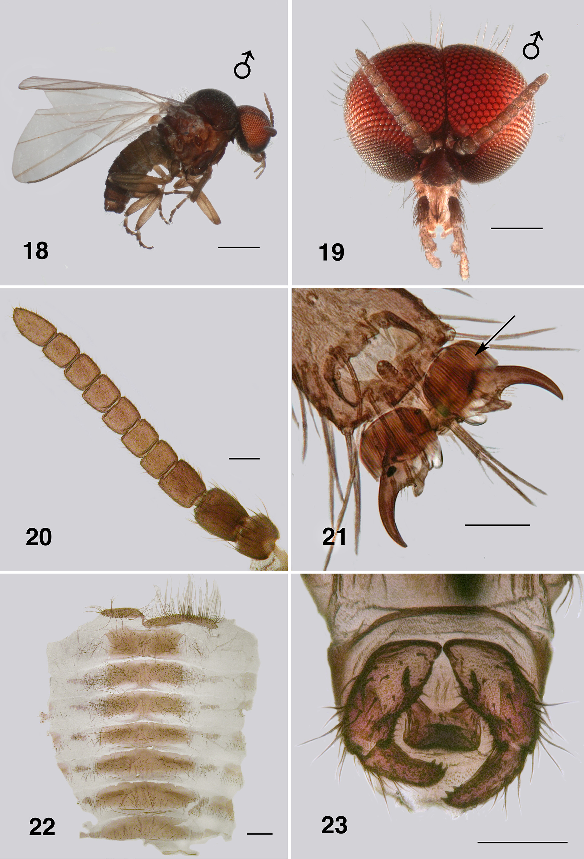
Adult male (reared specimens and others). Body ( Fig. 18 View FIGURES 18 – 23 ): general colour evenly black; total length 2.2–2.6 mm. Head ( Fig. 19 View FIGURES 18 – 23 ): width 0.89 mm; depth 0.6 mm; wider than thorax. Eyes: upper ommatidia very dark red, 0.04 mm in diameter, ca. 14 across, 20 down; lower ommatidia almost black, 0.018 mm in diameter, ca. 35 across and 23 down. Clypeus: black; vestiture of very sparse black hairs; 0.15 mm wide. Antenna ( Fig. 20 View FIGURES 18 – 23 ): total length 0.51– 0.55 mm; pedicel longer and wider than other divisions; first flagellomere longer than wide, others subrectangular; non–tapered, markedly narrow in comparison to that of female; scape and pedicel black, first flagellomere dark brown remainder evenly brown; flagellum markedly not hirsute. Mouthparts: poorly developed; length 0.26x head depth; mandibles insubstantial, finely tapered with apical hairs; laciniae as for mandible; maxillary palpus dark brown, 0.4 mm long, proportional lengths of 3rd, 4th and 5th articles 1.0:1.1:2.0, sensory vesicle small, occupying 0.33x width of article, opening 0.5x width of vesicle. Cervical sclerites markedly developed. Thorax: length 0.9– 1.1 mm; width 0.8 mm; in alcohol, scutum evenly velvety black, vestiture of fine recumbent pale hairs; scutellum and postscutellum concolourous with scutum, coarse long black hairs. Wing: 2.1–2.4 mm in length, 1.0– 1.1 mm in width; otherwise as for female. Haltere: tan. Legs: blackish brown; hind basitarsus about 5.5x as long as its greatest breadth, lacking row of stout spines; tarsal claw ( Fig. 21 View FIGURES 18 – 23 ) partially covered by grappling pad of ca. 20 hooks, distinct small basal tooth and heel. Abdomen ( Fig. 22 View FIGURES 18 – 23 ): black; abdominal scale with long fine hairs, tergites markedly broad, on tergites II–V hirsute laterally, less so on posterior others. Genitalia: ventral view ( Fig. 23 View FIGURES 18 – 23 ), ventral plate directed ventrally giving appearance of broadly concave apex, 1.5–2.0x wider than long, median keel well developed, with vestiture of fine hairs, plate roughly sculpted laterally; anteromedial depression broadly U to V–shaped, slight central convexity, basal arms short and pigmented ( Fig. 24 View FIGURES 24 – 25 ); median sclerite poorly expressed, broad and slightly divided apically; parameres present, moderately expressed, plate–like, apical rows of small spicules; adeagal membrane with sparse rows of minute microtrichia; gonocoxa 1.7x longer than basal width, markedly coarse black hairs on distal half; gonostylus ( Fig. 25 View FIGURES 24 – 25 ), approximately 3.0x longer than basal width, apically with 3 substantial terminal spines—variable and occasionally with one spine markedly displaced to outer apex.
Pupa (based on numbers of specimens). (Fig. 26). Body: length, female 2.2–2.8 mm; male 2.2–3.3 mm. Head: frontal cephalic plate lacking dorsal depression; ratio of basal width to vertex width of female 1.0:1.5, for basal width to length 1.0:2.3, rounded apically (Fig. 28), male ratios 1.0:1.6 and 1.0:2.3 respectively (Fig. 29); evenly tuberculate, frontal setae absent, facial setae present, but insubstantial, ocular spine absent. Thorax. Dorsum with very small tubercles, no pattern ( Fig. 30 View FIGURES 30 – 31 ); dorsocentral setae spine–like and curled apically. Gill (Fig. 26, 27): basically a single expanded tapered tube directed anteroventrally, 1.7–2.1 mm, full length 0.3 mm at greatest width with ca. 19 annulations on the longer anterior portion with 2 or 3 on a stub–like posterior lobe (Fig. 27), fenestra normal ( Fig. 30 View FIGURES 30 – 31 ), fine filaments absent. Abdominal armature: sternal hooks absent, but sternite VIII with sparse multi–pointed scales; tergites I & II with 4–6 moderately expressed setae; tergite III with four hooks posteriorly per side, 3 or 4 other fine setae, no lateral hook; tergite IV as for III, but with single small hook laterally; tergites V– VIII with markedly poorly–expressed spine comb anteriorly, laterally morphing into low multi–pointed scales, four to five fine setae posteriorly; tergite IX with poorly–expressed broad row of low spines and scales anterad, terminal spines short, not markedly sharp, grapnel hooks well expressed ( Fig. 31 View FIGURES 30 – 31 ). Cocoon (Fig. 26). Surface smooth, fabric coarsely woven, silk filaments obvious, medium brown; distinctly slipper–shaped fully covering pupa with well defined anterior edge, slight median projection, complete ventral floor, close fitted to pupa.
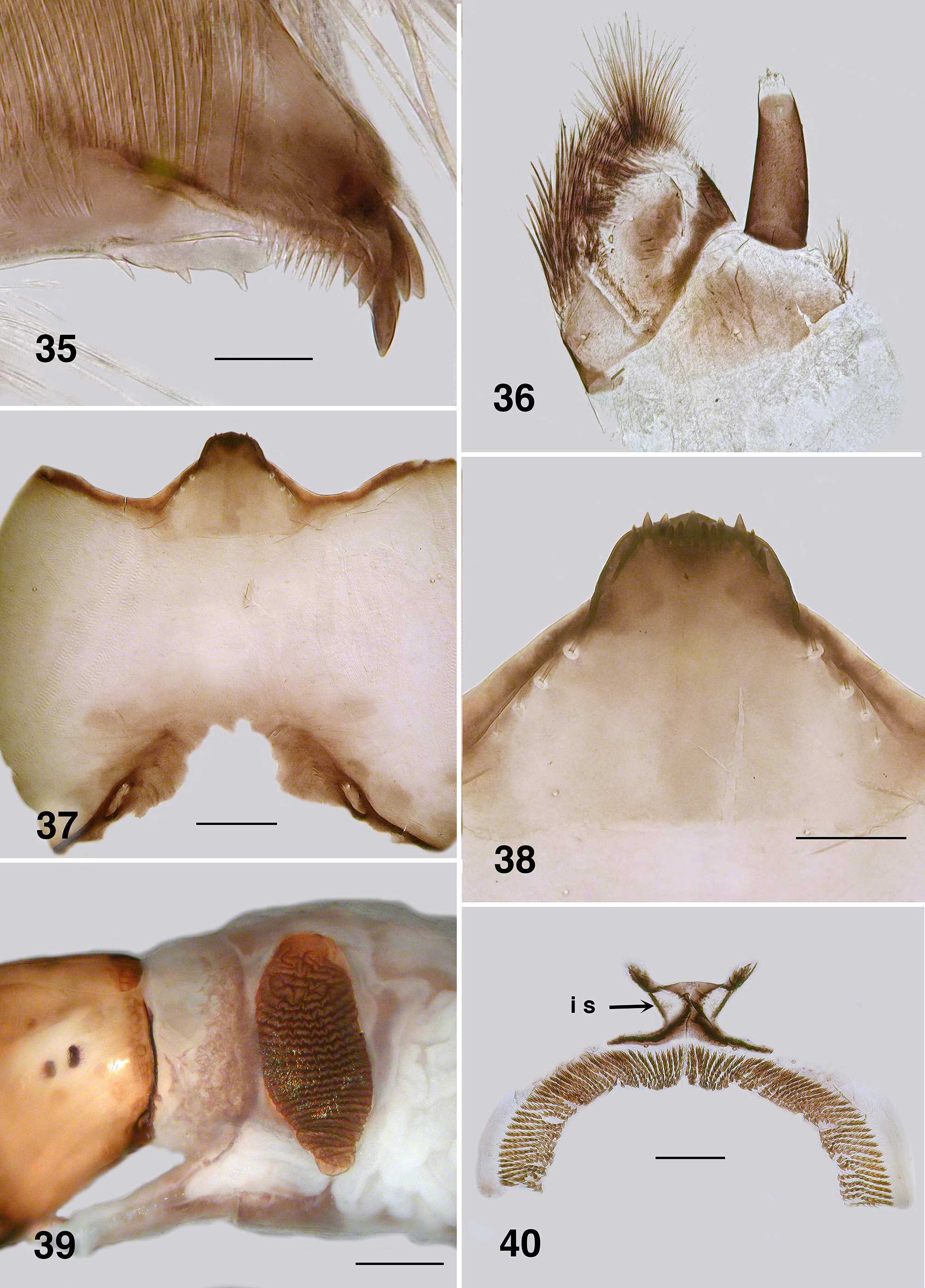
Larva (based on numbers of last instar larvae). Body (Fig. 32): overall grayish brown, stubby (head large in relation the body), thorax and anterior abdomen subequal in diameter, expanded smoothly posteriorly; total length 4.0– 4.9 mm. Head (Figs. 33): distinctly bicolourous, background pale translucent yellowish brown, head spots marked, but not strongly pigmented, medial and posterior spots form distinct inverted T, mediolateral spots distinct; most spots with negative centre; probable males have a lighter head pattern than females; head length 0.69–0.70 mm, width 0.50–0.60 mm; distance between antennal bases 0.30–0.38 mm; lateral margins of head smoothly convex, more so posteriorly; cervical sclerites well developed and pigmented, rounded posteriorly but flared anteriorly, not fused to postocciput; anterolateral edges of apotome sharply pigmented; genae with darker 'eye brow' above stemmata. Antenna (Fig. 34): overall clear pale brown; total length 0.4 mm; well extended beyond labral fan stem; distal article slightly longer than other two articles combined; proportions of basal, median and distal article 1.0: 0.6: 1.8. Labral fan: stem markedly translucent; 62–64 fine rays, 0.56 mm in length, 0.01 mm in width; microtrichia as long as ray width, distinct pattern with ca. 5 microtrichia decreased rapidly in length. Mandible ( Fig. 35 View FIGURES 35 – 40 ): apical brush well developed with distinct pigmented base: apical teeth not markedly developed; subapical teeth small, 8–9 spinous teeth; 2 distinct serrations widely separated, sensillum distinct and finely pointed; blade region long, smooth and slightly concave. Maxilla ( Fig. 36 View FIGURES 35 – 40 ): lobe markedly cone–shaped, asymmetrical, palp longer than lobe, closely applied, 3x as long as basal width. Postgenal cleft ( Fig. 37 View FIGURES 35 – 40 ): small, but U–shaped with irregular anterior apex, sclerotized posterior tentorial pit cuticle extensive, pits ovoid; ratio of hypostoma, bridge and cleft 1.0:1.9:0.7; suboesophageal ganglion not pigmented. Hypostoma ( Fig. 38 View FIGURES 35 – 40 ): ventral edge as raised dome; 13 teeth, median tooth barely protruded beyond edge, two sublateral teeth smaller and subequal in length, other larger and protruded beyond edge, lateral teeth larger and well protruded, paralateral teeth smaller and sharp, variable expression, no others evident; no hypostomal serrations; four hypostomal setae per side. Postgenal bridge: pale and concolourous with genae. Thorax: ( Fig. 39 View FIGURES 35 – 40 ) anterior prothorax dark brown, remainder paler; pharate pupal gill as black, paddle–shaped horn; annulations of gill concertinaed. Abdomen: evenly medium brown, darkened posteriorly; abdominal segments expanded smoothly; posteroventral tubercles not markedly developed. Anal papillae: three simple lobes. Posterior proleg ( Fig. 40 View FIGURES 35 – 40 ): rectal scales absent; anal sclerite X– shaped with median region poorly expressed, anterior arms slightly flared, shorter than posterior arms, interarm struts distinct, posterior arms short; accessory sclerite absent, pigmented semicircular sclerite absent, but clear cuticle evident in that position. Posterior circlet: ca. 76–80 rows of 11–13 hooks (total ca. 930).
Etymology. Named by Davies & Györkös (1988) after Murray H. Colbo.
Types. The original type material as designated by Davies & Györkös (1988: 111) was stated to be a holotype pharate female with parts mounted on slides. Paratypes of 5 pupae, a pupal exuviae in alcohol with filaments on slides and 2 mature larvae mounted on slides. Label data as "Halls' Gap (37º 07´S/ 142º 07´E) in a slow flowing drain in a grassy forest, 26.viii.1958. I. M. Mackerras". As partially explained by Craig (2011), the dissected materials were never permanently mounted on slides and, when recovered, were still in depression slides, in glycerine, of which most had crept out of the depression. Alcohol material was dry—not surprising after some 20 years, or so. Much of the described material by Davies & Györkös (loc. cit.) was not recovered. That which was, was badly bleached and essentially useless. Some, however, was stained in Chlorozoal Black and examined to confirm it was of P. colboi , then placed in microvials, pinned, labeled as below and deposited in the Australian National Insect Collection (ANIC), CSIRO, Canberra.
Holotype. Female head and hind legs. Label data:– [ Holo –/ type (red edges)] [ Austrosimulium colboi / Davies & Györkös 1988 / Victoria, Hall’s Gap / (37° 07’S/142° 07’E)/ 26.viii. l958/ I. M. Mackerras] [Exam / prep./ DAC, 2011]. Stained with Chlorazol Black, in glycerine microvial on pin. ( ANIC).
Paratypes. Labels as for holotype, but with [Para–/ type (yellow edges)]. One mature last instar larva, head and pupal gill histoblasts separate, stained with Chlorazol Black, in glycerine microvial on pin. Female hind legs, ex– pupa, not stained, as above. Pupal gills and thoracic cuticle, stained with Chlorazol Black, as above. Four separate pupal gills, as above. ( ANIC).
Additional material. Numerous specimens of larvae, pupae and reared adults. Two reared females and pupal exuviae. ETOH. Label data:– [ Paraustrosimulium / colboi ] [ Australia, Vic., Grampians Nat. Prk.,/ Syphon Rd Bdg, Glenelg Rv, "Big Cord",/ S37.31232, E142.36711, 263 mabs, / 26–xi–2014, Coll. D.A.& R.E.G. Craig./ OZV14b] [ UASM #/ 370849]. One reared female and pupal exuviae, as above, but with [ ANIC Database No./ 29 026696].
Two tubes, ETOH of larvae, pupae, reared male and female. Label data:– [ Paraustrosimulium / colboi ] [ Australia, Vic., Grampians Nat. Park,/ Glenelg Rd, upstream of culvert,/ S37.18138, E142.37428, 233 mabs,/ 10–ix–2014, Coll. D.A.& R.E.G. Craig./ OZV12.], one with [ UASM /370850] the other [ ANIC Database No./ 29 026697]. One tube, ETOH, larvae, pupae, reared females and males. Label data:– [ Paraustrosimulium / colboi ] [ Australia, Vic., Grampians Nat. Park,/ Lodge Rd, Earl's culvert. S37.16957,/ E142.34879, 244 mabs, 11–vii–2011,/ Coll. D.A.& R.E.G. Craig. #8a.] [ UASM #/ 370851]. One tube, ETOH, larvae and pupae. Label data:– [ Paraustrosimulium / colboi ] [ AUSTRALIA / Victoria / Grampians N. P./ small stream X–ing/ Glenelg River Rd./ 28 September 1996 / JK Moulton] [ ANIC Database No./ 29 026698]. Twelve slides of all stages. Label data:- [ Paraustrosimulium / colboi / Aust. Victoria, Grampians/ Glenelg River/ 28-ix-1966 / Coll. JK Moulton] [ UASM #/ 370868 -370879].
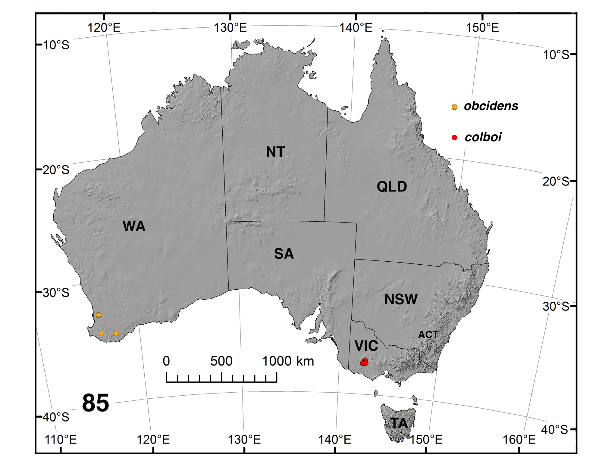
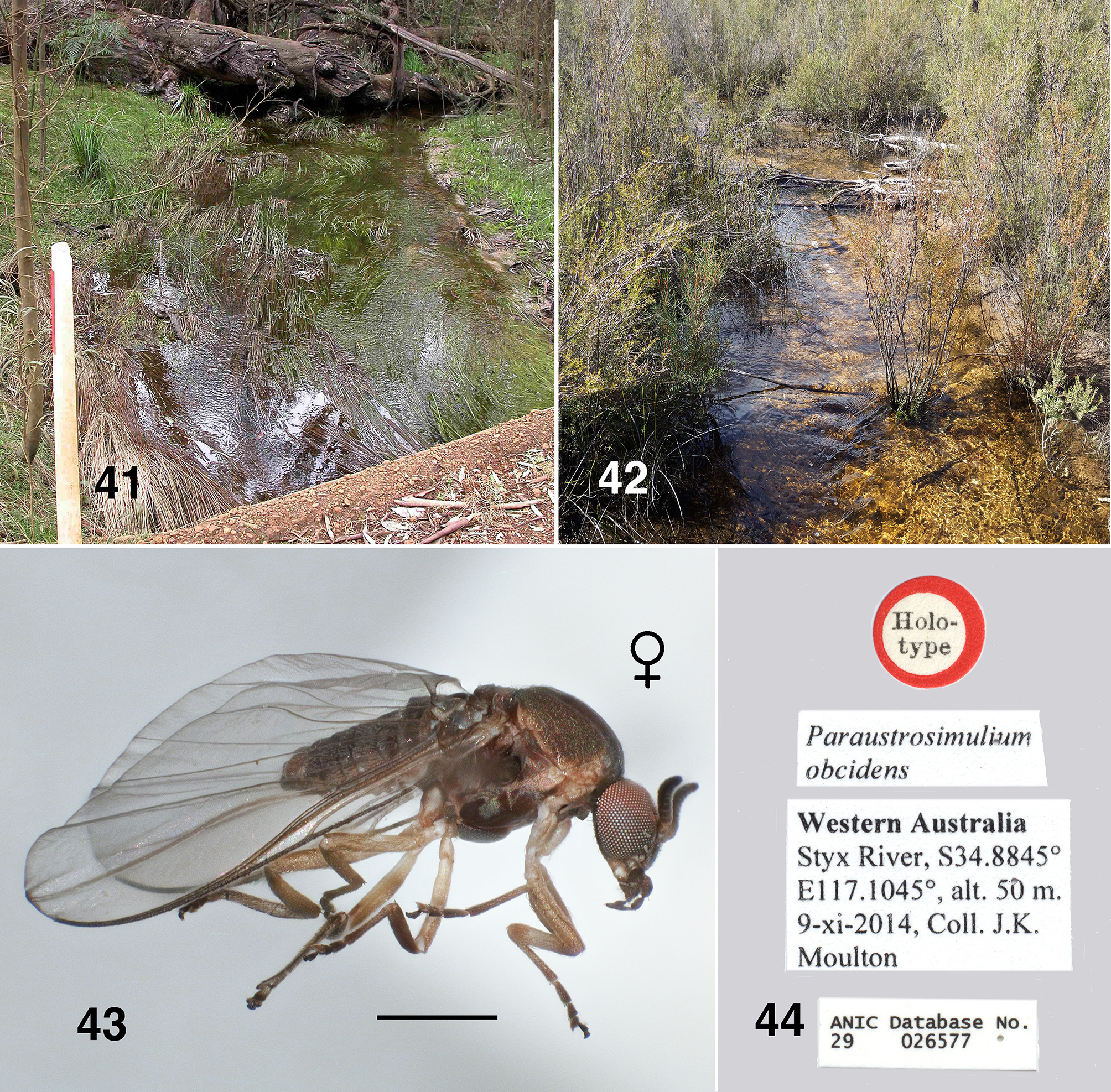
Distribution. Details given by Davies & Györkös (1988: 105) for the original material of P. colboi was that it had been collected in 1958 by Ian Mackerras from a site near Halls Gap, Grampians Nation Park, Victoria. However, map coordinates given locate that site in the Black Range State Park, some 30 km to the west of the Grampians and on checking (DAC pers. obs. 2011), the locality was not associated with running water. We are of the opinion that the longitude minutes given for the type locality is a typographical error, on the basis that it is the same as for the latitude, (i.e. 07'). For Halls Gap it should of ca. 32', but the exact location is unknown and original labeling missing. Other P. colboi material was found by JKM in 1996 in a stream to the southwest of Halls Gap and two other sites in that general area were established by DAC in 2011 and 2014 —a markedly precinctive distribution ( Fig. 85 View FIGURE 85 ):
Victoria: Grampians National Park, small stream crossing Glenelg River Road . 28–viii–1996. Coll. J.K. GoogleMaps Moulton. Grampians N. P., Lodge Road, Earl's Culvert , S37.16957º E142.34879º, 244 mabs, 10–vii–2011. Coll. D.A. & R.E.G GoogleMaps . Craig. Grampians N. P., Glenelg Road, culvert , S37.18138º E142.37428º, 228 mabs, 10–ix–2014. Coll. D.A. & R.E.G. Craig. GoogleMaps
Bionomics. Clearly an early spring species and univoltine, Paraustrosimulium colboi larvae and pupae occur in ephemeral, slowly–flowing streams (velocity 30–44 cm /sec) with generally colder water (temperature 9.6– 14.5ºC) and sandy substrate. Larvae are restricted to trailing vegetation and woody debris. At the Earl's Culvert locality ( Fig. 41 View FIGURES 41, 42 ), larvae of P. colboi occurred with those of Austrosimulium furiosum (Skuse) , A. montanum Makerras & Mackerras and Paracnephia umbratora (Tonnoir) —the later, too, almost an exclusively moderate– flow simuliid. At the Glenelg Road culvert stream ( Fig. 42 View FIGURES 41, 42 ), P. colboi was the only simuliid present and in considerable numbers. Nothing is known about behaviour of the adults.
Remarks. There is little doubt that the material discovered since the original collection by Mackerras and described by Davies & Györkös (loc. cite.) is of P. colboi . There is good concordance of morphological character states with, however, some exceptions. They made the point that female adults possess mandibular teeth only on the inner surface, similar to mandibles of New Zealand Austrosimulium . Our material of colboi (and obcidens below) has definite teeth on the outer surface ( Fig. 5 View FIGURES 1 – 6 ), albeit they are small and were perhaps not noticed by Davies & Györkös? Known Australian Austrosimulium (A.) and Novaustrosimulium all lack outer mandibular teeth (Craig, pers. obs.). The anterior arm of the female genital fork is illustrated as being narrow (their Fig. 25 View FIGURES 24 – 25 ), however, membranous areas to the side are not easily observed even in fresh material ( Fig. 15 View FIGURES 12 – 17 ), so in newer material the arm can be seen to resemble those of Austrosimulium , generally assumed to be unique in Simuliidae . Other character states of the fork are of similar expression. Then, too, is a discrepancy in the shape of the larval postgenal cleft— our material has a deeper cleft and is irregular (cf. Fig. 37 View FIGURES 35 – 40 with their Fig. 29). Of further concern is the number of hooks comprising the larval posterior circlet—our material has more, as well there are differences in the illustration (their Fig. 33) of the anal sclerite and our material ( Fig. 40 View FIGURES 35 – 40 ), where the sclerite is markedly more substantive. Indicative of minor variation between populations? The colour of the larvae as described by Davies & Györkös (1988: 111) is paler than our recent material (Fig. 32)—perhaps not surprising given the age of their material.


No known copyright restrictions apply. See Agosti, D., Egloff, W., 2009. Taxonomic information exchange and copyright: the Plazi approach. BMC Research Notes 2009, 2:53 for further explanation.
|
Kingdom |
|
|
Phylum |
|
|
Class |
|
|
Order |
|
|
Family |
|
|
Genus |
Paraustrosimulium colboi ( Davies & Györkös), 1988
| Craig, Douglas A., Moulton, John K. & Currie, Douglas C. 2017 |
Austrosimulium colboi Davies & Györkös, 1988 : 111
| Davies & Gyorkos 1988: 111 |
Austrosimulium (Austrosimulium) colboi
| Davies & Gyorkos 1988 |
Austrosimulium colboi
| Davies & Gyorkos 1988 |
Austrosimulium (Austrosimulium) colboi
| Davies & Gyorkos 1988 |
Austrosimulium colboi
| Davies & Gyorkos 1988 |
Austrosimulium colboi
| Davies & Gyorkos 1988 |
Austrosimulium colboi
| Davies & Gyorkos 1988 |
Austrosimulium colboi
| Davies & Gyorkos 1988 |
Paraustrosimulium colboi
| , Davies & Gyorkos 1988 |