
Salina anhuiensis, Ma, 2012
|
publication ID |
https://doi.org/ 10.11646/zootaxa.3389.1.4 |
|
DOI |
https://doi.org/10.5281/zenodo.5256055 |
|
persistent identifier |
https://treatment.plazi.org/id/9D4287A3-6167-FFDE-49C5-FBCCBCB352D0 |
|
treatment provided by |
Felipe |
|
scientific name |
Salina anhuiensis |
| status |
sp. nov. |
Salina anhuiensis View in CoL , sp. nov.
Figures 1 – 21 View FIGURES 1–5 View FIGURES 6–12 View FIGURES 13–18 View FIGURES 19–21 , Table 1
Type material. Holotype female, CHINA, Anhui, Tongluo Village , 15. X 2009, collection number 1122, in leaf litter . Paratypes: 8 females, same data as holotype. All deposited in School of Life Science , Nantong University, China .
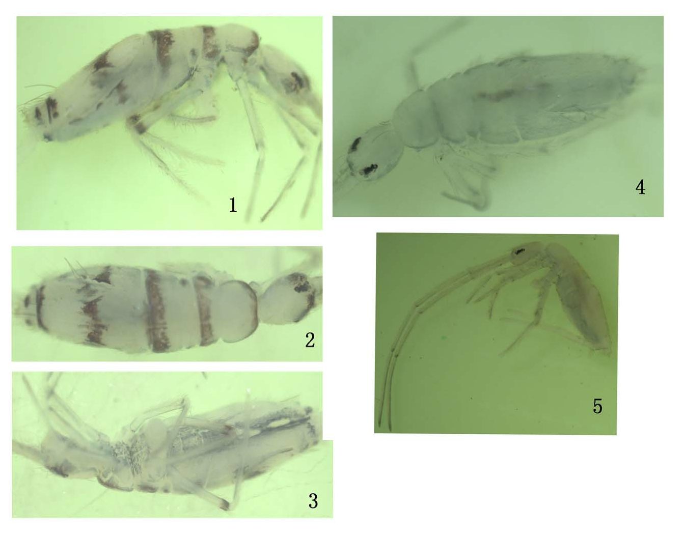
Description. Body length up to 2.3mm. Ground colour pale yellow; eye patches dark blue, a blue stripe between eye patches; anterior and lateral margin of Th. II with similar pigments; an irregular transverse blue stripe on Th. III, Abd. II – III, middle and posterior parts of Abd. IV and posterior part of Abd. V; Abd. I with an irregular blue spot on lateral margin; blue pigment also on distal part of hind femora ( Fig. 1&2 View FIGURES 1–5 ). Ventral side of body with a medial blue stripe from Th. II to Abd. IV ( Fig. 3 View FIGURES 1–5 ). Colour uniformly pale yellow and lacking blue stripe or spot on dorsal body in some specimens ( Figs 4, 5 View FIGURES 1–5 ).
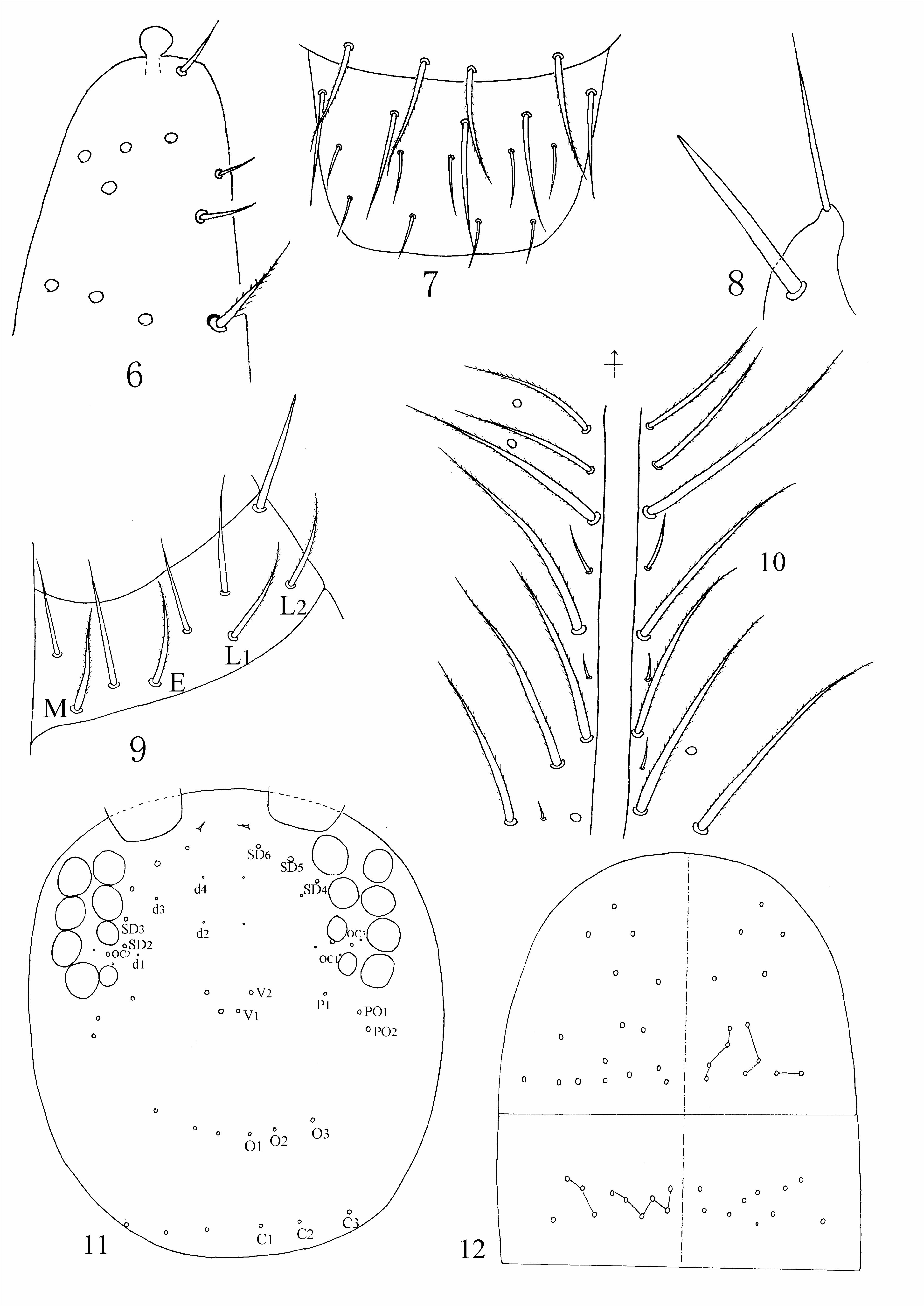
Head. Ant. 1.7 times length of body. Ratio of Ant. segments I – IV as 1.0/1.3 – 1.4/1.2 – 1.3/1.7 – ₁․9. Ant. IV apically with a bulb ( Fig. 6 View FIGURES 6–12 ). Ocelli 8+8, G & H smaller than others. Frontal spines 1+1. Prelabral setae 4, ciliate; labral setae 5, 5, 4, all smooth; labral margin not clearly seen ( Fig. 7 View FIGURES 6–12 ). Basal seta of maxillary palp blunt ( Fig. 8 View FIGURES 6–12 ). Labium with ABCDF setae ( Chen 1993), all smooth, seta F apically blunt; setae of labial base MEL 1 L 2, all ciliate ( Fig. 9 View FIGURES 6–12 ). Setae along cephalic groove long, acuminate and ciliate, short smooth setae present between ciliate setae ( Fig. 10 View FIGURES 6–12 ). Dorsal chaetotaxy of head as fig. 11, seta SD3 sometimes absent.
Thorax. Macrochaetae of Th. II – III as shown in Fig. 12 View FIGURES 6–12 . Median part of Th. II with 4 – 5 (rarely with 6) macrochaetae, posterior part with 9 – 13 macrochaetae on each side. Th. III with 11 – 13 macrochaetae on each side. Trochanteral organ with 29 – 55 setae ( Fig. 13 View FIGURES 13–18 ). Tibiotarsus with three types of setae: (1) type A, long and finely ciliate; (2) type B, normal ciliate; (3) type C, ciliate and broad ( Fig. 14 View FIGURES 13–18 ). Tenent hair spatulate and finely ciliate, 1.6 – 2.0 as long as inner side of unguis; unguiculus strongly truncate; unguis with a pair of lateral teeth, a pair of inner teeth, 1 – 2 unpaired teeth ( Fig. 15 View FIGURES 13–18 ) .
Abdomen. Macrochaetae and bothriotricha of Abd. I – V as shown in Fig. 16 View FIGURES 13–18 . Abd. I with 5 – 6 macrosetae on each side. Chaetal pattern on Abd. II as 2 central and 2 lateral macrochaetae. Abd. III with 2 bothriotricha medially and 1 bothriotrichum and 2 macrochaetae laterally. Abd. IV with 30 – 38 macro – mesochaetae medially and 11 – 18 macro – mesochaetae laterally. Chaetal pattern on Abd. V as 3/3. Anterior face of ventral tube with 4 distal macrochaetae and 15 – 25 meso – microchaetae on each side ( Fig. 17 View FIGURES 13–18 ); posterior face of ventral tube with 16 – 24 setae and 4 – 5 spines ( Fig. 18 View FIGURES 13–18 ). Tenaculum with 1 large finely ciliate seta ( Fig. 19 View FIGURES 19–21 ). Manubrial plaque with 3 ciliate macrochaetae ( Fig. 20 View FIGURES 19–21 ). Proportion of dens:manubrium=1.2 – 1.3. Proportion of vesicle: mucro=0.7 – 0.8. Mucro tridentate ( Fig. 21 View FIGURES 19–21 ).
Remarks. Salina anhuiensis n. sp. is similar to S. yosii Salmon, 1964 in chaetotaxy of Abd. I & II, but the two species differ in colour pattern (transverse blue stripes in S. anhuiensis sp. nov., but longitudinal blue spots from dorsal to Abd. V in S. yosii ) and chaetotaxy of Th. II & III (Th. II & III have more macrochaetae in S. anhuiensis sp. nov. than in S. yosii . Salina anhuiensis n. sp. is also similar to S. transversalis Yosii, 1961 , in that both species have a blue stripe on Th. II. However, there are other morphological between the species ( Table 1).


No known copyright restrictions apply. See Agosti, D., Egloff, W., 2009. Taxonomic information exchange and copyright: the Plazi approach. BMC Research Notes 2009, 2:53 for further explanation.