
Baiocis, Browne, 1962
|
publication ID |
https://doi.org/ 10.11646/zootaxa.4434.3.5 |
|
publication LSID |
lsid:zoobank.org:pub:3002EE95-60AC-4294-AE5A-62008E54ADB2 |
|
DOI |
https://doi.org/10.5281/zenodo.5970641 |
|
persistent identifier |
https://treatment.plazi.org/id/9E1C5030-FFCE-485B-FF60-70E0FA4FFE7E |
|
treatment provided by |
Plazi |
|
scientific name |
Baiocis |
| status |
|
Key to Baiocis View in CoL Males
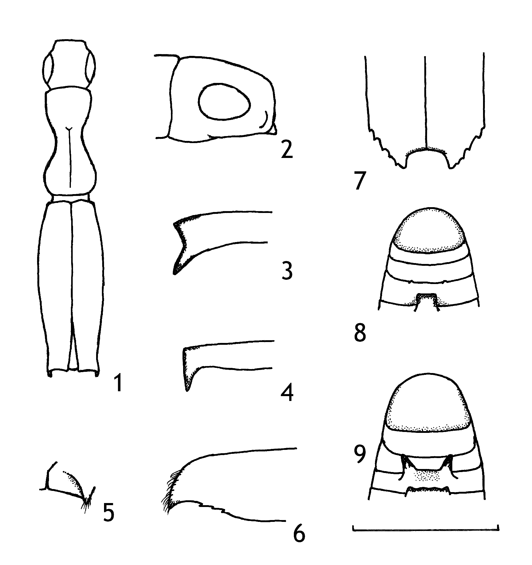
1 Elytra vase-shaped, constricted anterior to declivity ( Fig. 1 View FIGURES 1–9 ), with an elongate posterolateral process extending ventrally at nearly a right angle to plane of elytra ( Figs 3, 4 View FIGURES 1–9 ). Head as long as or longer than high. Eye oval with long axis parallel to body ( Fig. 2 View FIGURES 1–9 ).............................................................................................. 2
- Elytra not obviously constricted anterior to declivity. Posterolateral process shorter and more broadly triangular, sometimes extending almost horizontally. Head rounded, higher than long. Eye round or almost so.............................. 3
2 Head about as long as high, not flattened; vertex separated from frons by a distinct, evenly curved carina, below which frons is impressed. Posterolateral process of elytron vertical ( Fig. 4 View FIGURES 1–9 ). Posterior margins of ventrites 2‒4 not raised and thickened, and without teeth. 2.0‒ 2.2 mm long............................................................. anaticeps (Schedl) View in CoL
- Head flattened, longer than high ( Fig. 2 View FIGURES 1–9 ); frons very short without a carina separating it from vertex. Posterolateral process of elytron directed somewhat posteriorly ( Figs 3 View FIGURES 1–9 , 18 View FIGURES 18–25 ). Posterior margins of ventrites 2‒4 raised and thickened to form transverse ridges projecting as blunt teeth at the sides of the ridge. 2.4 mm long.............................. crassiventris n. sp.
3 First abdominal ventrite with a median process extending ventrally and posteriorly over base of second ventrite, and apically truncate, weakly bifid, or irregularly rounded................................................................ 4
- First abdominal ventrite without a median process, although posterior part may bear a small, median, triangular spine, or be raised to form a curved ridge............................................................................. 7
4 Frons and vertex angularly separated. Median process on first abdominal ventrite weakly bifid at apex. Posterolateral processes of elytra very long, the apical emargination U-shaped ( Fig. 29 View FIGURES 26–32 ). Larger species, 3.3‒3.5 mm long... sumatranus n. sp.
- Frons rounded into vertex. Median process on first abdominal ventrite truncate or irregularly rounded apically. Posterolateral processes of elytra short. Smaller species, 2.5‒2.8 mm long.................................................... 5
5 Fifth abdominal ventrite with a median conical spine in apical half ( Fig. 20 View FIGURES 18–25 ). Median process on first abdominal segment with posterior margin irregularly rounded, and bearing two pairs of minute denticles. 2.5‒2.6 mm long........... spicatus n. sp.
- Fifth abdominal ventrite without a spine or process. Median process on first abdominal segment not gibbous ventrally, truncate posteriorly........................................................................................... 6
6 Second ventrite with a pair of small, rounded protuberances, separated by about the width of the process on ventrite 1 ( Fig. 8 View FIGURES 1–9 ). Elytra strongly emarginate at apex, the emargination broadly U-shaped with base of emargination almost straight ( Fig. 7 View FIGURES 1–9 ). 2.5‒2.7 mm long......................................................................... incisus (Sampson) View in CoL
- Second ventrite without protuberances. Elytral apex shallowly V-shaped ( Fig. 11 View FIGURES 10–17 ). 2.8 mm long.......... inimicus (Schedl) View in CoL
7 Second ventrite gibbous medially, with a strong, pointed, posteroventrally directed tooth at each end of the gibbosity ( Fig. 2 2 View FIGURES 1–9 ), the teeth separated by about the width of a curved ridge extending across about one third of the posterior part of the first ventrite ( Fig. 9 View FIGURES 1–9 ). 2.7‒2.8 mm long............................................................. spiniventris n. sp.
- Abdomen usually not or weakly gibbous; if gibbous, then without teeth on second ventrite........................... 8
8 Third abdominal ventrite with a small median triangular raised area near the posterior margin, its apex posterior, fourth ventrite with a larger median conical tooth ( Fig. 32 View FIGURES 26–32 ). 2.8‒3.0 mm long.......................................... laosi n. sp.
- Third and fourth abdominal ventrites without raised area or tooth................................................ 9
9 Metanepisternum with a carina at the anterior margin of the metanepisternum and metaventrite depression. Elytral apex with emargination broadly, very shallowly U-shaped, with basal margin nearly straight ( Figs 10, 13 View FIGURES 10–17 ). Posterior part of elytra weakly curved ventrally so that apex of apicolateral spines is usually visible from above.................................. 10
- Metanepisternum with one or two small tubercles or spines at the anterior margin of the metanepisternum and metaventrite depression. Elytral apex with emargination crescentic or shallowly V-shaped ( Fig. 11 View FIGURES 10–17 ). Posterior part of elytra more strongly curved ventrally so that apex of apicolateral spines is visible only from sides, not from above........................ 12
10 Apex of elytral disc just above declivity and declivity matt except close to suture, with a row of small granules on each interstria, a larger granule at the end of interstriae 1 and 3 at top of declivity ( Fig. 10 View FIGURES 10–17 ). 2.0‒ 2.4 mm long.... angustiformis (Schedl) View in CoL
- Apex of elytral disc just above declivity shining, with only a few minute granules on interstriae, without larger granules on interstriae 1 and 3 at top of declivity ( Fig. 13 View FIGURES 10–17 )............................................................. 11
11 Larger species, 2.7‒2.8 mm long. Posterolateral tooth of elytra directed more strongly ventrally, barely visible from above......................................................................................... nubilosus Roberts View in CoL
- Smaller species, 1.9‒2.4 mm long. Posterolateral tooth of elytra directed less strongly ventrally, clearly visible from above....................................................................................... pernanulus (Schedl) View in CoL
12 First ventrite with a small median triangular spine extending horizontally over base of second ventrite. Apicolateral tooth of elytra truncate when viewed from behind. Large species, 4.7 mm long............................. sublunaris (Schedl) View in CoL
- First ventrite unarmed. Apicolateral tooth of elytra pointed when viewed from behind. Smaller species, not more than 3.0 mm long............................................................................................... 13
13 Frons very finely punctured, except close to epistoma. Upper rim of declivity weakly carinate laterally, the carina not extend- ing close to suture ( Fig. 5 View FIGURES 1–9 ). Apicolateral tooth of elytra forming an almost equilateral triangle, making an angle of 60°‒70° at its tip when viewed from side ( Fig. 6 View FIGURES 1–9 ). 2.3‒2.4 mm long....................................... perinimicus (Schedl) View in CoL - Frons coarsely punctured, especially on lower half. Upper rim of declivity more strongly carinate, carina extending close to suture. Apicolateral tooth of elytra more acute making an angle of 30°‒40° when viewed from side ( Fig. 15 View FIGURES 10–17 )............ 14
14 Apicolateral tooth of elytra short ( Fig. 13 View FIGURES 10–17 ). Declivital sulcus narrower vertically relative to width across elytra (23: 4), about 5 times wider than deep. Second ventrite without a transverse ridge. 2.1‒2.5 mm long. Thailand to Sulawesi ... orientalis n. sp.
- Apicolateral tooth of elytra longer ( Fig 15 View FIGURES 10–17 ). Declivital sulcus broader vertically relative to width across both elytra, about 4 times wider than deep (28: 7). Second ventrite sloping posteriorly at about 60° to the vertical from a humped transverse ridge running across the middle of the ventrite ( Fig. 15 View FIGURES 10–17 ). 2.8‒2.9 mm long. Papua New Guinea only........... unispineus Roberts


No known copyright restrictions apply. See Agosti, D., Egloff, W., 2009. Taxonomic information exchange and copyright: the Plazi approach. BMC Research Notes 2009, 2:53 for further explanation.