
Parapharyngodon hispidus, Ferreira & Vieira & Silva & Ribeiro & Ferreira & Muniz-Pereira, 2021
|
publication ID |
https://doi.org/ 10.11646/zootaxa.4980.1.12 |
|
publication LSID |
lsid:zoobank.org:pub:BCC6F4E1-C924-466F-8328-FFFAC42A9D6E |
|
DOI |
https://doi.org/10.5281/zenodo.4945108 |
|
persistent identifier |
https://treatment.plazi.org/id/9E2E3523-F22C-FF95-FF5B-FB2AFD8FFC2D |
|
treatment provided by |
Plazi |
|
scientific name |
Parapharyngodon hispidus |
| status |
sp. nov. |
Parapharyngodon hispidus n. sp.
( Figs. 1 View FIGURE 1 and 2 View FIGURE 2 )
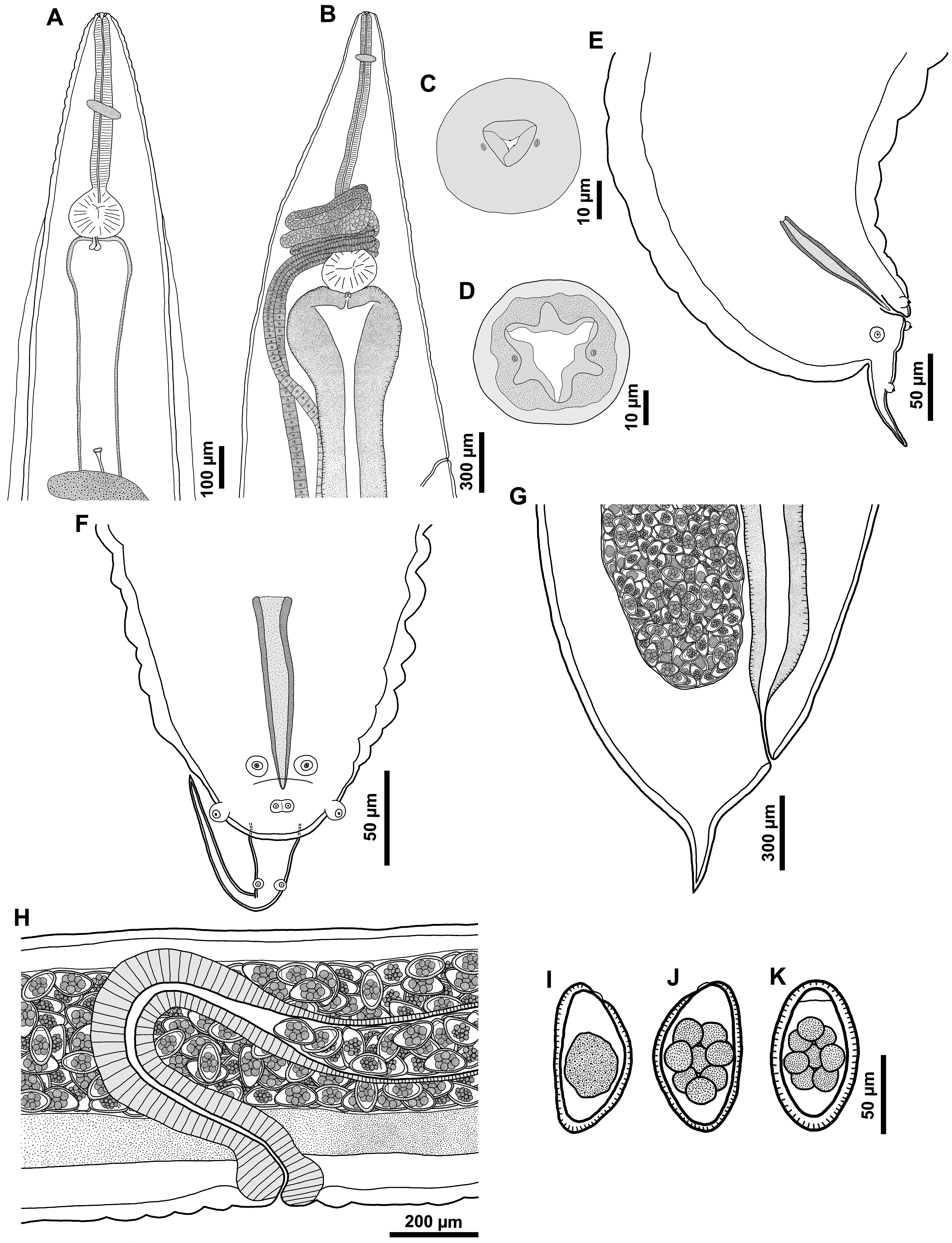
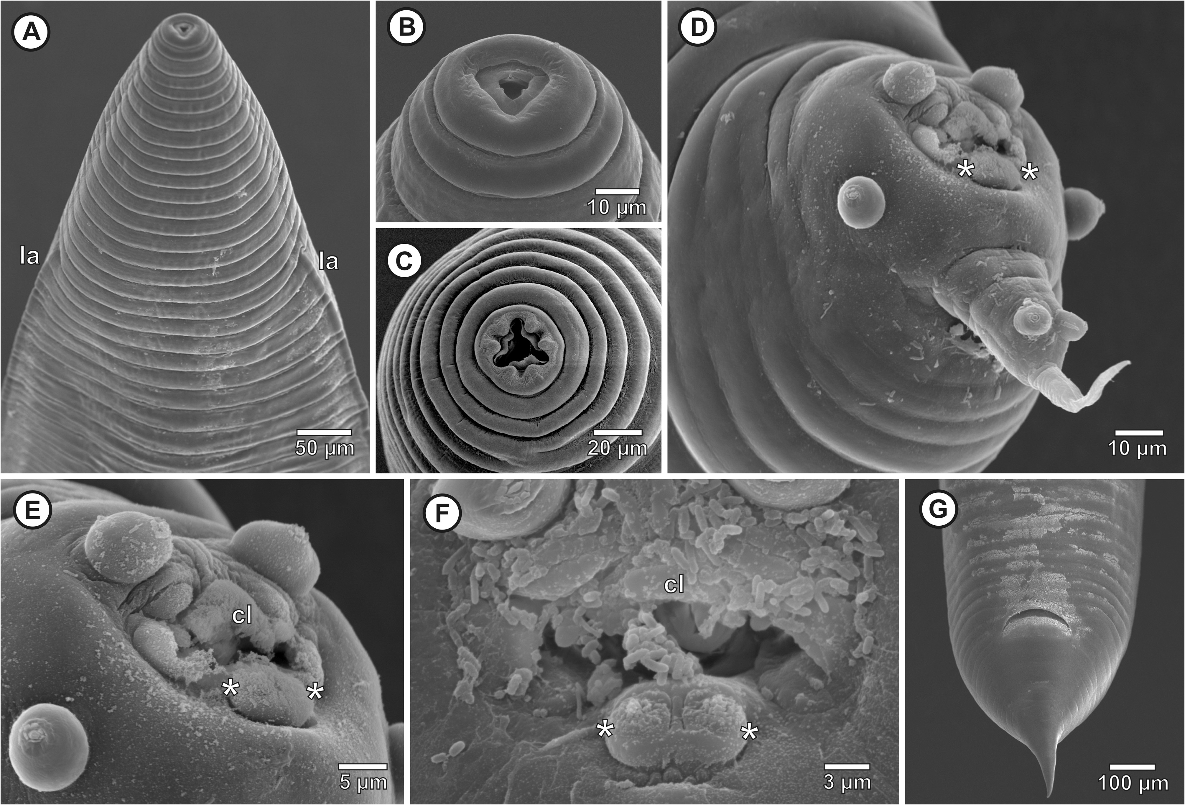
General: Small, robust, whitish nematodes. Cuticle with prominent annulations beginning just behind anterior extremity and continuing to anus or cloacal aperture. Sexual dimorphism evident, males with approximately 30% of total body length of females. Males with triangular mouth aperture, with internal cuticular projections, cephalic papillae not visible, two lateral amphids, near lateroventral side of mouth ( Figs. 1C View FIGURE 1 , 2B View FIGURE 2 ). Females with star shaped mouth aperture; surrounded by three bilobed lips, one dorsal and two lateroventral, two amphids each opening on lateral lips ( Figs. 1D View FIGURE 1 , 2C View FIGURE 2 ). Oxyuriform oesophagus, ending in valved oesophageal bulb ( Figs. 1A, B View FIGURE 1 ). Nerve ring at level of first third of oesophagus ( Figs. 1A, B View FIGURE 1 ). Excretory pore far posterior to oesophageal bulb in both males and females ( Figs. 1A, B View FIGURE 1 ). Males without caudal alae; caudal appendage directed dorsally ( Figs. 1E, F View FIGURE 1 , 2D View FIGURE 2 ). Lateral alae only in males ( Figs. 1A View FIGURE 1 , 2A View FIGURE 2 ).
Male (Based on 10 adult mature specimens, measurements of holotype in parentheses): Total body length 3.2–3.6 (3.2) mm, body width at level of oesophagus-intestinal junction 225–375 (270). Total oesophagus length 525–710 (525); corpus 420–530 (420) long, oesophageal bulb 110–140 (115) long, and 120–150 (125) wide. Nerve ring 138–163 (138) and excretory pore 1.2–1.5 (1.5) mm from anterior end. Lateral alae present ( Figs. 1A View FIGURE 1 , 2A View FIGURE 2 ) 1.7– 2.1 (1.7) mm long, beginning at the level of the oesophageal bulb and ending far distant to cloacal aperture. Cloacal lips smooth, not echinate ( Figs. 1F View FIGURE 1 , 2D–F View FIGURE 2 ). Posterior region with four pairs of caudal papillae ( Figs. 1E, F View FIGURE 1 , 2D View FIGURE 2 ); the first pair is subventral located immediately anterior to cloacal aperture; the second is lateral, located posterior to cloacal aperture; third pair is subventral, at level of the second pair and immediately posterior to cloacal aperture; and the fourth pair is located on caudal appendage Figs. 1E, F View FIGURE 1 , 2D–F View FIGURE 2 ). One spicule well sclerotized 110–123 (110) long, terminating in a sharp tip ( Figs. 1E, F View FIGURE 1 ). Gubernaculum absent. Tail conical 135–305 (175) (including caudal appendage), with a subdorsal caudal appendage 57–95 (75) long ( Figs. 1E, F View FIGURE 1 , 2E View FIGURE 2 ) .
Female (Based on 10 adult mature specimens, measurements of allotype in parentheses): Total body length 7.4–10.5 (10) mm, body width at level of oesophagus-intestinal junction 720–1,160 (900). Total oesophagus length 1.3–1.7 (1.6) mm; corpus 1–1.4 (1.3) mm, oesophageal bulb 220–250 (240) long, and 250–310 (310) wide. Nerve ring 192–250 (230) and excretory pore 2.2–3.3 (2.8) mm from anterior end. Vulva not prominent ( Fig. 1H View FIGURE 1 ), equatorial, 3.6–5.5 (5) mm from anterior end, followed by short vagina and ovijector direct posteriorly. Didelphic, uteri amphidelphic, and ovaries directed anteriorly. Both ovaries encircling isthmus and anterior region of oesophageal bulb ( Fig. 1B View FIGURE 1 ). Eggs subovate, 67–100 long, 37–60 wide, flattened on one side, with one subapical operculum, without polar filament, early stages of cleavage in the ovijector ( Figs. 1H, J, K View FIGURE 1 ). Eggshell thick, with punctated surface. Tail 400–590 (590) long (including spike tip) ( Figs. 1G View FIGURE 1 , 2G View FIGURE 2 ), corresponding approximately to 5.5% of total body size (mean proportional size, based on the proportional tail sizes of all females analyzed), with short spiked tip, 190–245 (210) long.
Taxonomic summary
Type host: Tropidurus hispidus (Spix) ( Squamata : Tropiduridae ) (Neotropical Lava Lizard, calango-de-muro)
Type locality: Campus de Ciências Agrárias (Agrarian Sciences Campus) (CCA) (09°19’41”S, 40°32’59”W) of the Universidade Federal do Vale do São Francisco (Univasf), municipality of Petrolina, state of Pernambuco, Brazil.
Site of infection: large intestine.
Prevalence: 56.85% (112 infected hosts)
Mean intensity: 4.13 ± 11.96 parasites per infected hosts
Mean abundance: 2.35 ± 10.56 parasites per analyzed hosts
Range of infections: 1–21
Type material: Holotype male (CHIOC 38994a)), allotype female (CHIOC 38994b), 2 males and 4 females paratypes (CHIOC 38994c).
Etymology: The species name of the hosts species is used as a noun in apposition to Parapharyngodon ..
Remarks: The main feature considered for the differentiation of species in Parapharyngodon and Thelandros Wedl species ( Oxyurida : Pharyngodonidae ) is the egg morphology and development ( Bursey et al. 2013, Velarde-Aguilar et al. 2015, Rizvi et al. 2017). Therefore, the specimens evaluated in the present study were classified as Parapharyngodon since they present eggs with a subterminal operculum and in the early stages of cleavage in ovijector.
With the description of the new species the genus Parapharyngodon currently includes 55 valid species ( Bursey & Goldberg 2015, Velarde-Aguilar et al. 2015, Araujo Filho et al. 2015, Garduño-Montes de Oca et al. 2016, Ramallo et al. 2016, Rizvi et al. 2017, Pereira et al. 2017, Santos et al. 2018), which can be differentiated by zoogeographic region, the presence or absence of body alae in males and females; male characteristics, such as the number and arrangement of caudal papillae, the morphology of anterior border of cloacal aperture, and the size and type of distal end of spicules; and female characteristics, such as position of the ovaries relative to the oesophageal bulb, the morphology of eggshell, and tail ( Bursey et al. 2013, 2015, Velarde-Aguilar et al. 2015, Rizvi et al. 2017, Pereira, 2017).
In the Neotropical realm, ten valid species of Parapharyngodon have been described so far ( Table 1 View TABLE 1 ). Parapharyngodon hispidus n. sp. has four pairs of caudal papillae, as do four other Neotropical species, namely, P. bainae Pereira, Sousa & Souza Lima , P. silvoi , P. sanjuanensis Ramallo, Bursey, Castillo & Acosta , and P. sceleratus . Parapharyngodon hispidus n. sp., P. silvoi , and P. sceleratus have a smooth anterior border of cloacal aperture, differing from the echinate anterior border of the cloaca of P. bainae and P. sanjuanensis ( Pereira et al. 2011, 2018, Ramallo et al. 2016). Parapharyngodon hispidus n. sp. differs from P. silvoi , and P. sceleratus as the latter two have an unpaired post cloacal papilla ( Freitas 1957, Vicente et al. 1993, Araujo Filho et al. 2015, Velarde-Aguilar et al. 2015), which is absent in P. hispidus n. sp.
We highlight some considerations about two Neotropical Parapharyngodon species. Rizvi & Bursey (2013), Bursey & Goldberg (2015), and Rizvi et al. (2017) considered the females of P. alvarengai as featuring a conical tail with no spike. However, the original description of this species (see Freitas 1957) clearly shows a stout spike, which was also observed by Velarde-Aguilar et al. (2015) and Pereira et al. (2017). In the description of P. silvoi, Araujo Filho et al. (2015) describe females having ovaries extending beyond the oesophageal bulb (ovaries pre bulbar). However, we analyzed the species’ drawings in the original description and observed that the drawn female had post bulbar ovaries. Thus, we consider here the characteristics of P. silvoi demonstrated in the graphic representations.
Among the species of other zoogeographic distribution, P. hispidus n. sp. belongs to a group with four pairs of caudal papillae, without unpaired post cloacal papilla, and smooth cloacal lips ( Table 2 View TABLE 2 ). The species composing this group are Parapharyngodon baueri Bursey & Goldberg (Afrotropical realm), P. californiensis Read & Amrein (Nearctic realm), P. jairaipurii Rizvi & Bursey (Oriental realm), P. skrjabini Vakker (Palearctic realm), and P. striatus Singh & Malhotra (Oriental realm) ( Table 2 View TABLE 2 ). Parapharyngodon jairaipurii , described for Hemidactylus flaviviridis Ruppell ( Squamata , Gekkonidae ) from India, is the only species presenting the same set of characteristics of spicule tip (sharp pointed), female tail end (stout spike), and eggshell morphology (punctate) ( Rizvi & Bursey 2013) ( Table 2 View TABLE 2 ).
Although P. hispidus n. sp. and P. jairaipuri share key characteristics used to diagnose species, including sharppointed spicules, female tail ending with a stout spike, and eggs with punctated shells, other morphological and morphometric characteristics differentiate these species ( Table 3 View TABLE 3 ). The main characteristics differentiating these species are the size of spicules (spicules of P. jairaipuri are around 55% of the size spicules of P. hispidus n. sp.) and the arrangement of the caudal papillae (two pairs of adcloacal papillae in P. jaraipuri which are absent in P. hispidus n. sp.) ( Rizvi & Bursey 2013) ( Table 3 View TABLE 3 ).
No known copyright restrictions apply. See Agosti, D., Egloff, W., 2009. Taxonomic information exchange and copyright: the Plazi approach. BMC Research Notes 2009, 2:53 for further explanation.