
Kirbyana projecta, Chen & Zhi, 2023
|
publication ID |
https://doi.org/ 10.11646/zootaxa.5347.1.1 |
|
publication LSID |
lsid:zoobank.org:pub:E9658506-5801-4B92-8140-A8FCE1EC8F40 |
|
DOI |
https://doi.org/10.5281/zenodo.8390946 |
|
persistent identifier |
https://treatment.plazi.org/id/AD31A0A5-D71A-47A4-95FD-3F73071CD614 |
|
taxon LSID |
lsid:zoobank.org:act:AD31A0A5-D71A-47A4-95FD-3F73071CD614 |
|
treatment provided by |
Plazi |
|
scientific name |
Kirbyana projecta |
| status |
sp. nov. |
Kirbyana projecta sp. nov.
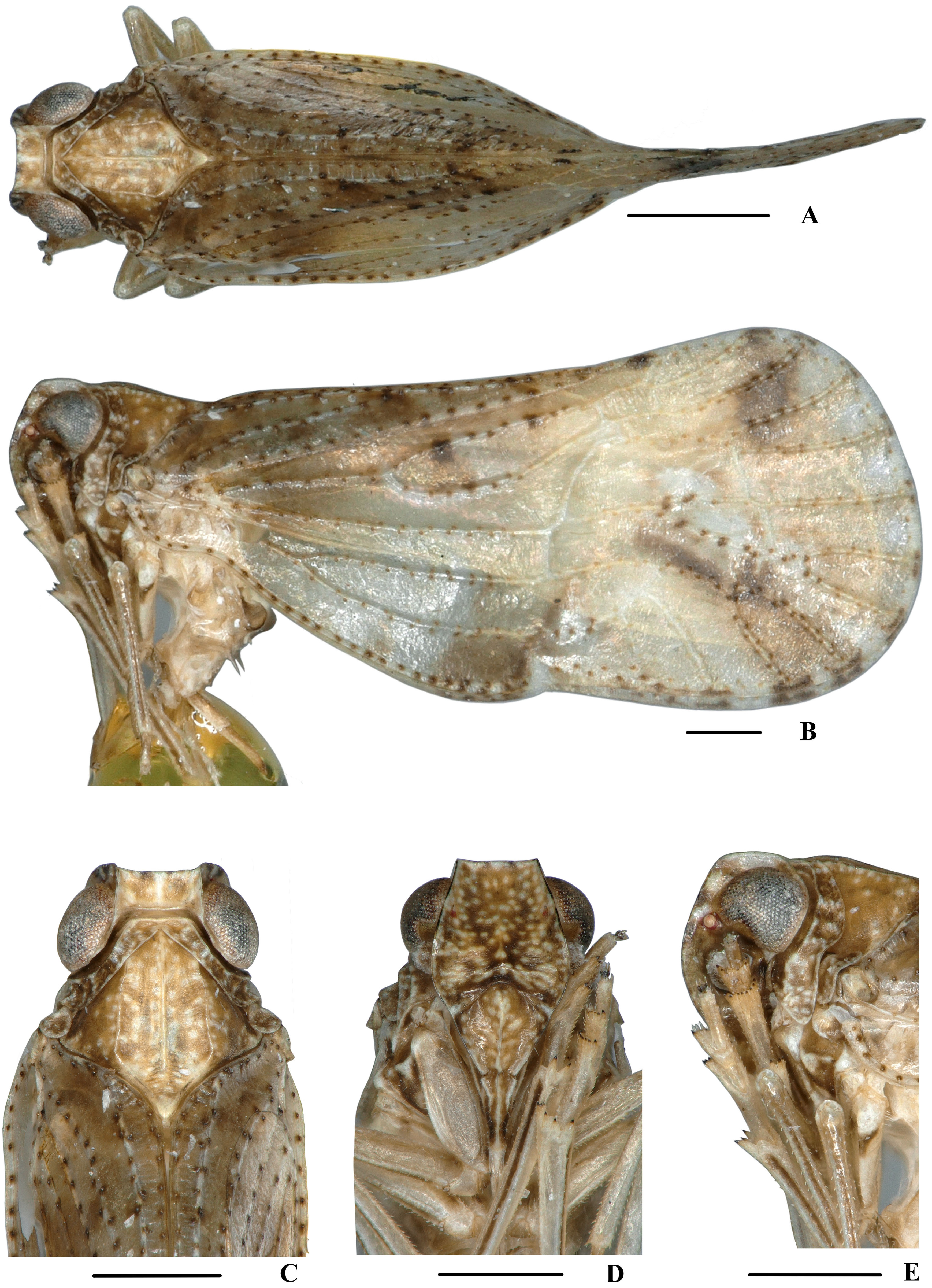
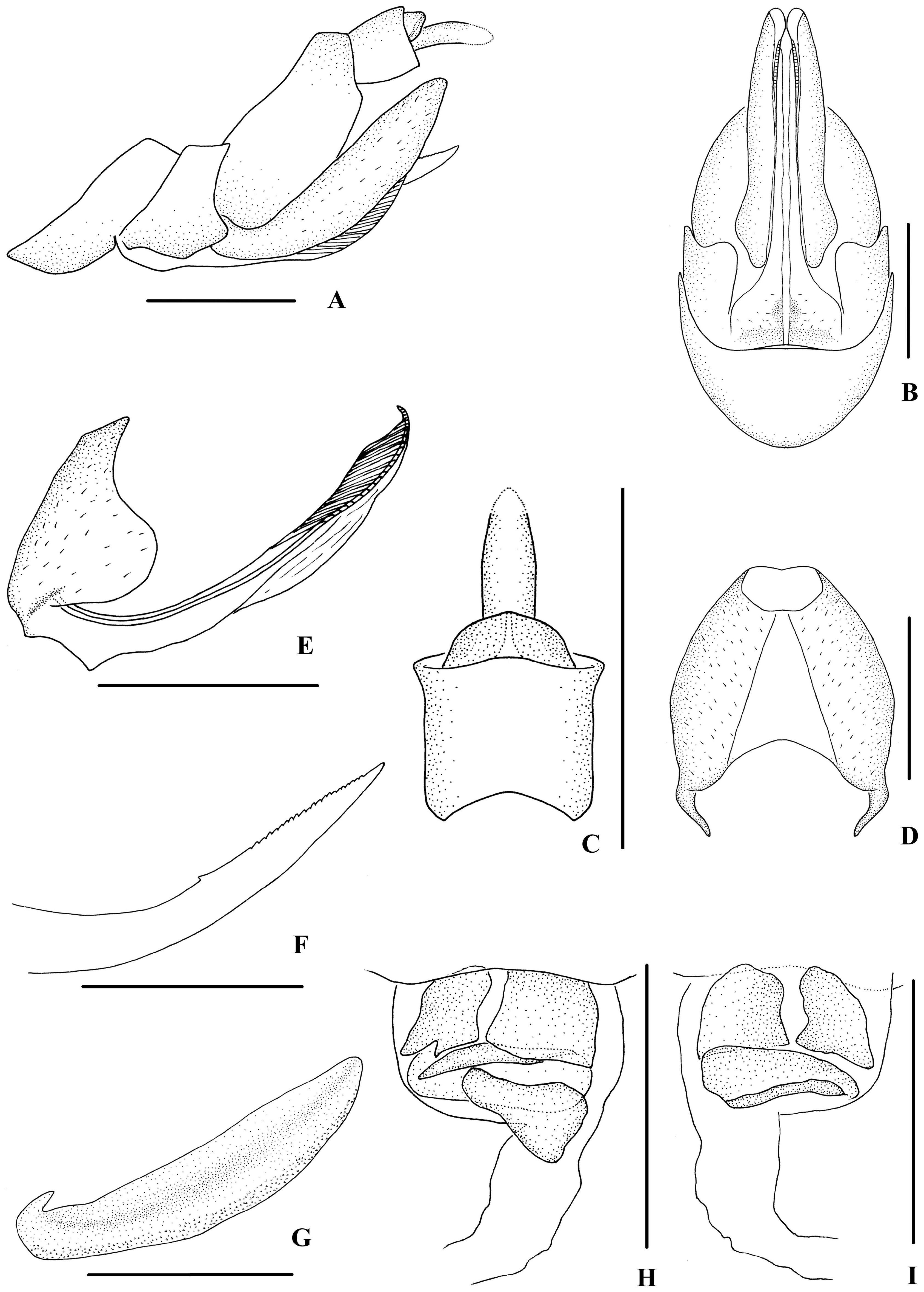
( Figs 43‒45 View FIGURE 43 View FIGURE 44 View FIGURE 45 )
Description. Body length: male 4.7‒5.0 mm (n = 3), female 5.2 mm (n = 1).
Coloration. General color brown ( Fig. 43A–E View FIGURE 43 ). Eyes dark brown, ocelli light yellow, semitransparent. Vertex, face, frons, pronotum and mesonotum brown, speckled with small pale spots. Rostrum yellowish brown. Forewing semi-translucent; pale brown, apical 2/3 yellowish brown, a large dark brown spot near Cu fork, and small brown spots on the ends of longitudinal veins, stigma yellowish brown. Hind tibiae brown and abdominal sternites dark brown.
Head and thorax. Vertex ( Figs 43C View FIGURE 43 , 44A View FIGURE 44 ) broad, 2.0 times wider than long; anterior margin truncated, posterior margin archedly recessed. Frons ( Figs 43D View FIGURE 43 , 44B View FIGURE 44 ) widest slight below the level of antennae, 1.2 times wider than long; frontoclypeal suture nearly concave into an arch; middle carina with basal half absent; lateral carinae distinct and slightly elevated. Pronotum ( Figs 43C View FIGURE 43 , 44A View FIGURE 44 )1.8 times longer than vertex; median carina distinct, posterior margin nearly at right angle. Mesonotum 1.4 times longer than pronotum and vertex combined. Forewing ( Fig. 44C View FIGURE 44 ) 2.3 times longer than wide, with 11 apical and 6 subapical cells; fork Sc+RP basad of fork CuA 1 +CuA 2, first crossvein r-m basad of fork MP, RP two branches, MP with five terminals: MP 11, MP 12, MP 2, MP 3, and MP 4, fork MP 1 +MP 2 basad of fork MP 3 +MP 4. Metatibiotarsal formula: 6/8‒9/9, second segment of hind tarsus with 4 platellae.
Male genitalia. Pygofer ( Fig. 44D, E View FIGURE 44 ) symmetrical, dorsal margin concave and U-shaped, slightly widened towards apex in ventral view; in lateral view, lateral lobes triangular, caudally extended, caudal margin with a bulged projection at dorsal 1/3, medioventral process round in ventral view. Anal segment ( Fig. 44D, F View FIGURE 44 ) broad, tubular, dorsal margin almost straight, ventral margin extremely extended, apical lobes round in lateral view; 1.5 times longer than wide in dorsal view; anal style strap-shaped, beyond anal segment. Gonostyli ( Fig. 44D, E, G View FIGURE 44 ) symmetrical in ventral view; in inner lateral view, dorsal margin bending inwards in a right angle in the middle, apical part acute. Aedeagus ( Fig. 44H–K View FIGURE 44 ) with total of five processes. Right apex of periandrium with a medium-sized spinous process, which basal 2/3 straight and apical 1/3 curved, apex left- dorsocephalically directed; the apex of the ventral margin of periandrium with a long spinous process, slightly curved and ventrocephalically directed; left side of periandrium with two spinous processes, one originating from basal 1/3, curved and dorsocaudally directed, the other one on the apex, slender, slightly curved and ventrocephalically directed. Endosoma (=flagellum) moderately sclerotised, relatively short, generally curved to the right, dorsal margin with a straight spinous process, cephalically directed.
Female genitalia. Posterior margin of pregenital sternite concave.Tergite IX( Fig. 45A, D View FIGURE 45 ) moderately sclerotised, with length almost equal to width in caudal view. Anal tube ( Fig. 45A, C View FIGURE 45 ) short, nearly rectangular, slightly widened towards apex, 1.1 times wider than long in dorsal view; dorsal and ventral margins slightly convex in lateral view, anal styles strap-shaped. Gonapophysis VIII ( Fig. 45E View FIGURE 45 ) elongate, and slightly curved upwards. Gonapophysis IX ( Fig. 45F View FIGURE 45 ) with one middle tooth, at a distance ratio, between middle tooth to apex and length of denticulate portion, of 1.6. Gonoplac ( Fig. 45G View FIGURE 45 ) rod-like, 4.3 times longer than wide. Posterior vagina ( Fig. 45H, I View FIGURE 45 ) elongate. The base of the ventral wall of posterior vagina each with a nearly quadrilateral sclerite at left and right sides and a nearly triangular sclerite in the middle aera; the dorsal wall with a large transverse nearly oval sclerite basally.
Type material. Holotype: ♂, CHINA: Laiyanghe (22°36’N, 101°0’E), Simao City , Yunnan Province, 23 August 2014, leg. Zheng-Xiang Zhou; paratypes: 2♂♂ 1♀, same data as holotype; 1♂, Menghai County (21°57’N, 100°27’E), Yunnan Province, 13 July 2013, leg. Ji-Chun Xing. GoogleMaps
Host plant. Unknown.
Distribution. China (Yunnan).
Remarks. Male genitalia of K. projecta sp. nov. are similar to those of K. spinata sp. nov., but differ in: (1) the spinous process on ventral margin of periandrium strong and broad (the spinous process on ventral margin of periandrium weakly sclerotized in K. spinata ); (2) the spinous process on right side of periandrium shorter than 1/2 length of periandrium (the spinous process on right side of periandrium longer than 1/2 length of periandrium); (3) the caudal margin of pygofer with a bulged projection (the latter without the same projection).
Etymology. The specific name refers to the caudal margin of the pygofer with a bulged projection.

No known copyright restrictions apply. See Agosti, D., Egloff, W., 2009. Taxonomic information exchange and copyright: the Plazi approach. BMC Research Notes 2009, 2:53 for further explanation.