
Dilacreon (Eluzalmon) falcatus, Chen & Zhi, 2023
|
publication ID |
https://doi.org/ 10.11646/zootaxa.5347.1.1 |
|
publication LSID |
lsid:zoobank.org:pub:E9658506-5801-4B92-8140-A8FCE1EC8F40 |
|
DOI |
https://doi.org/10.5281/zenodo.8390882 |
|
persistent identifier |
https://treatment.plazi.org/id/91B06B52-E9D7-4FE2-B248-7A618BBB2BE4 |
|
taxon LSID |
lsid:zoobank.org:act:91B06B52-E9D7-4FE2-B248-7A618BBB2BE4 |
|
treatment provided by |
Plazi |
|
scientific name |
Dilacreon (Eluzalmon) falcatus |
| status |
sp. nov. |
Dilacreon (Eluzalmon) falcatus sp. nov.
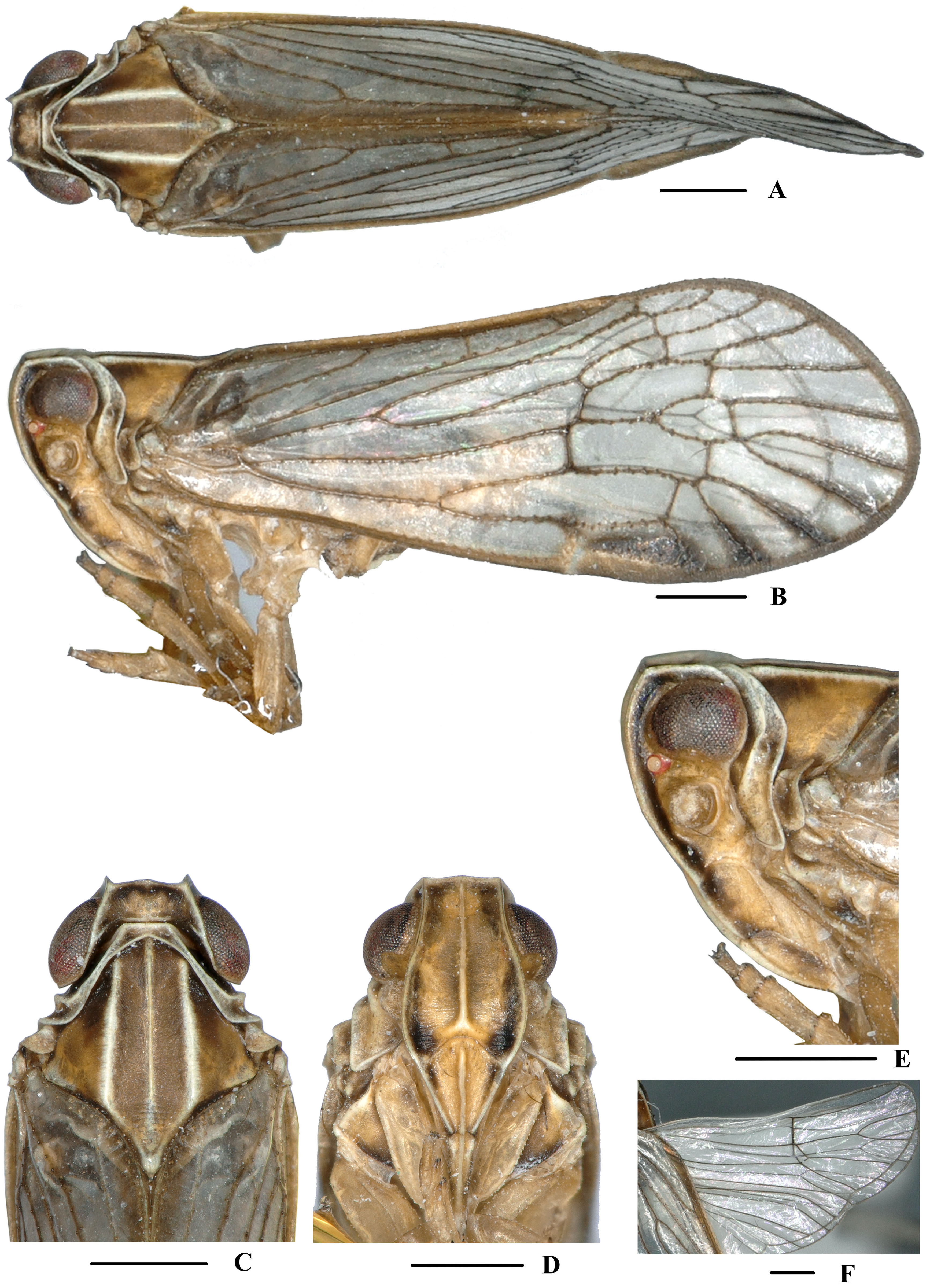
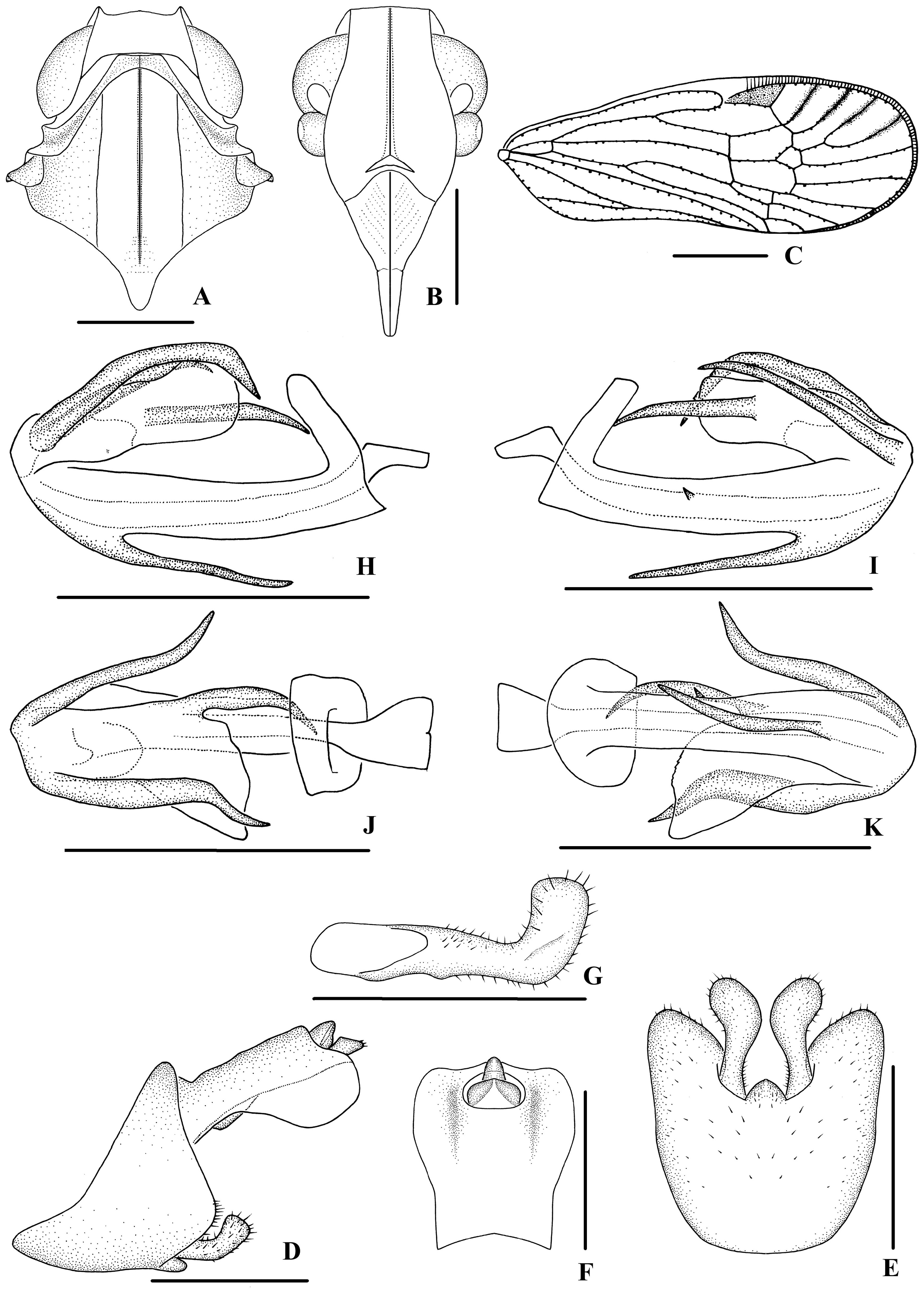
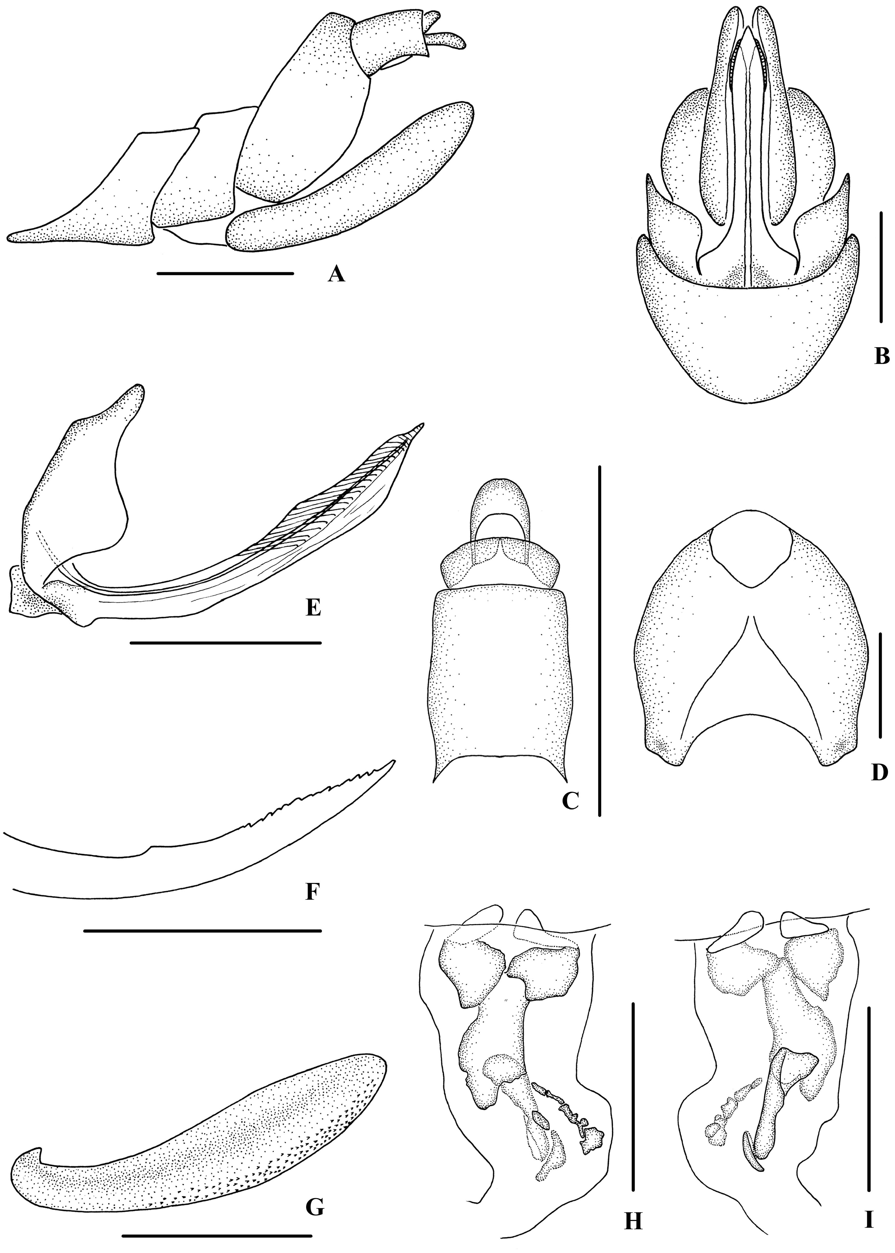
( Figs 19‒21 View FIGURE 19 View FIGURE 20 View FIGURE 21 )
Description. Body length: male 5.1‒5.4 mm (n = 5), female 6.1‒6.5 mm (n = 8).
Coloration. General color grayish brown ( Fig. 19A–F View FIGURE 19 ). Eyes blackish brown, ocelli light yellow, semitransparent. Vertex generally brown with lateral carinae yellowish brown. Face generally yellowish brown with lateral margins dark brown; rostrum yellowish brown. Pronotum and mesonotum brown with light yellow carinae. Forewing grayish brown, semi-translucent, veins darker; stigma dark brown. Hind tibiae and abdominal sternites yellowish brown.
Head and thorax. Vertex ( Figs 19C View FIGURE 19 , 20A View FIGURE 20 ) broad, 2.7 times wider than long; anterior margin slightly convex, median carina obsolete, posterior margin archedly recessed. Frons ( Figs 19D View FIGURE 19 , 20B View FIGURE 20 ) widest slightly below the level of antennae, 1.3 times as wide as long; frontoclypeal suture nearly concave into an arch; middle carina complete; lateral carinae distinct and slightly elevated. Pronotum ( Figs 19C View FIGURE 19 , 20A View FIGURE 20 ) 2.4 times longer than vertex; median carina indistinct, posterior margin nearly at right angle. Mesonotum 1.7 times longer than pronotum and vertex combined. Forewing ( Fig. 20C View FIGURE 20 ) 2.7 times longer than wide, with 10 apical and 6 subapical cells; fork Sc+RP distad of fork CuA 1 +CuA 2, first crossvein r-m slightly basad of fork MP, RP two branches, MP with five terminals: MP 11, MP 12, MP 2, MP 3, and MP 4, fork MP 1 +MP 2 basad of fork MP 3 +MP 4. Metatibiotarsal formula: 6/7/8, second segment of hind tarsus with three platellae.
Male genitalia. Pygofer ( Fig. 20D, E View FIGURE 20 ) symmetrical, dorsal margin concave and U-shaped, slightly widened towards apex in ventral view; in lateral view, lateral lobes triangularly extended caudally, medioventral process triangular in ventral view. Anal segment ( Fig. 20D, F View FIGURE 20 ) broad, dorsal margin almost straight, apical half of ventral margin convex, apical lobes round in lateral view; 1.2 times longer than wide in dorsal view; anal style strap-shaped, slightly beyond anal segment. Gonostyli ( Fig. 20D, E, G View FIGURE 20 ) symmetrical in ventral view; in inner lateral view, base of ventral margin slightly concave, dorsal margin bending inwards in a right-angle arc in the middle, apical part slightly extended, apical margin round. Aedeagus ( Fig. 20H–K View FIGURE 20 ) with total of five processes. The ventral margin of periandrium with a slender spinous process near apex, relatively straight, apex ventrocephalically directed; left side in the middle with a very short spinous process, dorsocephalically directed. Endosoma (=flagellum) moderately sclerotised, medium-sized, generally dorsally curved. Base of right side with a long and broad falcate spinous process, which basal half pointed upward and then right-ventrocephalically directed. Left side with two spinous processes, one originated from base, left-cephalically directed, the other one on the apex, medium-sized, slightly curved and right-ventrocephalically directed.
Female genitalia. Posterior margin of pregenital sternite concave. Tergite IX ( Fig. 21A, D View FIGURE 21 ) moderately sclerotised, 1.2 times longer than wide in caudal view. Anal tube ( Fig. 21A, C View FIGURE 21 ) short, nearly rectangular, 1.4 times longer than wide in dorsal view; dorsal margin nearly straight and ventral margin slightly concave in lateral view, anal styles strap-shaped. Gonapophysis VIII ( Fig. 21E View FIGURE 21 ) elongate, and slightly curved upwards. Gonapophysis IX ( Fig. 21F View FIGURE 21 ) with one middle tooth, at a distance ratio, between middle tooth to apex and length of denticulate portion, of 1.8. Gonoplac ( Fig. 21G View FIGURE 21 ) rod-like, 4.0 times longer than wide. Posterior vagina ( Fig. 21H, I View FIGURE 21 ) elongate. The ventral wall of posterior vagina with three large sclerites, two nearly round at base and one long longitudinal sclerite in the middle aera; several small sclerites on the right distal half, elliptic or irregular in shape, arranged in an oblique row, and a nearly oval sclerite located on the left side of the queue. The dorsal wall with a long longitudinal large sclerite and a small elliptic sclerite on distal half.
Type material. Holotype: ♂, CHINA: Bakaxiaozhai (21°58’N, 101°13’E), Mengla County, Yunnan Province, 15 June 2016, leg. Ying-Jian Wang and Qiang Luo; paratypes: 3♂♂ 3♀♀, same data as holotye; 1♂ 5♀♀, Mohan Town, Mengla County , Yunnan Province, 20‒22 June 2016, leg. Ying-Jian Wang, Qiang Luo and Liang-Jing Yang. GoogleMaps
Host plant. Unknown.
Distribution. China (Yunnan).
Remarks. Male genitalia of D. (E.) falcatus sp. nov. are similar to those of D. (E.) chionomus Fennah, 1980 , but differ in: (1) the ventral margin of periandrium with a spinous process (the latter without spinous process in the same position); (2) left side of periandrium at middle with a very short spinous process (the latter with one spinous process on dorsal margin and left side); (3) right side of endosoma with one spinous process (right side of endosoma with two spinous processes in D. (E.) chionomus ).
Etymology. The specific name refers to apex of right side of endosoma with a long and broad falcate spinous process.
No known copyright restrictions apply. See Agosti, D., Egloff, W., 2009. Taxonomic information exchange and copyright: the Plazi approach. BMC Research Notes 2009, 2:53 for further explanation.