
Dilacreon ( Eluzalmon ) biprocessus, Chen & Zhi, 2023
|
publication ID |
https://doi.org/10.11646/zootaxa.5347.1.1 |
|
publication LSID |
lsid:zoobank.org:pub:E9658506-5801-4B92-8140-A8FCE1EC8F40 |
|
DOI |
https://doi.org/10.5281/zenodo.8390862 |
|
persistent identifier |
https://treatment.plazi.org/id/E54F0F8A-17B5-45FC-A5E3-0E80EF50177B |
|
taxon LSID |
lsid:zoobank.org:act:E54F0F8A-17B5-45FC-A5E3-0E80EF50177B |
|
treatment provided by |
Plazi |
|
scientific name |
Dilacreon ( Eluzalmon ) biprocessus |
| status |
sp. nov. |
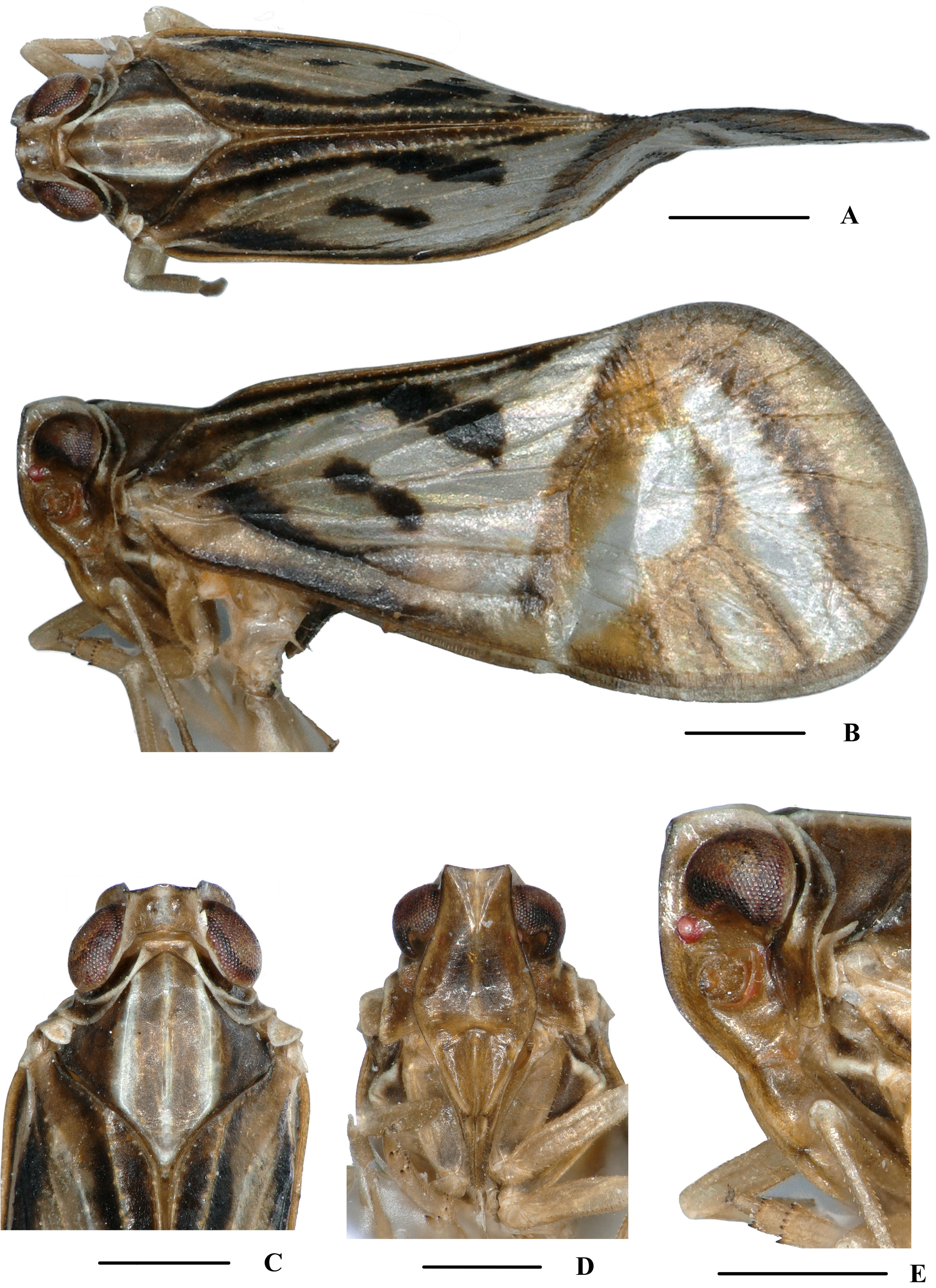
Dilacreon ( Eluzalmon) biprocessus sp. nov.
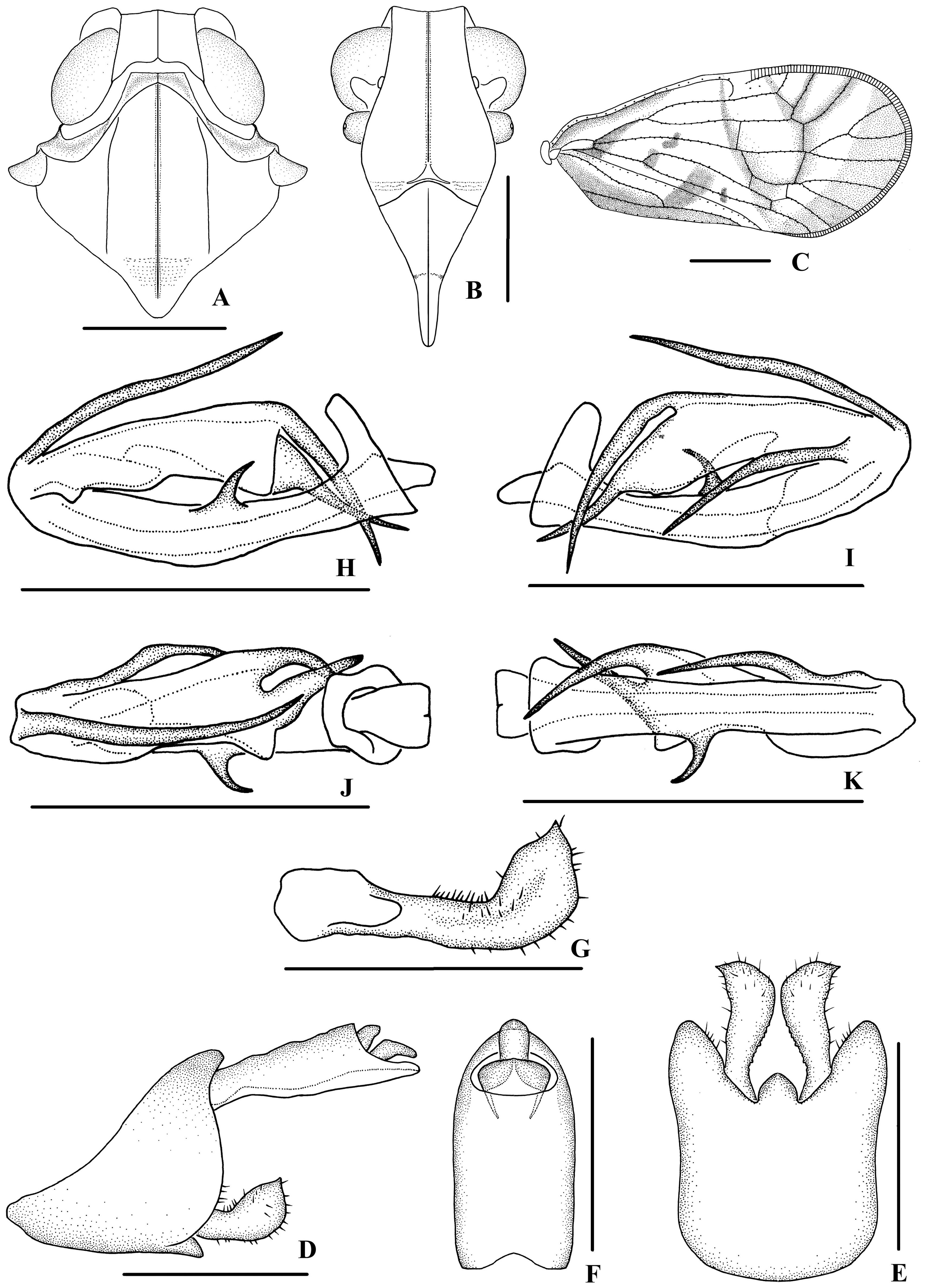
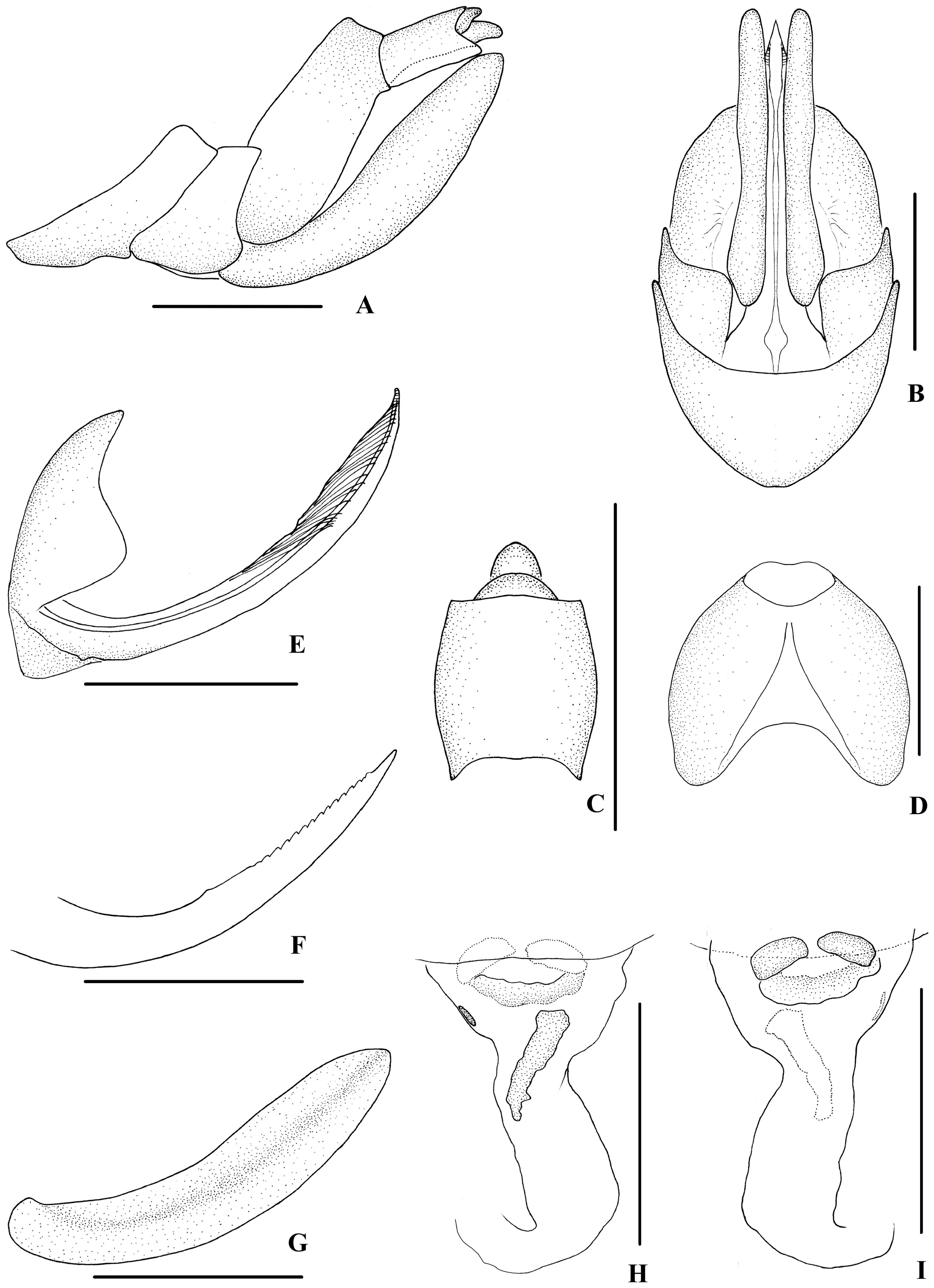
( Figs 8‒10 View FIGURE 8 View FIGURE 9 View FIGURE 10 )
Description. Body length: male 4.6‒4.8 mm ( n = 4), female 5.0‒ 5.3 mm ( n = 3).
Coloration. General color brown ( Fig. 8A–E View FIGURE 8 ). Eyes dark brown, ocelli brown, semitransparent. Vertex generally brown with carinae yellowish brown. Face generally brown; rostrum light brown. Pronotum and mesonotum brown with yellowish white carinae. Forewing yellowish brown, translucent, the basal half with dorsal and ventral margins dark brown, an oblique dark brown stripe near basal cell and another arising from Y fork to middle of CuA 1, three small dark brown spots located on CuA fork, the middle of CuA 2 and M veins, the apical half of forewing with a broad V-shaped stripe light yellowish brown and other stripes along transverse veins, RP 1 and RP 2; stigma yellowish brown. Hind tibiae yellowish brown and abdominal sternites dark brown.
Head and thorax. Vertex ( Figs 8C View FIGURE 8 , 9A View FIGURE 9 ) broad, 2.1 times wider than long; anterior margin slightly convex, posterior margin archedly recessed. Frons ( Figs 8D View FIGURE 8 , 9B View FIGURE 9 ) widest slightly below the level of antennae, 1.2 times as wide as long; frontoclypeal suture nearly concave into an arch; middle carina complete; lateral carinae distinct and slightly elevated. Pronotum ( Figs 8C View FIGURE 8 , 9A View FIGURE 9 ) 2.0 times longer than vertex; median carina indistinct, posterior margin nearly at right angle. Mesonotum 1.7 times longer than pronotum and vertex combined. Forewing ( Fig. 9C View FIGURE 9 ) 2.1 times longer than wide, with 9 apical and 5 subapical cells; fork Sc+RP slightly distad of fork CuA 1 +CuA 2, first crossvein r-m slightly basad of fork MP, RP two branches, MP with five terminals: MP 11, MP 12, MP 2, MP 3, and MP 4, fork MP 1 +MP 2 distad of fork MP 3 +MP 4. Metatibiotarsal formula: 6/7/8, second segment of hind tarsus with three platellae.
Male genitalia. Pygofer ( Fig. 9D, E View FIGURE 9 ) symmetrical, dorsal margin concave and U-shaped, slightly concaved medially in ventral view; in lateral view, lateral lobes arched extended caudally, medioventral process round in ventral view. Anal segment ( Fig. 9D, F View FIGURE 9 ) tubular, dorsal margin almost straight, apical 3/4 of ventral margin slightly convex, apical lobes finger-like in lateral view; 2.0 times longer than wide in dorsal view; anal style strap-shaped, slightly beyond anal segment. Gonostyli ( Fig. 9D, E, G View FIGURE 9 ) symmetrical in ventral view; in inner lateral view, base of ventral margin nearly straight, dorsal margin bending inwards in an obtuse arc in the middle, apical part with a small spike dorsocaudally directed. Aedeagus ( Fig. 9H–K View FIGURE 9 ) with total of five processes. Periandrium with a short spinous process positioning slightly to dorsal margin of its right side in the middle, slightly curved and right-dorsocephalically directed; apex with a long spinose process positioning slightly to dorsal margin of its left side, curved and ventrocephalically directed. Endosoma (=flagellum) moderately sclerotised, relatively long, generally dorsally curved. Dorsal margin with a long spinous process basally, slightly curved, apex dorsocephalically directed; apex with two long spinous processes, the upper one ventrocephalically directed and the lower one right-ventrocephalically directed.
Female genitalia. Posterior margin of pregenital sternite concave. Tergite IX ( Fig. 10A, D View FIGURE 10 ) moderately sclerotised, with length almost equal to width in caudal view. Anal tube ( Fig. 10A, C View FIGURE 10 ) short, nearly rectangular, 1.1 times longer than wide in dorsal view; dorsal and ventral margins nearly straight in lateral view, anal styles strap-shaped. Gonapophysis VIII ( Fig. 10E View FIGURE 10 ) elongate, and slightly curved upwards. Gonapophysis IX ( Fig. 10F View FIGURE 10 ) with one middle tooth, at a distance ratio, between middle tooth to apex and length of denticulate portion, of 2.4. Gonoplac ( Fig. 10G View FIGURE 10 ) rod-like, 4.5 times longer than wide. Posterior vagina ( Fig. 10H, I View FIGURE 10 ) elongate. The ventral wall of posterior vagina with a long sclerite in the middle area and a very small sclerite the left side basally.
Type material. Holotype: ♂, CHINA: Wuzhishan National Nature Reserve ( 18°54’N, 109°41’E), Wuzhishan City, Hainan Province, 18 May 1997, leg. Mao-Fa Yang; GoogleMaps 3♂♂ 3♀♀, Beibeng Township ( 29°14’N, 95°11’E), Motuo County, Tibet Autonomous Region, 15 August 2020, leg. Yong-Jin Sui. GoogleMaps
Host plant. Unknown.
Distribution. China ( Hainan, Tibet).
Remarks. Male genitalia of D. ( E.) biprocessus sp. nov. are similar to those of D. ( E.) chionomus Fennah, 1980 , but differ in: (1) the left side of periandrium without spinous process in the middle (in D. ( E.) chionomus , the left side of periandrium with a spinous process in the middle); (2) dorsal margin of endosoma with a long spinous process basally (the latter with two spinous processes on the right side of periandrium basally); (3) endosoma without spinous process in the middle and with two spinous processes at apex (the latter with one spinous process in the middle and at apex).
Etymology. The specific name refers to the apex of endosoma with two spinous processes.
No known copyright restrictions apply. See Agosti, D., Egloff, W., 2009. Taxonomic information exchange and copyright: the Plazi approach. BMC Research Notes 2009, 2:53 for further explanation.