
Dilacreon ( Eluzalmon ) congjiangensis, Chen & Zhi, 2023
|
publication ID |
https://doi.org/10.11646/zootaxa.5347.1.1 |
|
publication LSID |
lsid:zoobank.org:pub:E9658506-5801-4B92-8140-A8FCE1EC8F40 |
|
DOI |
https://doi.org/10.5281/zenodo.8390866 |
|
persistent identifier |
https://treatment.plazi.org/id/A24F07CC-3ACF-411C-B69F-8FD8780B839A |
|
taxon LSID |
lsid:zoobank.org:act:A24F07CC-3ACF-411C-B69F-8FD8780B839A |
|
treatment provided by |
Plazi |
|
scientific name |
Dilacreon ( Eluzalmon ) congjiangensis |
| status |
sp. nov. |
Dilacreon ( Eluzalmon) congjiangensis sp. nov.
( Figs 11‒13 View FIGURE 11 View FIGURE 12 View FIGURE 13 )
Description. Body length: male 5.2 mm ( n = 1), female 5.1–5.3 mm ( n = 7).
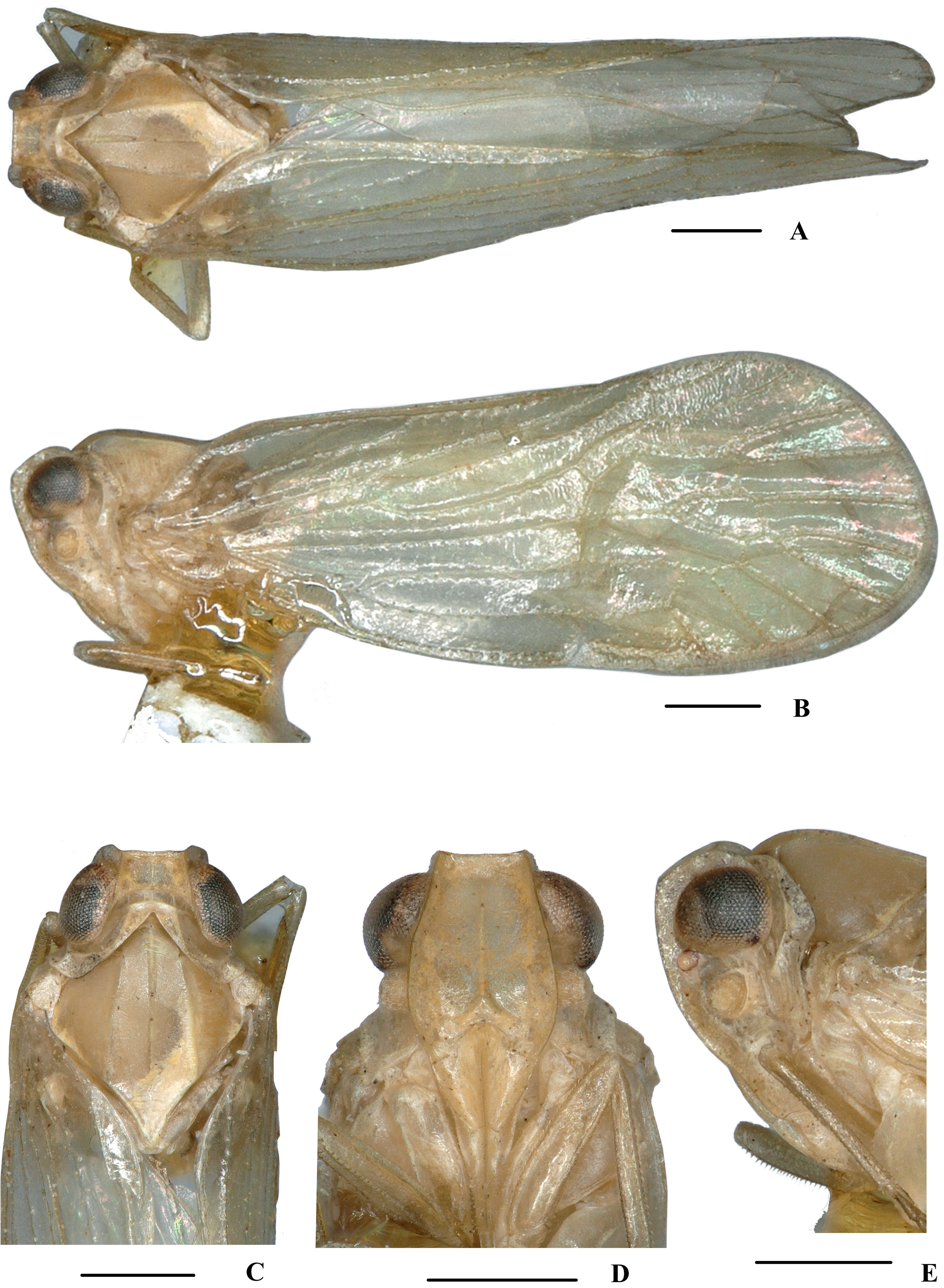
Coloration. General color yellowish brown ( Fig. 11A–E View FIGURE 11 ). Eyes dark brown, ocelli light yellow, semitransparent. Vertex generally yellowish brown, carinae brown (except median carina yellowish brown). Face generally yellowish brown; rostrum yellowish brown. Pronotum and mesonotum yellowish brown. Forewing semi-translucent, light yellowish brown, stigma yellowish brown. Hind tibiae and abdominal sternites yellowish brown.
Head and thorax. Vertex ( Figs 11C View FIGURE 11 , 12A View FIGURE 12 ) broad, 2.1 times wider than long; anterior margin truncated, posterior margin archedly recessed. Frons ( Figs 11D View FIGURE 11 , 12B View FIGURE 12 ) widest slightly below the level of antennae, 1.1 times as long as wide; frontoclypeal suture nearly concave into an arch; middle carina not attaining basal margin; lateral carinae distinct and elevated. Pronotum ( Figs 11C View FIGURE 11 , 12A View FIGURE 12 ) 2.3 times longer than vertex; median carina indistinct, posterior margin nearly at right angle. Mesonotum 1.7 times longer than pronotum and vertex combined. Forewing ( Fig. 12C View FIGURE 12 ) 2.6 times longer than wide, with 10 apical and 6 subapical cells; fork Sc+RP slightly basad of fork CuA 1 +CuA 2, RP two branches, MP with five terminals: MP 11, MP 12, MP 2, MP 3, and MP 4, fork MP 1 +MP 2 basad of fork MP 3 +MP 4. Metatibiotarsal formula: 6/6/8, second segment of hind tarsus with three platellae.
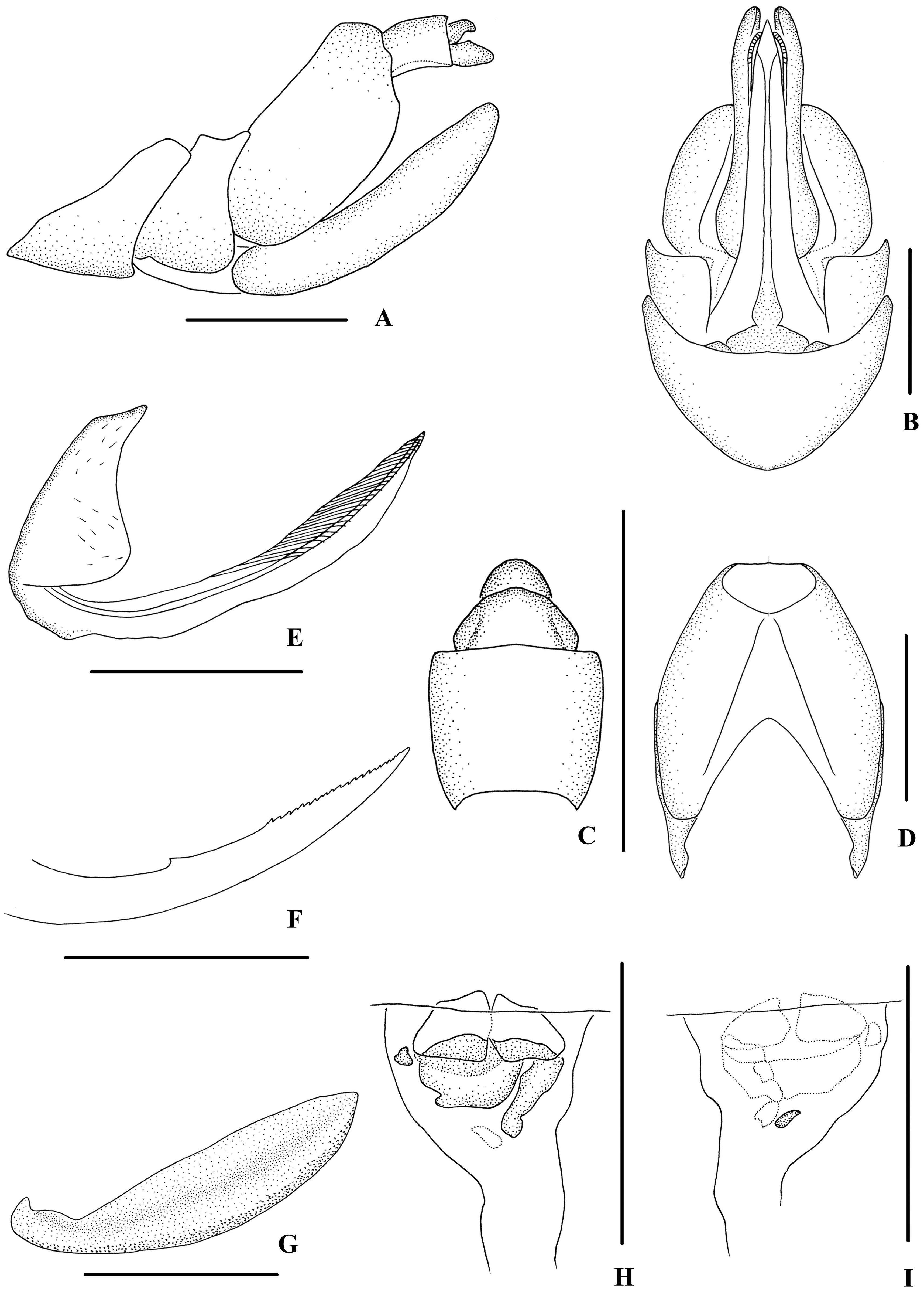
Male genitalia. Pygofer ( Fig. 12D, E View FIGURE 12 ) symmetrical, dorsal margin concave and U-shaped, slightly widened towards apex in ventral view; in lateral view, lateral lobes arched, caudally extended, medioventral process round in ventral view. Anal segment ( Fig. 12D, F View FIGURE 12 ) tubular, dorsal and ventral margins almost straight, apical lobes pointed in lateral view; 1.6 times longer than wide in dorsal view; anal style strap-shaped, beyond anal segment. Gonostyli ( Fig. 12D, E, G View FIGURE 12 ) symmetrical in ventral view; in inner lateral view, dorsal margin bending inwards in the middle, apical part extended, apical margin round. Aedeagus ( Fig. 12H–K View FIGURE 12 ) with total of four processes. In right side, periandrium with a spinose process apically, curved and ventrocephalically directed. Basal 1/3 of periandrium with a curved spinose process on left side, apex dorsally directed. Base part of periandrium densely covered with denticles at left, right and ventral surface. Endosoma (=flagellum) moderately sclerotised, long, generally dorsally curved. Base with a straight spinose process positioning slightly to dorsal margin of its left side, cephalically directed; left apex tapering into a short spinose process, ventrocephalically directed.
Female genitalia. Posterior margin of pregenital sternite concave. Tergite IX ( Fig. 13A, D View FIGURE 13 ) moderately sclerotised, slightly shorter than wide in caudal view. Anal tube ( Fig. 13A, C View FIGURE 13 ) short, nearly square, slightly widened towards apex in dorsal view; dorsal margin convex and ventral margin concave in lateral view, anal styles strap-shaped. Gonapophysis VIII ( Fig. 13E View FIGURE 13 ) elongate, and slightly curved upwards. Gonapophysis IX ( Fig. 13F View FIGURE 13 ) with one middle tooth, at a distance ratio, between middle tooth to apex and length of denticulate portion, of 1.8. Gonoplac ( Fig. 13G View FIGURE 13 ) rod-like, 3.8 times longer than wide. Posterior vagina ( Fig. 13H, I View FIGURE 13 ) elongate. In ventral view, base with a large nearly round sclerite in the middle area, left side with a small sclerite and right side with a longitudinal narrow sclerite; in dorsal view, middle area with a small oblong sclerite.
Type material. Holotype: ♂, CHINA: Guanghui Town ( 25°37’N, 108°20’E), Congjiang County, Guizhou Province, 21‒30 July 2016, leg. Ying-Jian Wang, Zheng-Xue Zhao and Niang Gong; GoogleMaps paratypes: 7♀♀, same data as holotype. GoogleMaps
Host plant. Unknown.
Distribution. China ( Guizhou).
Remarks. Male genitalia of D. ( E.) congjiangensis sp. nov. are similar to those of D. ( E.) lobatus Zhang & Chen, 2013 , but differ in: (1) basal 1/3 of periandrium with a spinose process on left side (the latter without spinose process in the same position); (2) the left side of apex of periandrium without spinose process (in D. ( E.) lobatus , the left side of apex of periandrium with a spinose process); (3) base of endosoma with a straight spinose process (the latter without spinose process in the same position); (4) the forewings without any markings (the forewings with markings in D. ( E.) lobatus ).
Etymology. The species name is derived from the name of the type locality, Congjiang County in Guizhou Province.

No known copyright restrictions apply. See Agosti, D., Egloff, W., 2009. Taxonomic information exchange and copyright: the Plazi approach. BMC Research Notes 2009, 2:53 for further explanation.