
Neocarpia trispina, Chen & Zhi, 2023
|
publication ID |
https://doi.org/10.11646/zootaxa.5347.1.1 |
|
publication LSID |
lsid:zoobank.org:pub:E9658506-5801-4B92-8140-A8FCE1EC8F40 |
|
DOI |
https://doi.org/10.5281/zenodo.8390989 |
|
persistent identifier |
https://treatment.plazi.org/id/6B38339B-7E9E-4358-A346-416CE41E6E1E |
|
taxon LSID |
lsid:zoobank.org:act:6B38339B-7E9E-4358-A346-416CE41E6E1E |
|
treatment provided by |
Plazi |
|
scientific name |
Neocarpia trispina |
| status |
sp. nov. |
Neocarpia trispina sp. nov.
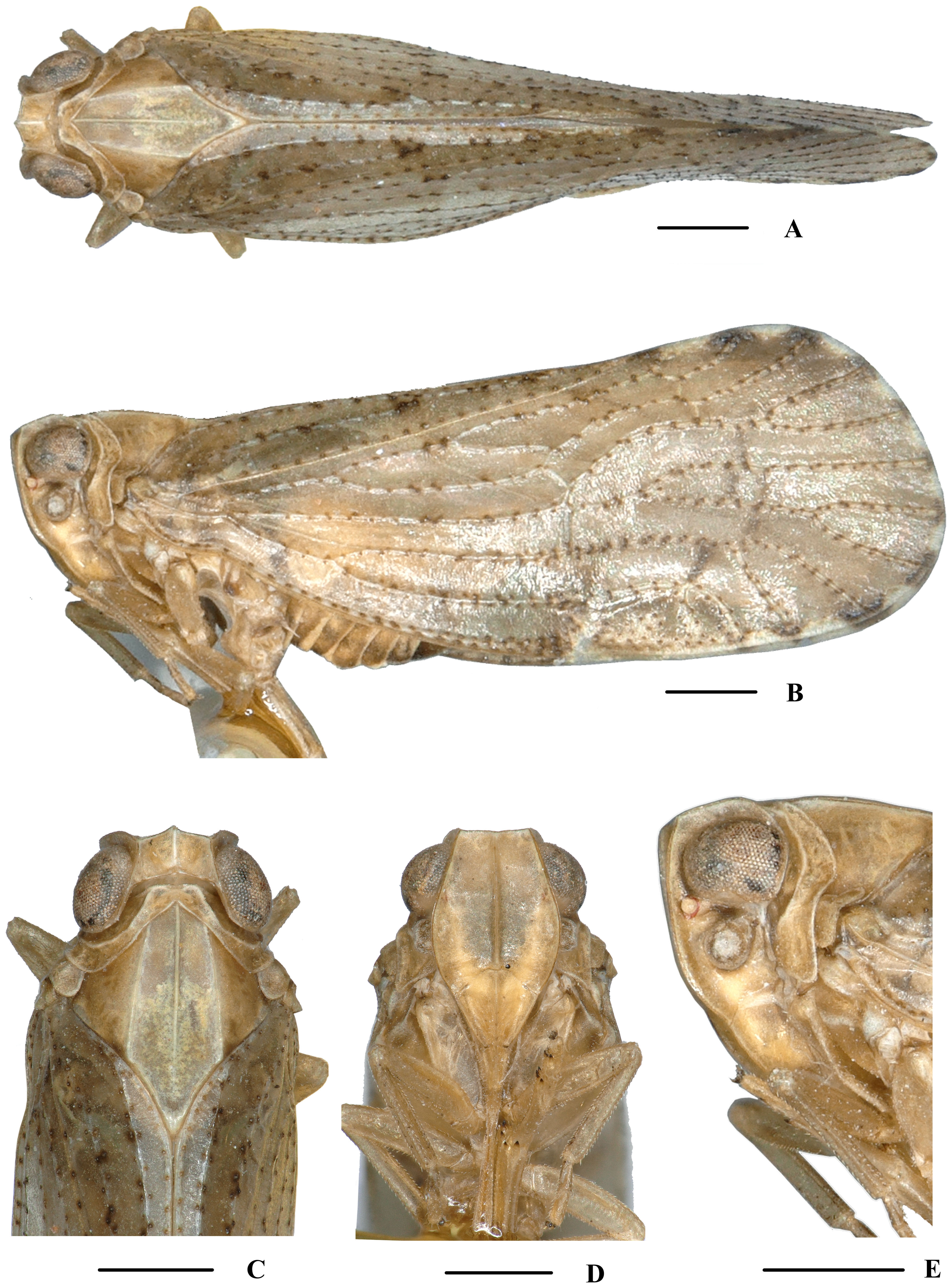
( Figs 65‒67 View FIGURE 65 View FIGURE 66 View FIGURE 67 )
Description. Body length: male 4.9‒5.9 mm ( n = 25), female 5.3‒6.4 mm ( n = 45).
Coloration. General color yellowish brown ( Fig. 65A–E View FIGURE 65 ). Eyes brown, ocelli light yellow. Vertex generally yellowish brown, carinae brown to dark brown (except median carina yellowish white). Face generally yellowish brown, carinae brown to dark brown; rostrum yellowish brown. Pronotum and mesonotum yellowish brown. Forewing semi-translucent, basal half yellowish brown and distal half darker, with blackish brown spots on end of longitudinal veins. Hind tibiae and abdominal sternites yellowish brown.
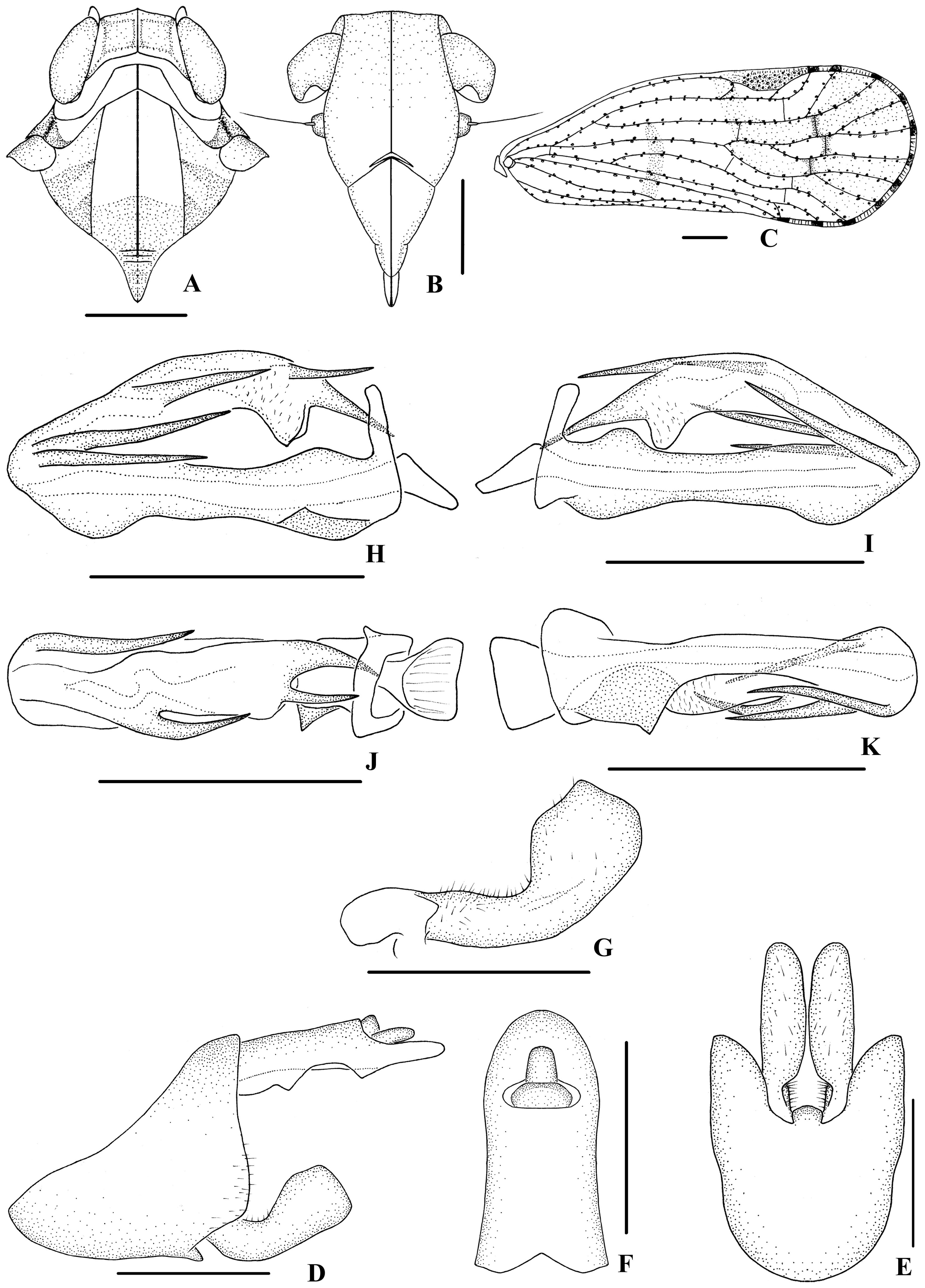
Head and thorax. Vertex ( Fig. 65C View FIGURE 65 , 66A View FIGURE 66 ) broad, 2.2 times wider than long; anterior margin slightly produced in the middle, posterior margin archedly recessed. Frons ( Fig. 65D View FIGURE 65 , 66B View FIGURE 66 ) widest at the level of antennae, 1.2 times as long as wide; frontoclypeal suture nearly concave into an arch; middle carina complete; lateral carinae distinct and elevated. Pronotum ( Fig. 65C View FIGURE 65 , 66A View FIGURE 66 ) 2.3 times longer than vertex; median carina indistinct, posterior margin nearly at right angle. Mesonotum 1.6 times longer than pronotum and vertex combined. Forewing ( Fig. 66C View FIGURE 66 ) 2.7 times longer than wide, with 10 apical and 6 subapical cells; fork Sc+RP basad of fork CuA 1 +CuA 2, first crossvein r-m slightly distad of fork MP; RP two branches, MP with five terminals: MP 11, MP 12, MP 2, MP 3, and MP 4, fork MP 1 +MP 2 basad of fork MP 3 +MP 4. Metatibiotarsal formula: 6/5/8, second segment of hind tarsus with three platellae.
Male genitalia. Pygofer ( Fig. 66D, E View FIGURE 66 ) symmetrical, dorsal margin concave and U-shaped ventrally, slightly widened towards apex; in lateral view, lateral lobes triangularly extended caudally, medioventral process round ventrally. Anal segment ( Fig. 66D, F View FIGURE 66 ), dorsal margin almost straight, ventral margin with two triangular processes; and 1.9 times longer than wide in dorsal view; anal style strap-shaped, not beyond anal segment. Gonostyli ( Fig. 66D, E, G View FIGURE 66 ) symmetrical in ventral view; in inner lateral view, apical margin of gonostyli with a small blunt process, dorsal margin bending inwards in the middle. Aedeagus ( Fig. 66H–K View FIGURE 66 ) in total with seven processes. In right side, apex of periandrium with two straight spinose processes, the upper one dorsocephalically directed and the other one slightly shorter and apex cephalically directed; base of periandrium with a transverse triangular laminal process, right side directed; left side of periandrium with a straight spinose process at apex, dorsocephalically directed. Endosoma (=flagellum) moderately sclerotised, generally dorsally curved; right side each with a straight spinose process in the middle and near apex, dorsocephalically directed; apex with a spinose process, ventrocephalically directed.
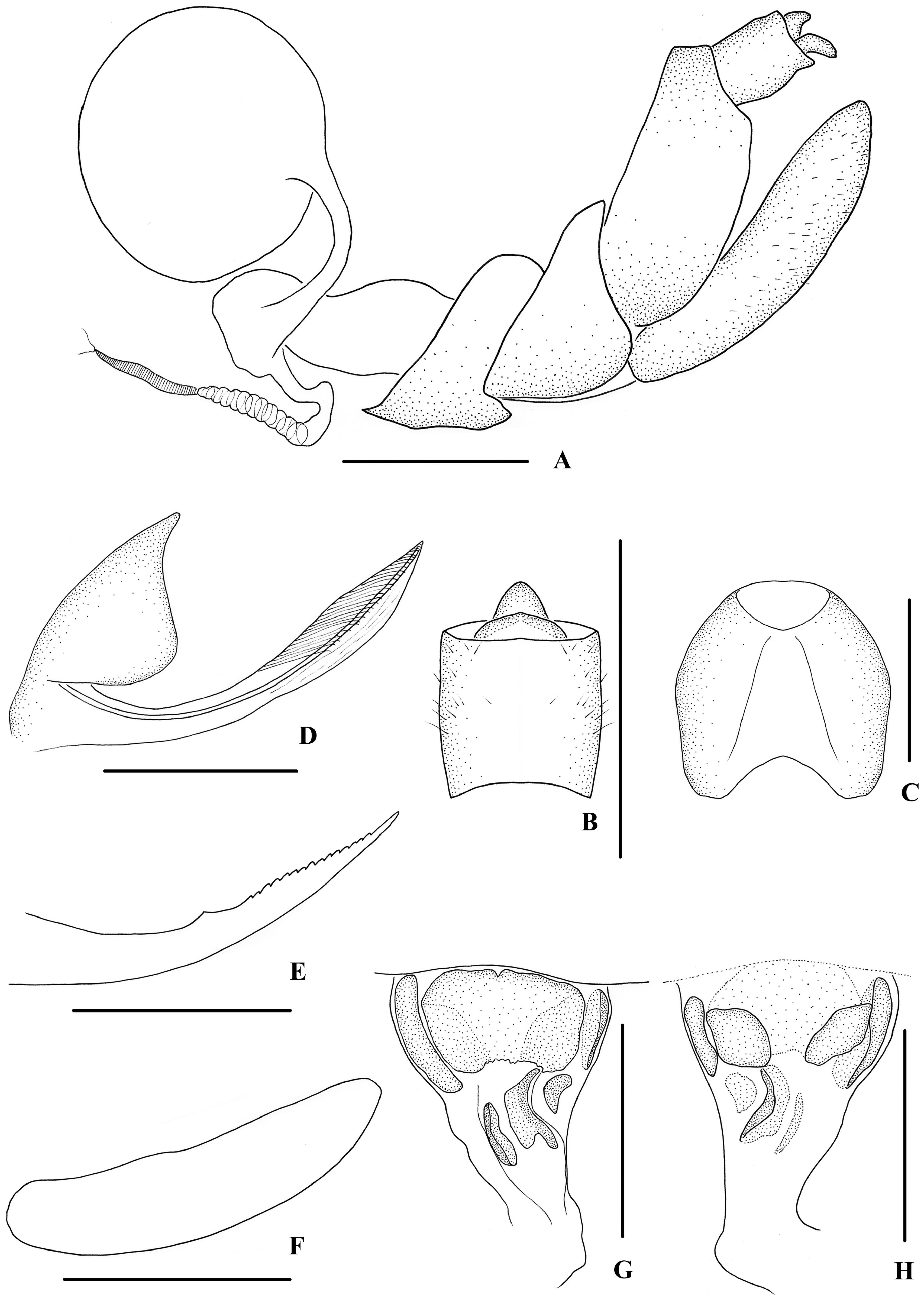
Female genitalia. Tergite IX ( Fig. 67A, C View FIGURE 67 ) moderately sclerotised, with length almost equal to width in caudal view. Anal tube ( Fig. 67A, B View FIGURE 67 ) short, nearly rectangular, slightly longer than wide in dorsal view; anal styles relatively short and small. Gonapophysis VIII ( Fig. 67D View FIGURE 67 ) elongate, and slightly curved upwards. Gonapophysis IX ( Fig. 67E View FIGURE 67 ) with one middle tooth, at a distance ratio, between middle tooth to apex and length of denticulate portion, of 1.6. Gonoplac ( Fig. 67F View FIGURE 67 ) rod-like, 4.3 times longer than wide. Posterior vagina ( Fig. 67G, H View FIGURE 67 ) elongate, in ventral view, with a large transverse oval sclerite and five longitudinal sclerites; in dorsal view, with two oval sclerites.
Type material. Holotype: ♂, CHINA: Mohan Town ( 21°14’N, 101°42’E), Mengla County, Yunnan Province, 22 June 2016, leg. Ying-Jian Wang; GoogleMaps paratypes: 21♂♂ 42♀♀, same collection area as holotype, 22–23 June 2016, leg. Liang-Jing Yang, Ying-Jian Wang and Qiang Luo; GoogleMaps 1♂, same collection area as holotype, 15 August 2013, leg. Mei-Na Guo; GoogleMaps 2♂♂ 3♀♀, Mei Village ( 24°8’N, 110°6’E), Jinxiu Town, Jinxiu County, Guangxi Zhuang Autonomous Region, 30 April 2011, leg. Xiao-Fei Yu, Rong Huang and Xin-Feng Zhang. GoogleMaps
Distribution. China ( Guangxi, Yunnan).
Host plant. Unknown.
Remarks. This species is similar to N. bidentata Zhang & Chen, 2013 , but differs in: (1) right side of periandrium with two spinose processes at apex and a transverse triangular laminal process at base (the latter with one spinose process at apex and without laminal process at base); (2) neither dorsal nor ventral margins of periandrium with process (in N. bidentata , dorsal margin of periandrium with a shovel-shaped process and ventral margin with a small triangular spine); (3) endosoma with three spinose processes (the latter with one spinose process).
Etymology. The specific name is derived from the Latin prefix “ tri- ” and noun “ spina ”, referring to endosoma of aedeagus with three spinose processes.
No known copyright restrictions apply. See Agosti, D., Egloff, W., 2009. Taxonomic information exchange and copyright: the Plazi approach. BMC Research Notes 2009, 2:53 for further explanation.