
Neocarpia Tsaur & Hsu, 2003
|
publication ID |
https://doi.org/10.11646/zootaxa.5347.1.1 |
|
publication LSID |
lsid:zoobank.org:pub:E9658506-5801-4B92-8140-A8FCE1EC8F40 |
|
DOI |
https://doi.org/10.5281/zenodo.8408644 |
|
persistent identifier |
https://treatment.plazi.org/id/9E48011E-4274-C24B-FF65-31CCE2F7FEFA |
|
treatment provided by |
Plazi |
|
scientific name |
Neocarpia Tsaur & Hsu, 2003 |
| status |
|
Genus Neocarpia Tsaur & Hsu, 2003 View in CoL View at ENA
Neocarpia Tsaur & Hsu, 2003: 440 View in CoL ; Löcker et al., 2010: 17 View Cited Treatment ; Zhang & Chen, 2013b: 42 View Cited Treatment ; Zhi et al., 2017: 20 View Cited Treatment .
Type species: Neocarpia maai Tsaur & Hsu, 2003 View in CoL , by original designation.
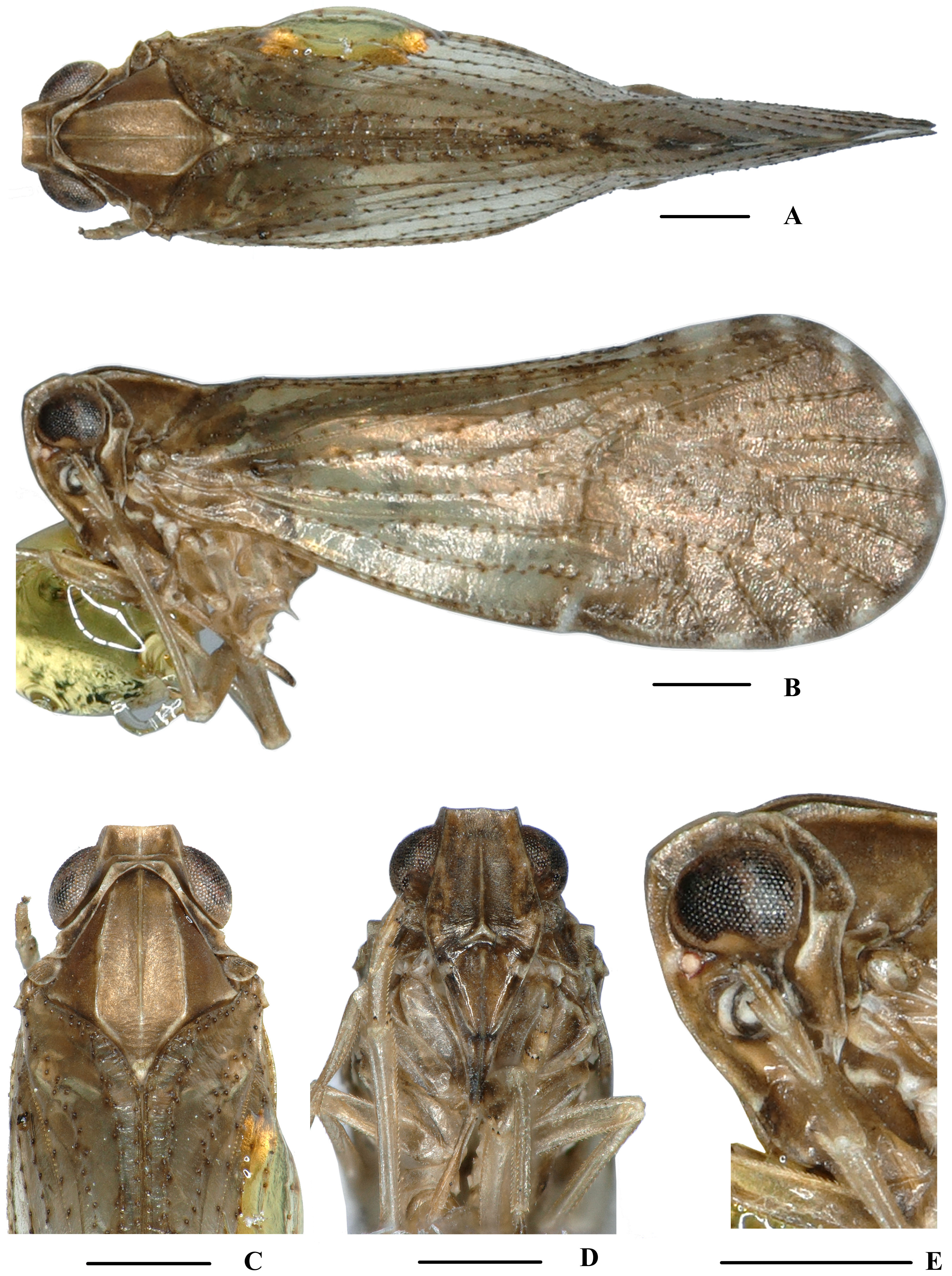
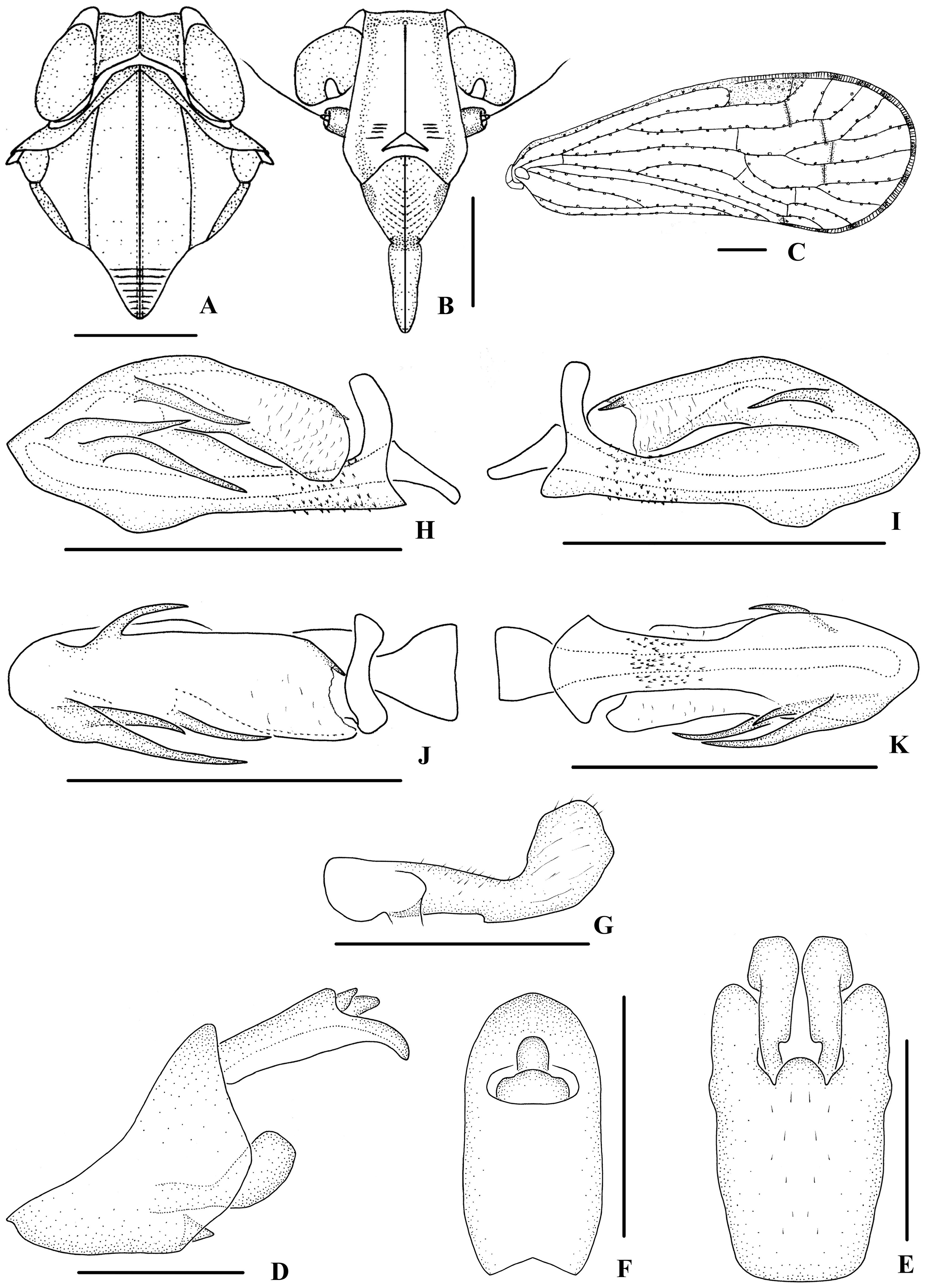
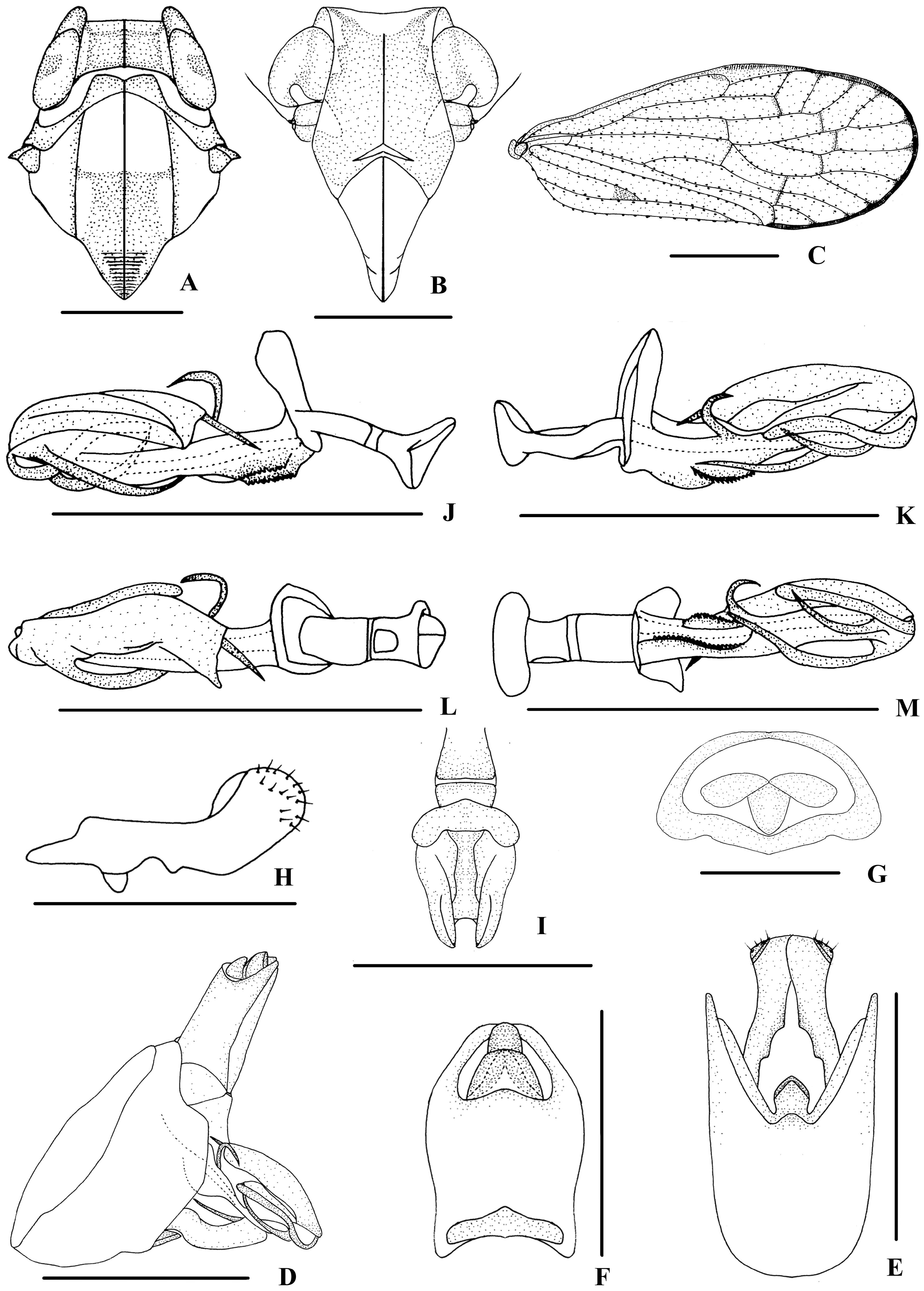
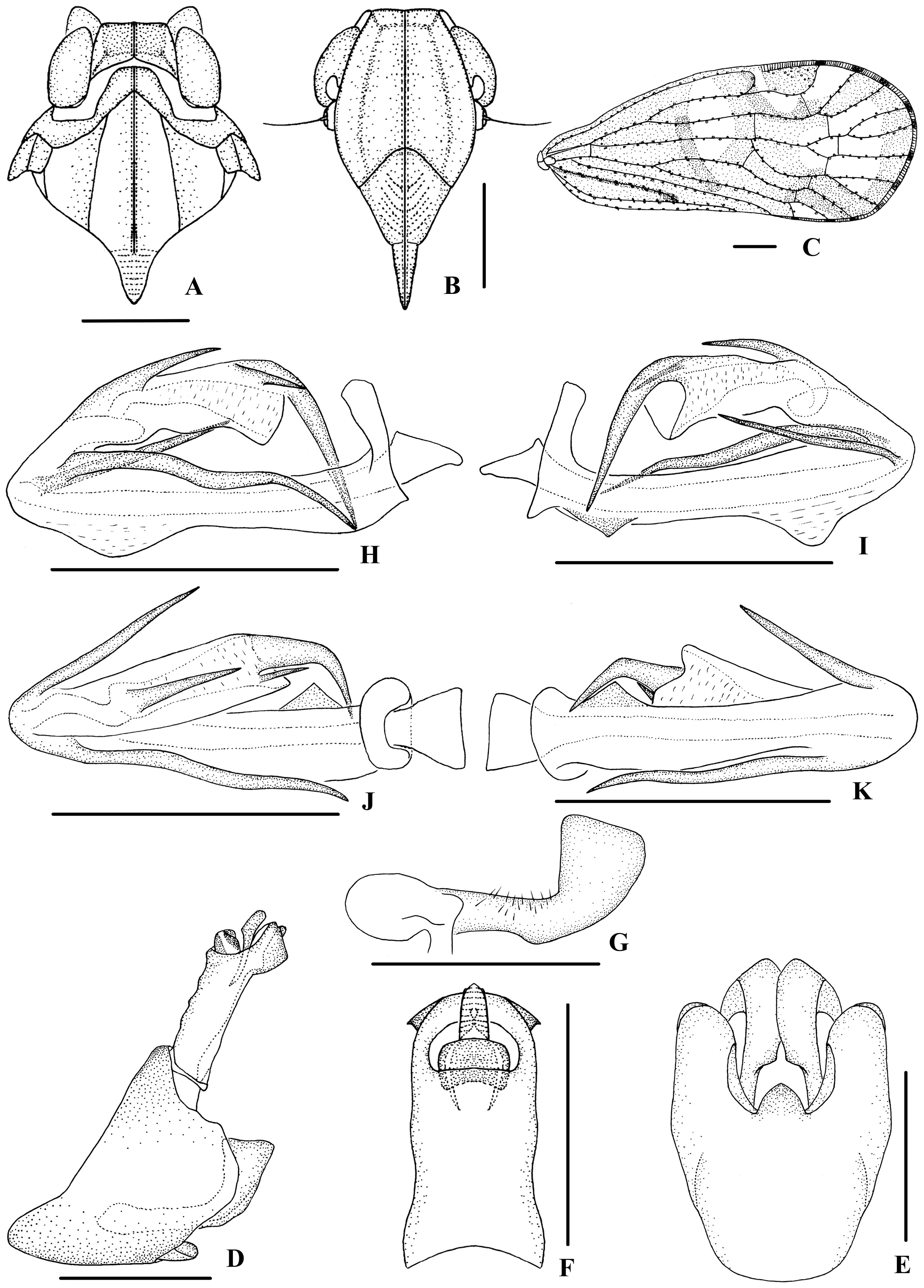
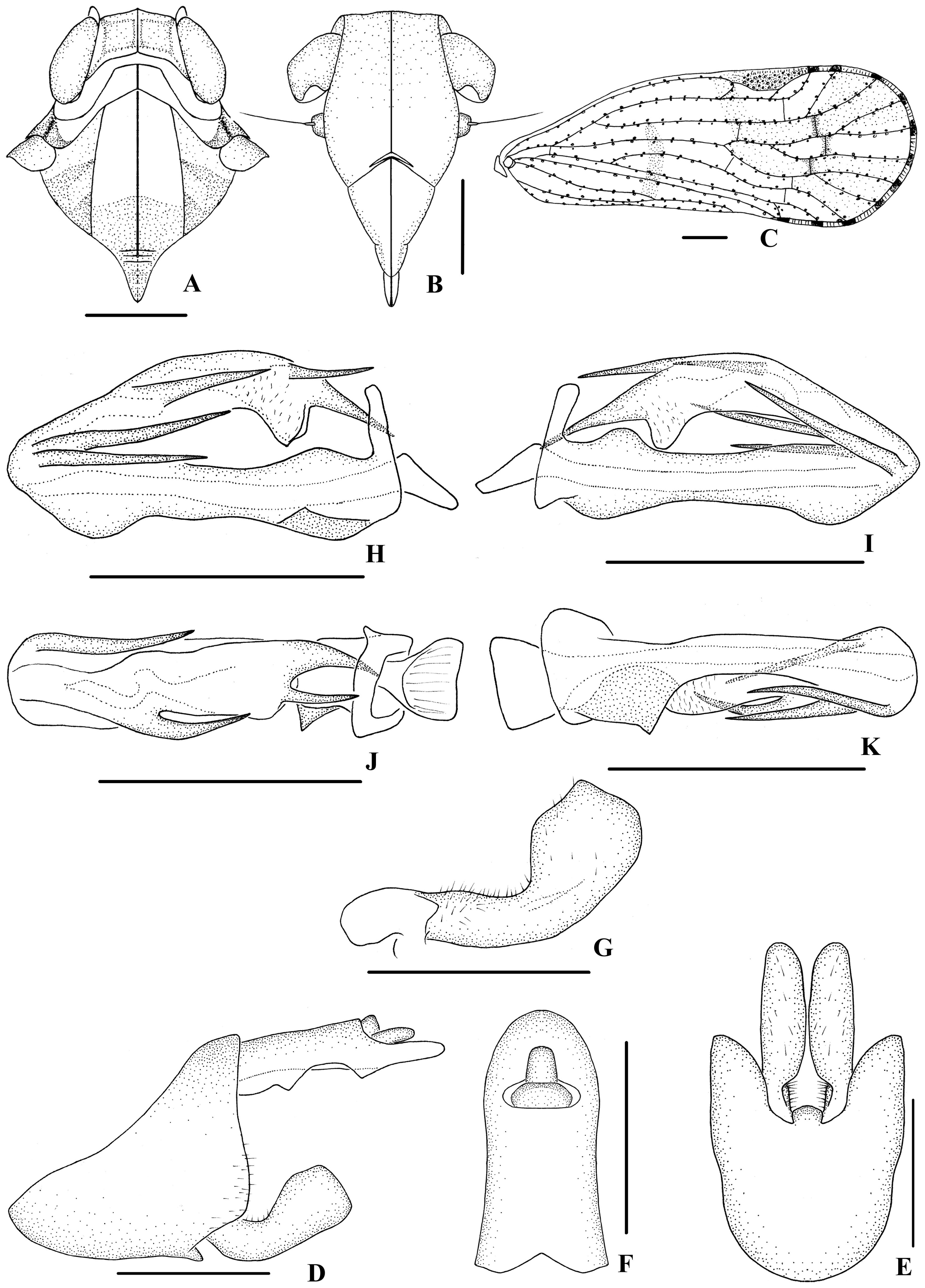
Description. Body size. Moderate-sized species. Body strongly compressed laterally.
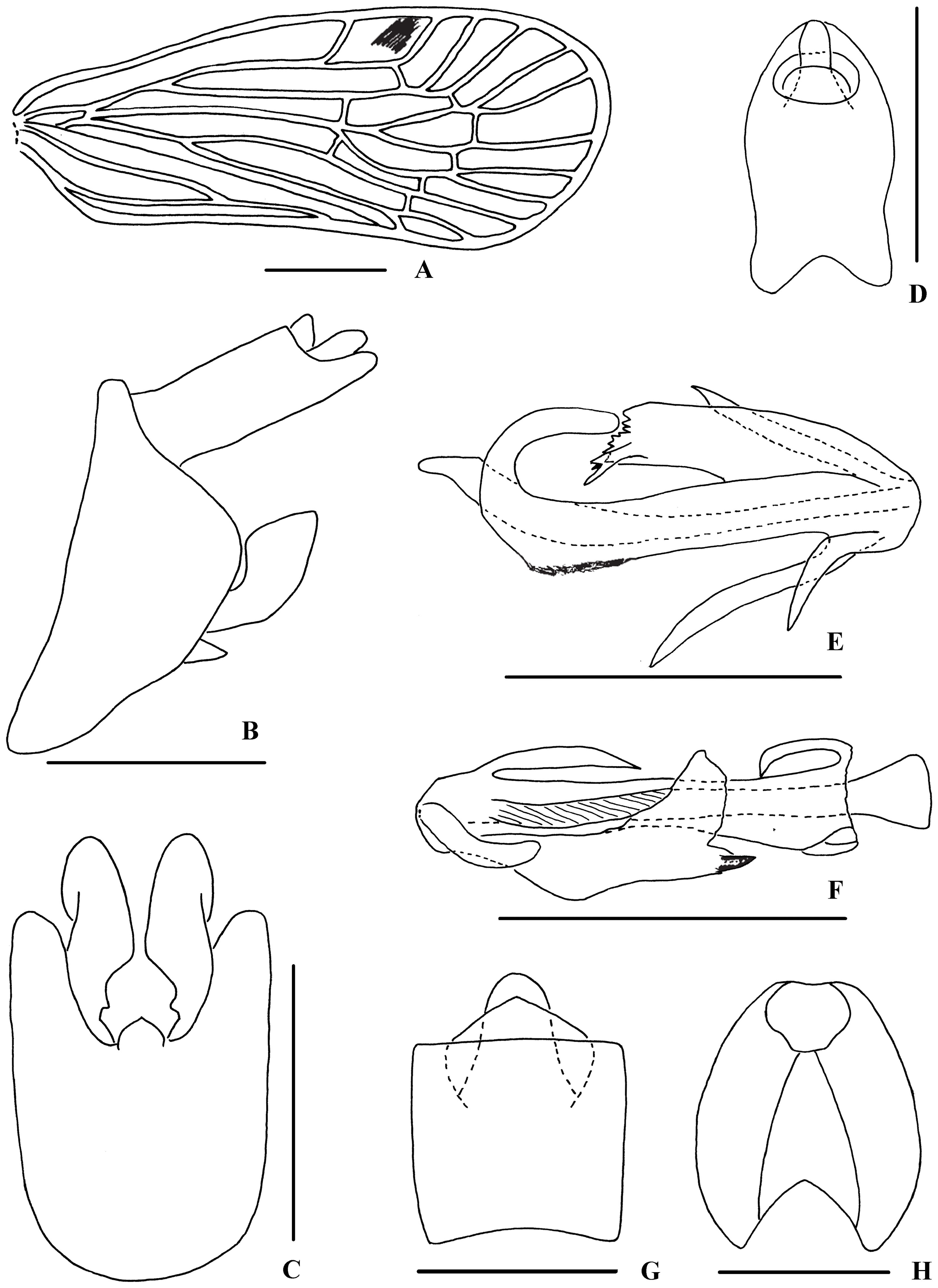
Head and Thorax. Head slightly narrower than pronotum in dorsal view. Vertex slightly widened to posterior emargination, broader than long and without subapical carina, lateral carinae moderately elevated. Frons with median carina; frontoclypeal suture generally angled or semicircular. Clypeus with well-developed median carina. Rostrum distinctly surpassing hind coxae. Pronotum short with intermediate carinae curved along posterior margins of eyes. Mesonotum tricarinate. Forewing in resting position steeply tectiform, widened towards apex, with rounded apical margin; Sc+R forming a common stem and M emerging separately from basal cell; position of fork Sc+R slightly basal or at the same level as fork CuA 1 +CuA 2; first crossvein MP -CuA 1 at least as long as MP 3+4 from MP fork to this crossvein, crossvein MP 3+4 -CuA 1 almost at same level as crossvein r-m, subapical cell MP 3+4 with upper margin (vein MP 3+4) fine concave, no crossvein between CuA 1 and CuA 2. Apical cells 10–11. Hind tibia lacking lateral spines.
3+4
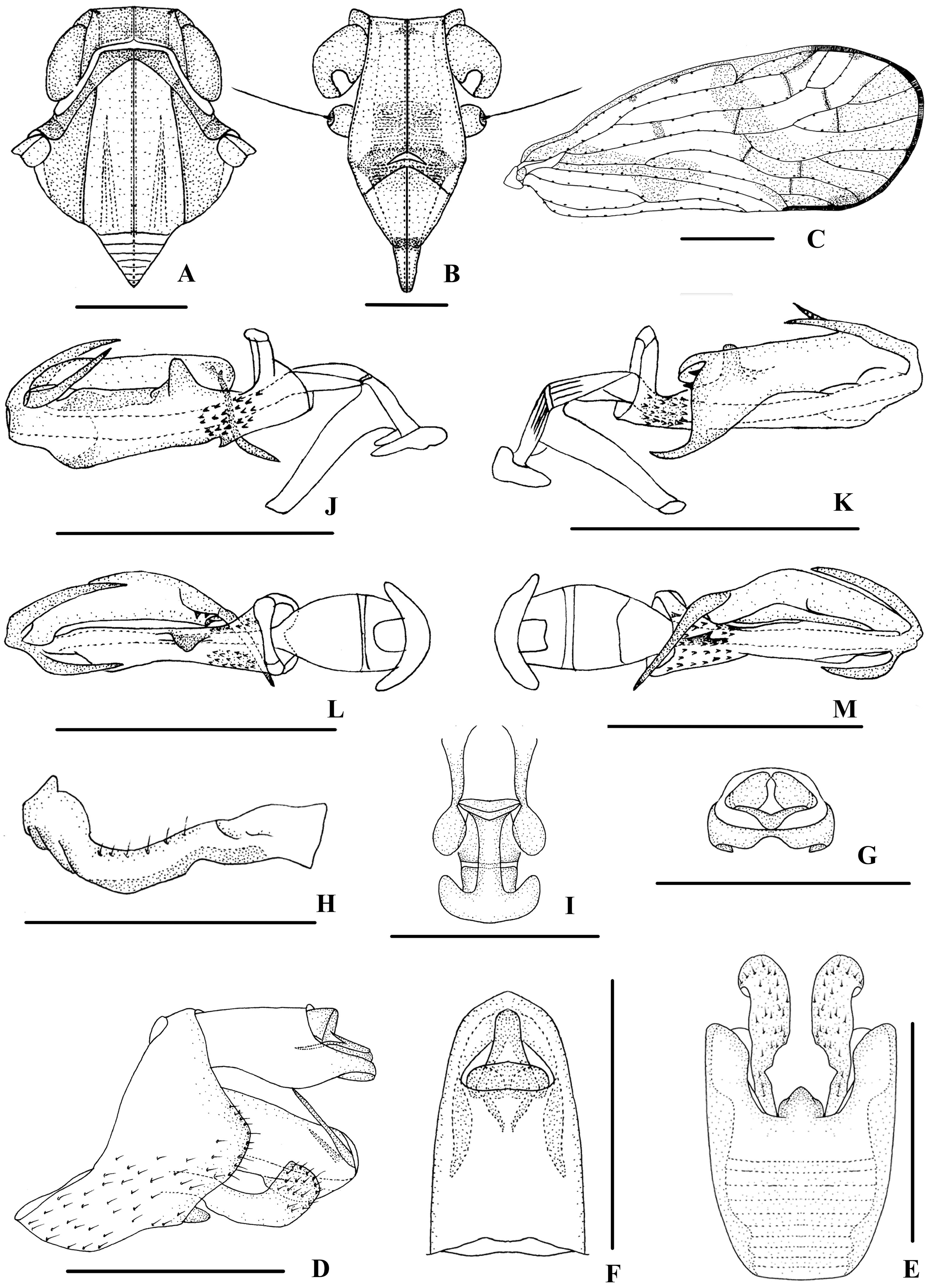
Male genitalia. Pygofer symmetrical and prolonged with symmetrical lateral lobes in lateral view. Medioventral process thumb-like in lateral view.Anal segment tubular, short and stout. Gonostyli relatively small and symmetrical. Aedeagus slender and endosoma (=flagellum) of aedeagus with spinose processes.
Female genitalia. Ovipositor elongate, orthopteroid and slightly curved upwards; anal segment square or rectangular in dorsal view; tergite IX without wax plate. Gonapophysis VIII slightly sclerotised, blade-like posteriorly. Gonapophysis IX single, blunt and strongly sclerotised, between middle tooth and apex with a row of denticles. Gonoplac slightly sclerotised, with many spinules on ventral edge in inner lateral view. Posterior vagina with sclerites.
Distribution. Sino-Japanese, Oriental and Australian regions.
Remarks. According to the original description of Neocarpia Tsaur & Hsu, 2003 and figure of the type species N. maai , in particular the feature of the forewing venation namely “transverse veinlet M 3+4 -Cu la (MP 3+4 -CuA 1) much longer than vein M 3+4 (MP 3+4) from M (MP) fork to this veinlet, subapical cells M 3+4 (MP 3+4) with upper margin (vein M 3+4) weakly concave, no transverse vein between Cu l and Cu 2 ” and relative position of crossveins MP 3+4 -CuA 1 and r-m, it is highly likely that Neocarpia Tsaur & Hsu is a synonym of Eucarpia Walker. However , since we have not seen the type specimen, in this study we do not formally synonymise the two genera until the types become available for study.
Key to species of Neocarpia Tsaur & Hsu View in CoL of the world
1. Ventral margin of periandrium without spinose process....................................................... 2
- Ventral margin of periandrium with one or two spinose process(es).............................................. 6
2. Right side of periandrium with two spinose processes apically.................................................. 3
- Right side of periandrium with one spinose process apically................................................... 4
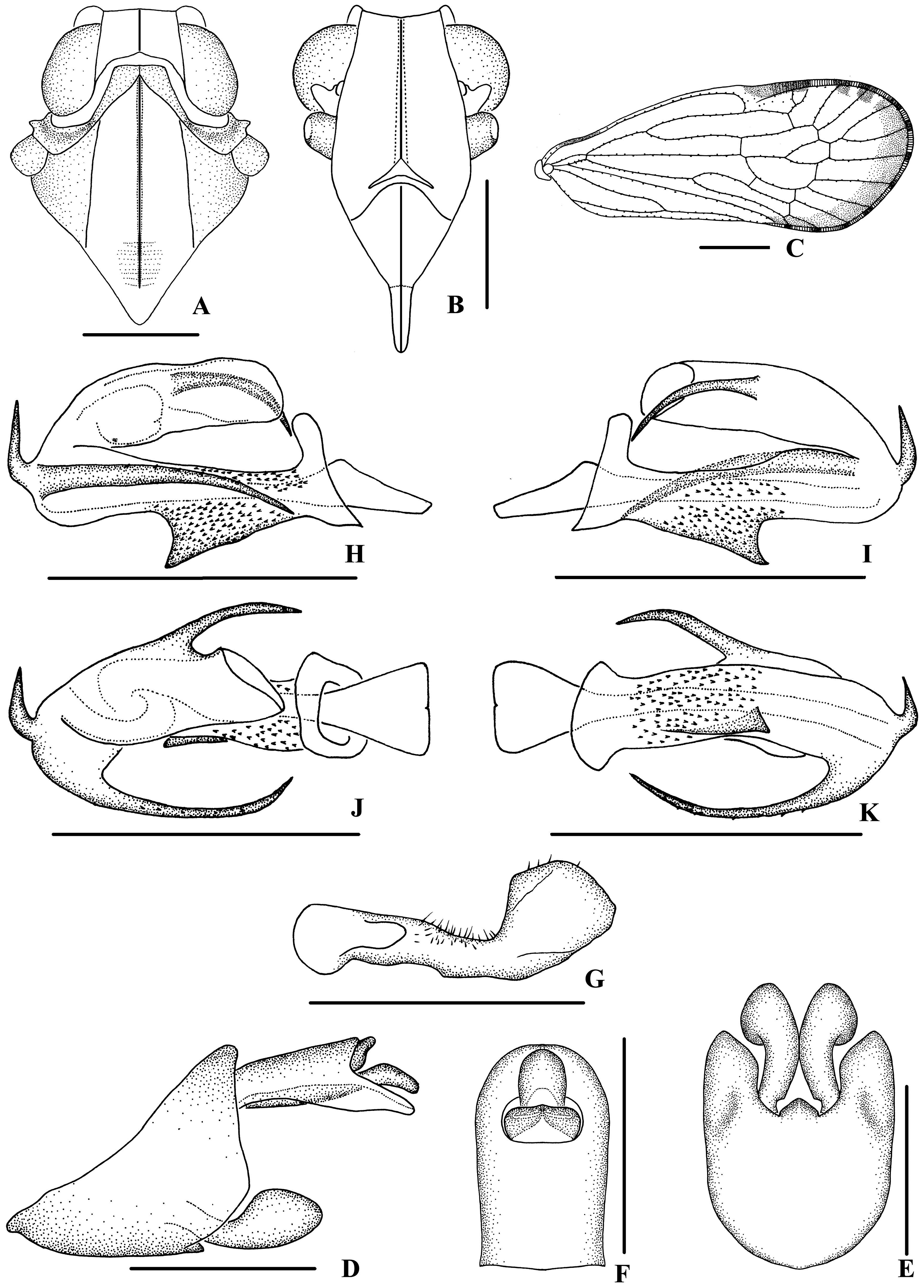
3. Right side of periandrium with a transverse triangular laminal process basally and left side with a spinose process ( Fig. 66H, I View FIGURE 66 )................................................................................... N. trispina sp. nov.
- Right side of periandrium without laminal process basally and left side without spinose process ( Fig. 55H, I View FIGURE 55 )............................................................................................... N. brevispina sp. nov.
4. Dorsal margin of periandrium with one process ( Emeljanov and Hayashi, 2007: Figs 23 View FIGURE 23 , 24 View FIGURE 24 )................................................................................................ N. okinawana Emeljanov & Hayashi View in CoL
- Dorsal margin of periandrium without process.............................................................. 5
5. Right side of endosoma (=flagellum) with a long spinose process basally, left base of periandrium with a spinose process ( Fig. 63H–K View FIGURE 63 )........................................................................... N. reversa Zhi & Chen View in CoL
- Right side of endosoma without process basally, left base of periandrium with a triangular laminal process ( Fig. 60H–K View FIGURE 60 )...................................................................................... N. longispina sp. nov.
6. Ventral margin of periandrium with one small triangular process at basal 1/3...................................... 7
- Ventral margin of periandrium without triangular process at base, while with one or two process(es) near or at apex....... 8
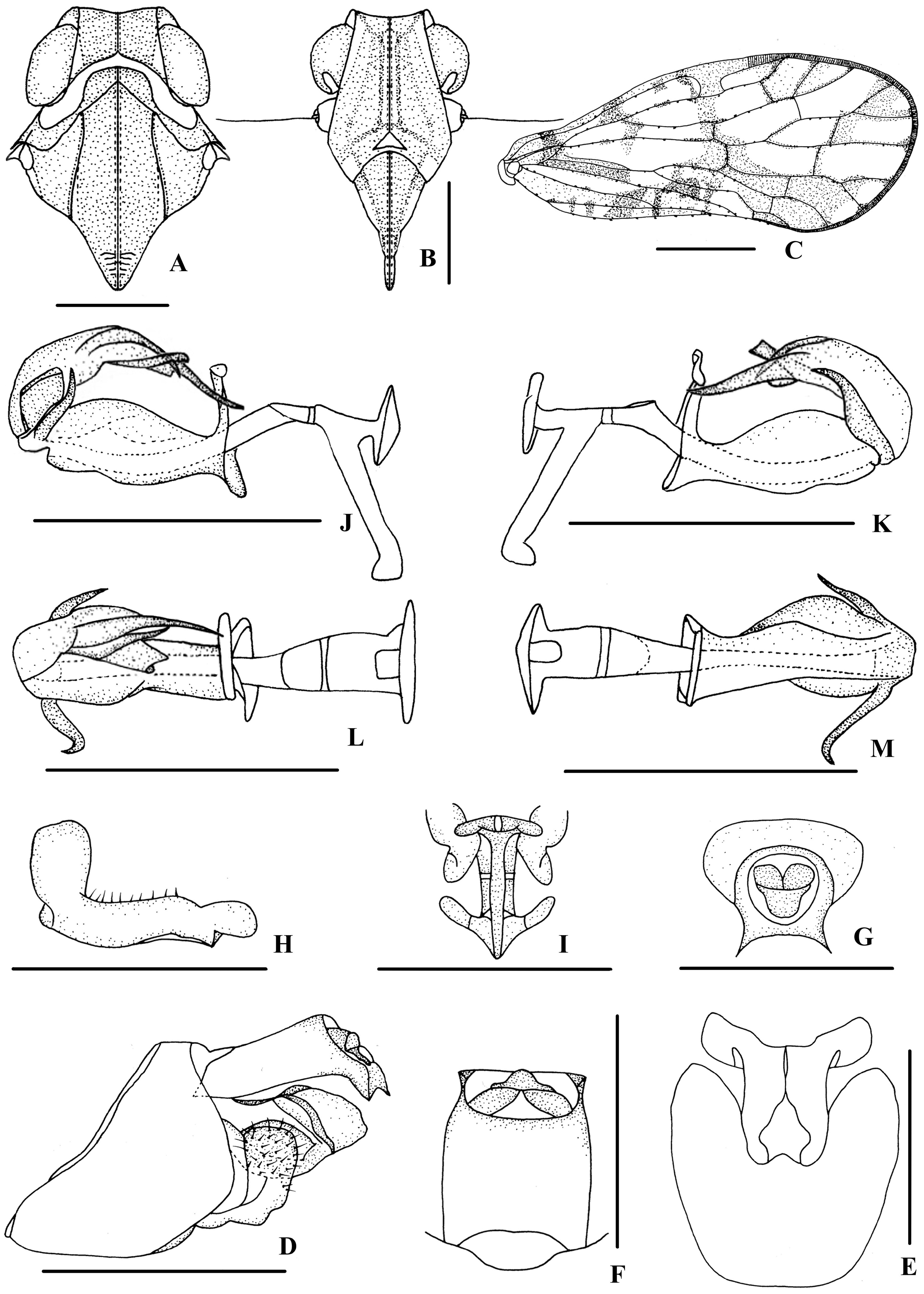
7. Left side of periandrium with a process near apex, dorsal margin with a shovel-shaped process, right side without process in the middle, base of process near apex of endosoma with two denticulations ( Fig. 52J–M View FIGURE 52 ); forewing without stripe......................................................................................... N. bidentata Zhang & Chen View in CoL
- Left side of periandrium without process, dorsal margin without process, right side with a short acute process in the middle, base of process near apex of endosoma without denticulation ( Fig. 49H–K View FIGURE 49 ); forewing with yellow stripes along the Y-veins................................................................................... N. acutata Zhi & Chen View in CoL
8. Endosoma with a prominent long process in the middle ( Löcker et al., 2010: Fig. 17A View FIGURE 17 )............ N. rhizophorae Löcker View in CoL
- Endosoma without process in the middle................................................................... 9
9. Dorsal margin of periandrium with a hook-shaped process, ventral margin of periandrium with one spinose process, endosoma with smooth apical margin ( Fig. 57J–M View FIGURE 57 ).............................................. N. hamata Zhang & Chen View in CoL
- Dorsal margin of periandrium without process, ventral margin of periandrium with two spinose processes, endosoma with sinuate apical margin ( Fig. 61E, F View FIGURE 61 )...................................................... N. maai Tsaur & Hsu View in CoL


No known copyright restrictions apply. See Agosti, D., Egloff, W., 2009. Taxonomic information exchange and copyright: the Plazi approach. BMC Research Notes 2009, 2:53 for further explanation.
|
Kingdom |
|
|
Phylum |
|
|
Class |
|
|
Order |
|
|
Family |
|
|
SubFamily |
Cixiinae |
|
Tribe |
Eucarpiini |
Neocarpia Tsaur & Hsu, 2003
| Chen, Xiang-Sheng & Zhi, Yan 2023 |
Neocarpia
| Zhi, Y. & Yang, L. & Zhang, P. & Chen, X. - S. 2017: 20 |
| Zhang, P. & Chen, X. - S. 2013: 42 |
| Locker, B. & Fletcher, M. J. & Gurr, G. M. 2010: 17 |
| Tsaur, S. - C. & Hsu, T. - C. 2003: 440 |