
Planopilumnus Balss, 1933
|
publication ID |
https://doi.org/ 10.5281/zenodo.275841 |
|
DOI |
https://doi.org/10.5281/zenodo.5664428 |
|
persistent identifier |
https://treatment.plazi.org/id/9E5387EE-FFE6-D343-84AA-F97AFED5A584 |
|
treatment provided by |
Plazi |
|
scientific name |
Planopilumnus Balss, 1933 |
| status |
|
Planopilumnus Balss, 1933 View in CoL
Planopilumnus Balss, 1933: 39 View in CoL .
Type species. Pilumnus spongiosus Nobili, 1905 , by original designation.
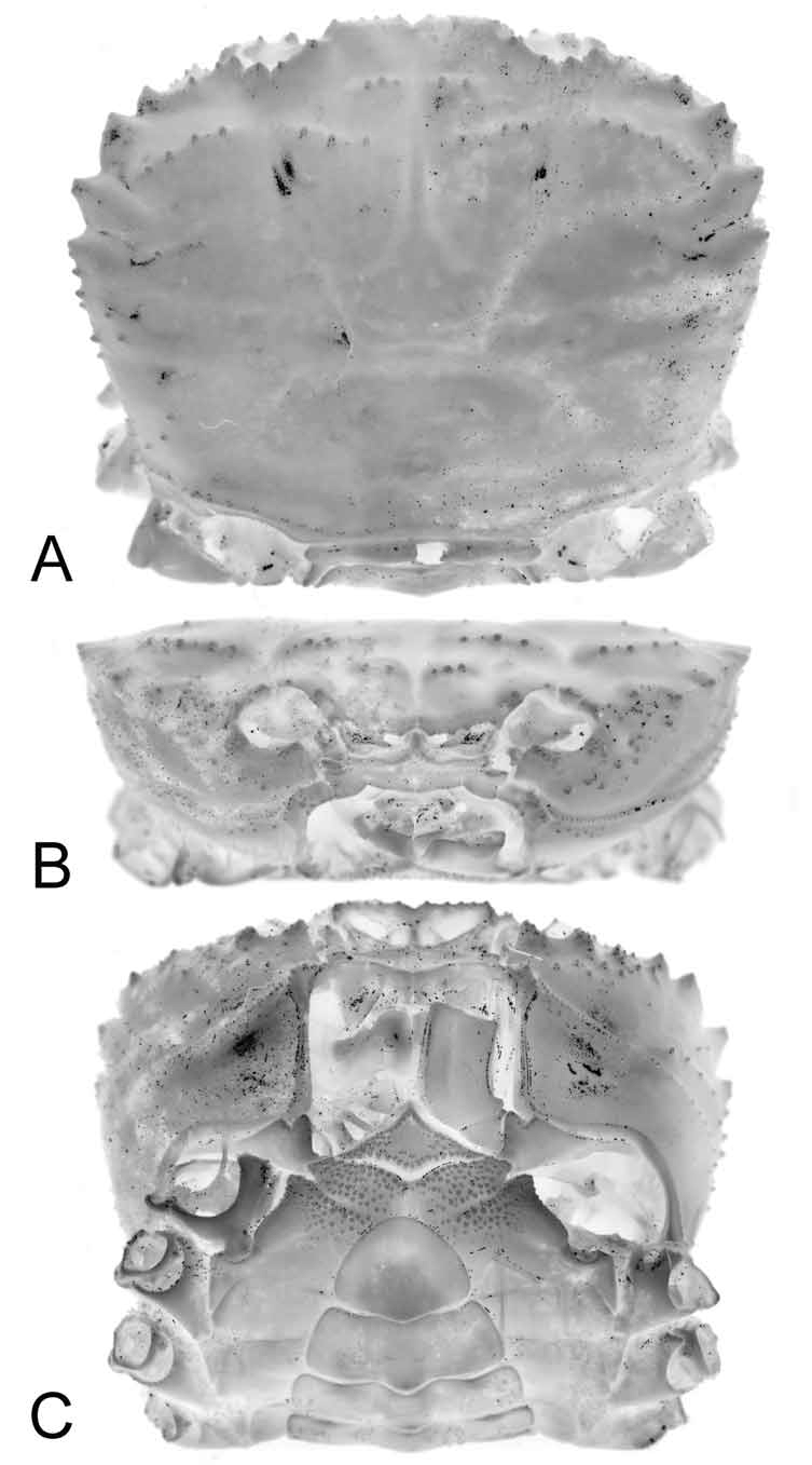
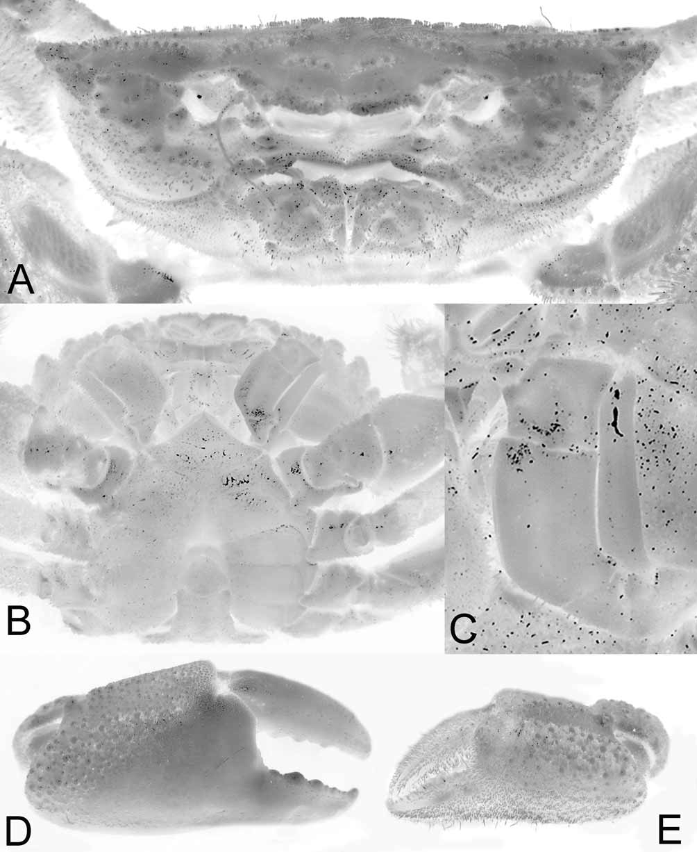
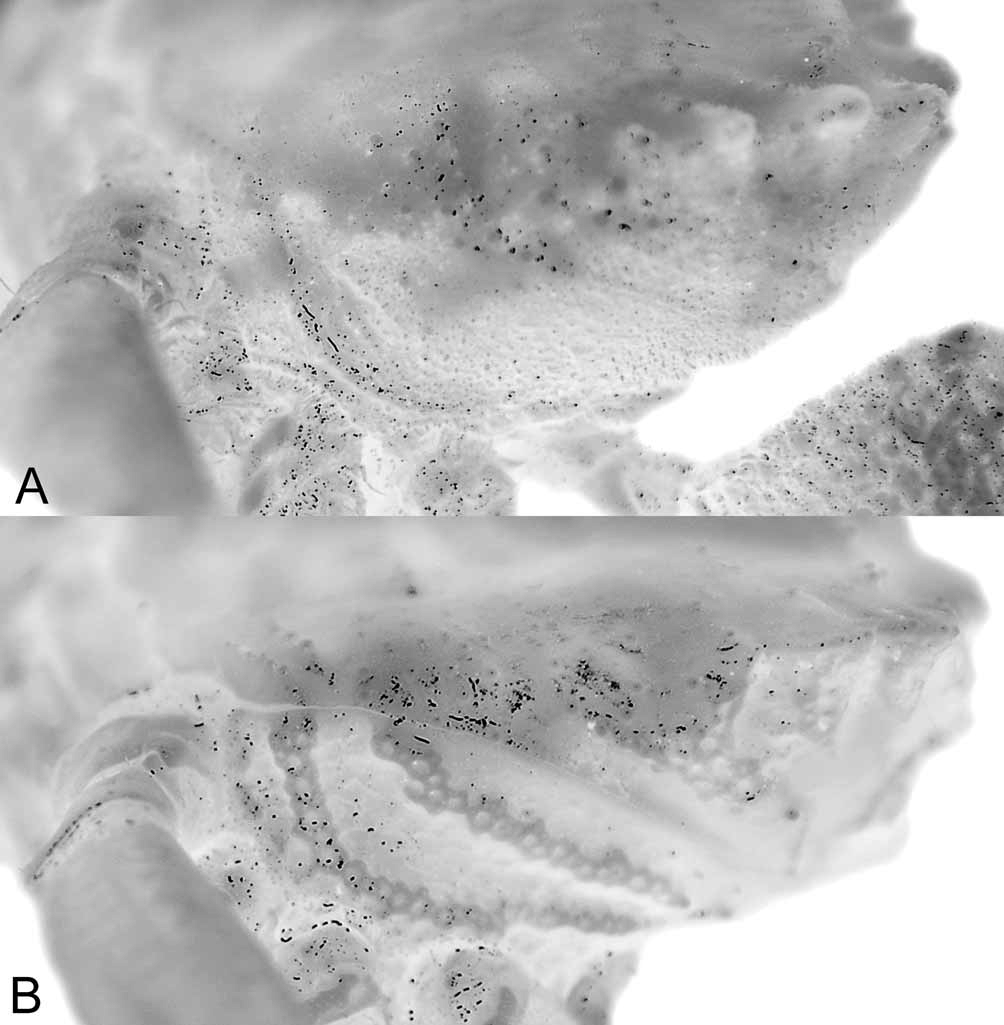
Diagnosis. Carapace distinctly broader than long ( Figs. 1 View FIGURE 1 A, 2, 4A, 8A, B); carapace, pereiopods covered with dense, short, soft pubescence obscuring most surfaces, teeth; setae usually arranged in distinct patterns ( Fig. View FIGURE 2
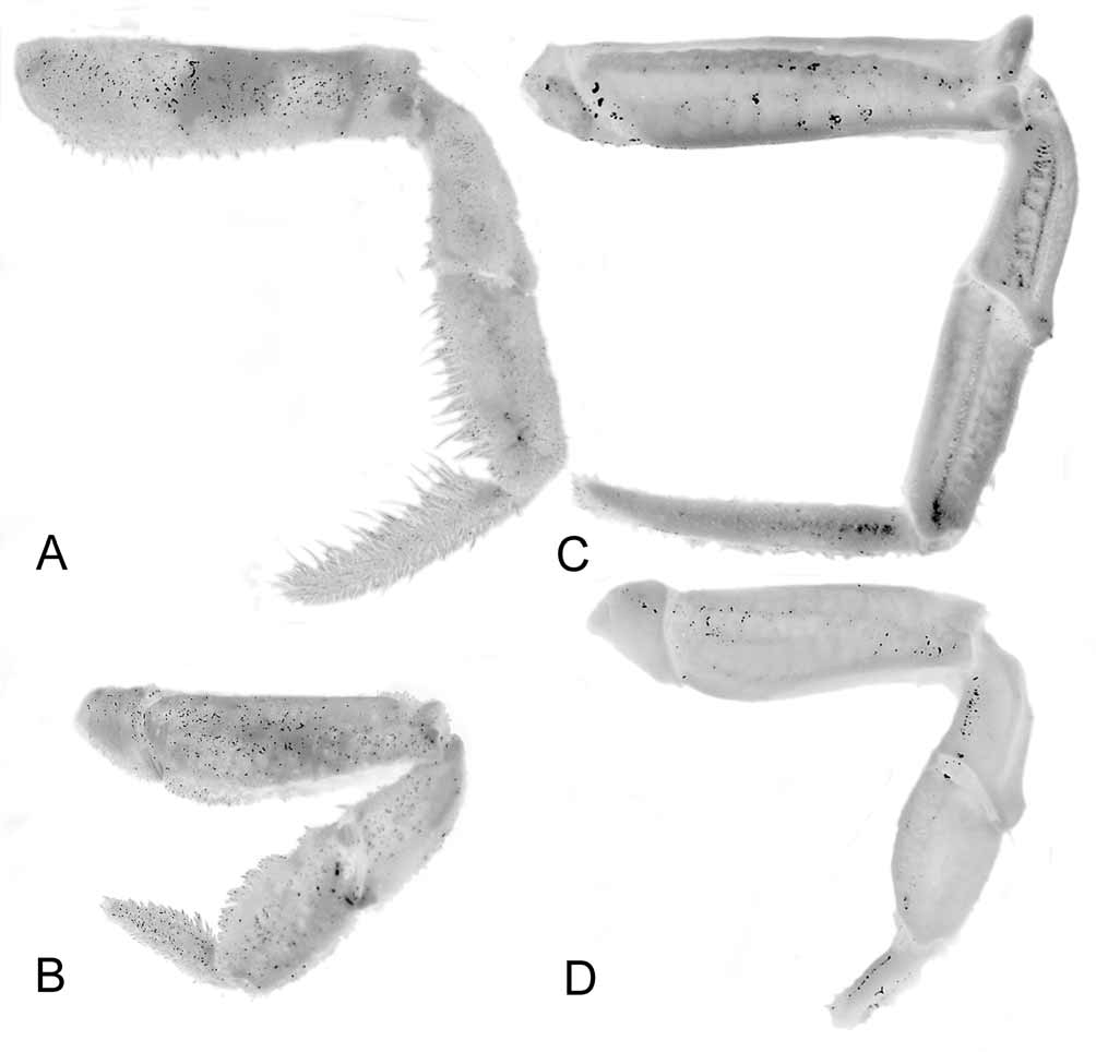
2A); dorsal carapace regions gently convex ( Figs. 1 View FIGURE 1 B, 3A); epigastric cristae distinct, lined with granules, separated by Y-shaped groove; mesogastric cristae distinct, lined with small granules, posterior to, not contiguous with epigastric cristae, gently sloping towards centre of carapace; postorbital cristae distinct, granular, not contiguous with mesogastric cristae, separated by distinct cervical groove, reaching only to near base of first anterolateral tooth ( Figs. 1 View FIGURE 1 A, 2, 8A, B); postorbital region not prominently sunken ( Fig. 1 View FIGURE 1 A, B, 2, 3A, 4A, 8A, B); posterolateral, posterior carapace regions with scattered granules but without clear transverse submarginal grooves ( Figs. 1 View FIGURE 1 A, 2, 8A, B); subhepatic region with distinct tubercles ( Figs. 1 View FIGURE 1 B, C, 3A, B); subbranchial region with scattered, indistinct groups of low granules, without obvious ridges or grooves ( Fig. 9 View FIGURE 9 A). Frontal margin with 2 truncate lobes separated by small, V-shaped cleft; separated from supraorbital margin by small fissure or cleft; inner angle of supraorbital margin low, well behind frontal margin, with median fissure dividing margin into 2 parts, the outer part longer ( Figs. 1 View FIGURE 1 A, 2, 4A, B). Suborbital margin concave with distinct inner, outer teeth, not cristate ( Figs. 1 View FIGURE 1 B, C, 3A, B, 4B). Surfaces of third maxilliped with pits, granules, not eroded; anteroexternal angle auriculiform ( Fig. 3 View FIGURE 3 A, C). External orbital tooth truncate, margin uneven to sinuous, usually divided into approximately 2 parts by cleft or fissure; first, second anterolateral teeth subequal with first tooth sometimes subtruncate in larger specimens, each with a low median longitudinal ridge; third anterolateral tooth small ( Figs. 1 View FIGURE 1 A, B, 2, 3A, 4A, B). Chelipeds in adult males, females distinctly unequal ( Figs. 2 View FIGURE 2 , 3 View FIGURE 3 D, E); dorsal margin of chela rounded without ridges although shallow subdorsal or dorsal longitudinal groove may be present; carpus, merus, varying parts of chela covered with scattered granules but without ridges, pits or eroded depressions; merus with low subdistal tooth; inner distal tooth of carpus low, dentiform; dense setae evenly covering, obscuring almost all surfaces except for tips of larger granules, outer, inner surfaces of palm, fingers ( Figs. 2 View FIGURE 2 , 3 View FIGURE 3 D, E); outer surfaces of larger palms in both sexes almost completely glabrous ( Fig. 3 View FIGURE 3 D, E). Surfaces of ambulatory leg articles without spines, ridges, pits or depressions; dorsal margins of merus, carpus, propodus rounded, without spines or crests; merus of first to third legs with low subdistal tooth; dense setae evenly covering, obscuring almost all surfaces ( Figs. 2 View FIGURE 2 , 10 View FIGURE 10 A, B). Surface of anterior thoracic sternum, outer surfaces of abdomen with scattered granules but without depressions ( Figs. 1 View FIGURE 1 C, 3B); sternites 1, 2 completely fused without trace of suture; s2/3 complete; s3/ 4 medially interrupted; s4/5, s5/6, s6/7 appears medially interrupted; s7/8 complete; longitudinal median groove present from sternites 6–8; male press button distinct, positioned on posterior margin of sternite 5, adjacent to s5/6; all abdominal somites, telson mobile. G1 relatively short, stout, almost straight, tip prominently dilated, appearing flared ( Fig. 4 View FIGURE 4 C, D). G2 about half length of G1 ( Fig. 4 View FIGURE 4 E, F).
Remarks. Although Planopilumnus has long been synoymised with Rathbunaria Ward, 1933 , the present study shows that the two are distinct (see remarks below for Rathbunaria ). Actually, Planopilumnus is closest to Platychelonion Crosnier & Guinot, 1969 (type and only species Platychelonion planissimum Crosnier & Guinot, 1969 ), originally described from relatively shallow waters off Congo, West Africa (see also Manning & Holthuis 1981). Although the carapace of Platychelonion was described as being only weakly pubescent, in contrast to Planopilumnus , their carapace features and pereiopodal characters are very similar. They share the same carapace form, with an almost identical frontal and anterolateral armature ( Figs. 11 View FIGURE 11 A, B, 12A, B), although the dorsal surface of Platychelonion is distinctly flatter ( Figs. 11 View FIGURE 11 A, B, 12A) than that of Platypilumnnus ( Figs. 1 View FIGURE 1 B, 3A). In addition to the degree of pubescence on the carapace and ambulatory legs (much more extensive in Planopilumnus ), other differences that separate Platychelonion from Planopilumnus include the junction of the antero- and posterolateral margins being distinct, the third anterolateral tooth been large and distinct, with the posterolateral margin lined only with small granules ( Figs. 11 View FIGURE 11 A, 12A) (third anterolateral tooth small, followed by progressively smaller granules towards posterolateral margin in Planopilumnus , Figs. 1 View FIGURE 1 A, 2, 4A, 8A, B), relatively weaker subhepatic tubercles ( Figs. 11 View FIGURE 11 B, C, 12B) (distinct in Planopilumnus , Figs. 1 View FIGURE 1 B, C, 3A, B), the dorsal margins of the ambulatory meri are armed with small spines ( Fig. 12 View FIGURE 12 E) (smooth or almost so in Planopilumnus , Figs. 2 View FIGURE 2 B, 10A, B), and the G1 is proportionately much stouter with the distal part tapering ( Fig. 12 View FIGURE 12 G–I) (relatively more slender with the tip flared in Planopilumnus , Fig. 4 View FIGURE 4 C, D).


No known copyright restrictions apply. See Agosti, D., Egloff, W., 2009. Taxonomic information exchange and copyright: the Plazi approach. BMC Research Notes 2009, 2:53 for further explanation.
|
Kingdom |
|
|
Phylum |
|
|
Class |
|
|
Order |
|
|
InfraOrder |
Brachyura |
|
Family |
Planopilumnus Balss, 1933
| Ng, Peter K. L. 2010 |
Planopilumnus
| Balss 1933: 39 |