
Chimarra levuensis, Johanson, Kjell Arne & Oláh, János, 2012
|
publication ID |
https://doi.org/10.5281/zenodo.210736 |
|
DOI |
https://doi.org/10.5281/zenodo.5664518 |
|
persistent identifier |
https://treatment.plazi.org/id/9F3E87DD-561D-FFE2-E89A-FC50FE50F919 |
|
treatment provided by |
Plazi |
|
scientific name |
Chimarra levuensis |
| status |
sp. nov. |
Chimarra levuensis , new species
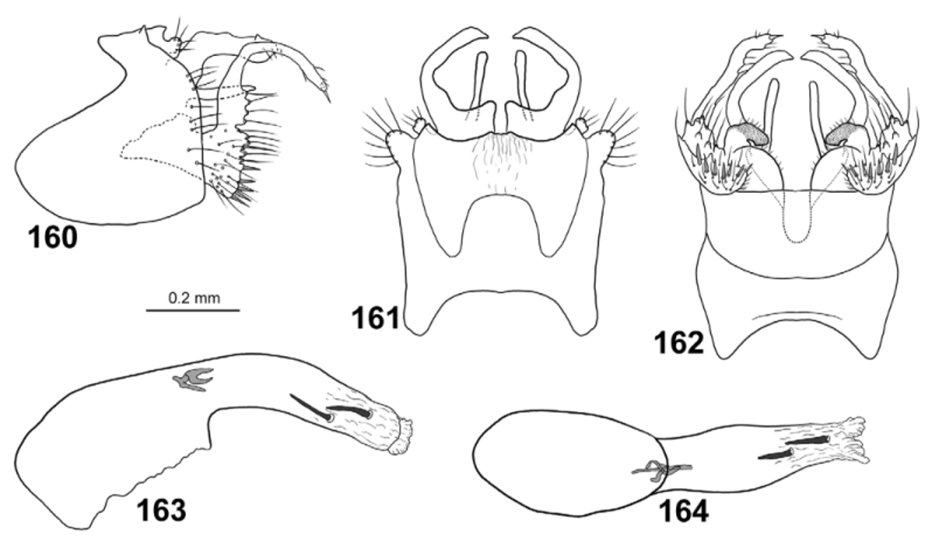
Figs. 30 View FIGURES 30 – 31 , 160–164 View FIGURES 160 – 164
Chimarra levuensis resembles C. psychodida , C. malickyi , and C. tipulida in having gonopods each with both a slender, strongly produced dorsal branch, and a shorter, plate-like ventral lobe. It is distinguished from all these species by the more rounded anteroventral margin of segment IX in lateral view, the shorter basoventral region of each gonopod, the presence of an apical megaseta on the dorsal branch of each gonopod, each lateral branch of tergum X being divided into dorsal and ventral branches, and the strongly mesad curved dorsolateral branch of each lateral branch of tergum X.
Male. Body pale brown, dorsal part of meso- and metathorax brown. Large dark area between lateral and anterior ocelli. Foreleg anterior claw as long as foreleg spur.
Wings ( Fig. 30 View FIGURES 30 – 31 ). Forewings 4.7 mm (n=1), brown; relatively broad, ratio of length to breadth 3.5; R1 nearly straight before crossvein r; radial sector not produced anterad immediately before discoidal cell; discoidal cell originating at mid-length of wing, 3x longer than wide; median cell as long as discoidal cell; crossvein r situated at base of fork I; fork I originating before crossvein s at distance equal to 1/2 length of crossvein s; nygma located near base of fork II; fork III 1 /7th as long as wing; fork V slightly longer than fork I, shorter than fork II; Cu2 well-separated from A at wing margin. Hind wings 3.9 mm (n=1), brown; broad, ratio of length to breadth about 3; margin slightly incurved at arculus, where Cu1 and Cu2 fused with margin; fork III as long as discoidal cell and1/10th as long as wing; fork V as long as fork I; 1A+2A 3x longer than 1A.
Male genitalia ( Figs. 160–164 View FIGURES 160 – 164 ). Segment IX as long as high; anterior plate nearly oval; posterior 1/2 of segment expanded dorsally and anteriorly parallelogram shaped plate; each anterodorsal margin deeply concave in lateral view; each ventral margin uniformly convex, without incision at vertical apodeme; each posterior margin nearly straight, curved anterad below cercus. In dorsal view segment IX with short, wide anterior lobes; in dorsal view anterodorsal margin forming deep, narrow, U-shaped incision without anterad-orienting processes on posteromesal margins. In ventral view segment IX nearly quadrangular, except incised at transverse apodeme; anterior margin widely concave; posterior margin straight; posterior margin without central projection. Tergum X divided into dorsal and ventral branches, surrounding phallic apparatus. In lateral view, dorsal branches parallel-sided along their length, angled nearly 90° posterad at mid-length; small dorsal process present immediately before apex; in dorsal view each lateral branch broad at base, slender distally, bent mesad at mid-length. Pair of ventral branches of tergum X smooth, shorter than dorsal branches, oriented posterad along their length. Sensillae on tergum X not visible. Cerci located near dorsal margin of segment IX and tergum X; slightly curved dorsad in lateral view and oriented posterolaterad in dorsal view; covered by long setae. Gonopods slender along their length, about as long as segment IX, 3-branched. Each dorsal branch clearly exceeding tergum X in lateral view; uniformly slender except with posterad produced setal tubercles basal posterior margin; bending posterad at mid-length before curved ventrad; in ventral view curved mesad; mesal megasetae absent. Ventral branch of each gonopod nearly triangular in lateral view, with undulating posterior margin and smooth ventral margin; each with mesal margin strongly irregular in ventral view. Mesal branches darkly pigmented, long, curved mesad in ventral view. Phallic apparatus slightly longer than rest of genitalia, curved ventrad along its length; phallotheca, in lateral view, with anterior part 2x thicker than posterior part; in ventral view anterior part about 1.5x wider than posterior part; apicoventral spine absent on phallotheca; small phallotremal sclerite in phallic apparatus complex constitutes of slender structures visible in lateral and ventral view; 2 short, nearly black endothecal spines present.
Female. Unknown.
Holotype male: VITI LEVU: Naitasiri Prov., 4 km WSW Colo-i-Suva Vlg., Mt. Nakobalevu, Malaise trap, 24.iv–12.v.2004, 18.055°S, 178.424°E, 372 m, leg E. Schlinger & M. Tokota’a [loc#11] [ FNIC].
Paratypes: Same data as holotype [loc#11] — 1 male [ BPBM]. Same data as holotype, except: 9–30.v.2003 [loc#11] — 1 male [ NHRS, DNA voucher IM9].
Etymology: Levuensis , named after Viti Levu, the type locality of the species.
Distribution: Viti Levu.

No known copyright restrictions apply. See Agosti, D., Egloff, W., 2009. Taxonomic information exchange and copyright: the Plazi approach. BMC Research Notes 2009, 2:53 for further explanation.
|
Kingdom |
|
|
Phylum |
|
|
Class |
|
|
Order |
|
|
Family |
|
|
Genus |