
Chimarra vanuensis, Johanson, Kjell Arne & Oláh, János, 2012
|
publication ID |
https://doi.org/10.5281/zenodo.210736 |
|
DOI |
https://doi.org/10.5281/zenodo.5664468 |
|
persistent identifier |
https://treatment.plazi.org/id/9F3E87DD-5629-FFD8-E89A-F9C8FB8BF929 |
|
treatment provided by |
Plazi |
|
scientific name |
Chimarra vanuensis |
| status |
sp. nov. |
Chimarra vanuensis , new species
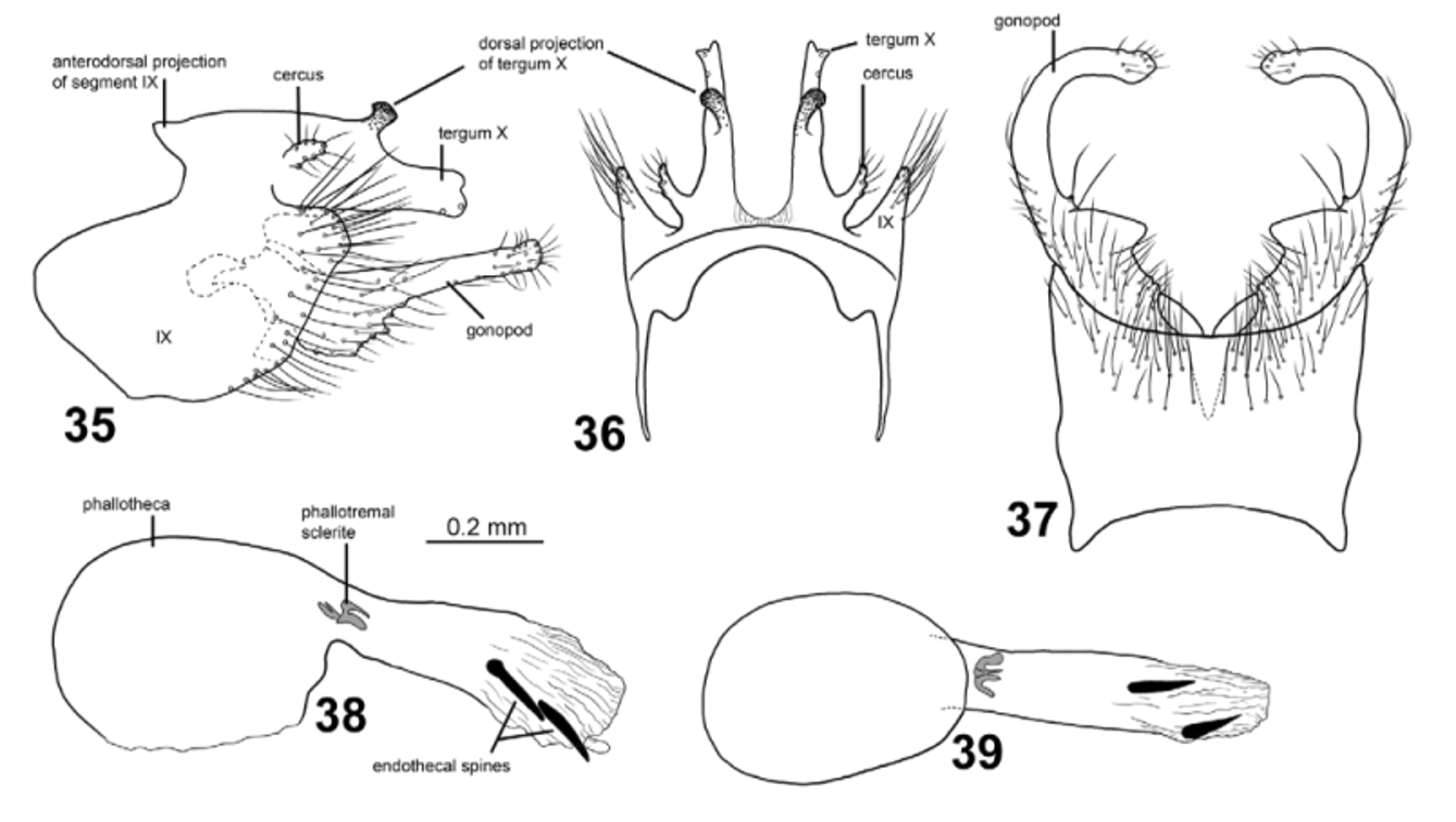
Figs. 6 View FIGURES 6 – 13 , 35–39 View FIGURES 35 – 39
This species can be separated from all other Fijian Chimarra by the presence of a large, pale hyaline spot at midlength in the forewings, located immediately anterior to the posterior wing margin. The genitalia, particularly tergum X, resembles those of C. devoensis and C. lavensis in the presence of a strongly dorsad-oriented dorsal branch. In C. vanuensis the ventral branch of tergum X is much longer than the dorsal branch, while in C. lavensis the ventral branch is shorter than the dorsal branch. The gonopods of C. vanuensis are shorter than segment IX, while in C. devoensis and C. lavensis the gonopods are longer than segment IX. The phallic apparatus of C. vanuensis has 2 endothecal spines, while those of C. devoensis and C. lavensis each have 4 endothecal spines.
Male. Head and thorax brown, dorsal part of meso- and metathorax dark brown. Area between ocelli nearly black. Foreleg anterior claw as long as foreleg spur.
Wings ( Fig. 6 View FIGURES 6 – 13 ). Forewings 5.4–5.6 mm (n=2), brown, with large, nearly round, pale hyaline spot between Cu1 and end of Cu2, immediately above posterior wing margin. Forewings broad, ratio of length to breadth 3.0; R1 slightly curved before crossvein r; radial sector slightly produced anterad immediately before discoidal cell; discoidal cell originating at mid-length of wing, about 3x longer than wide; median cell slightly longer than discoidal cell; crossvein r originating from base of fork I; fork I originating before crossvein s at distance equal to length of crossvein s; nygma located near base of fork II; fork III nearly 1/4th as long as wing; fork V slightly shorter than fork II; Cu2 ending in wing margin close to A. Hind wings 4.3–4.5 mm (n=2), brown, without pale, hyaline spot; ratio of length to breadth 2.9; margin weakly incurved at arculus, where Cu1b and Cu2 fused with margin; fork I originating slightly before anterodistal corner of discoidal cell; fork III 1.5x longer than discoidal cell and 1/6th as long as wing; fork V as long as fork I; 1A+2A about 3x as long as 1A.
Male genitalia ( Figs. 35–39 View FIGURES 35 – 39 ). Segment IX nearly as high as long in lateral view; each side anteroventrally produced into large, hyperbolic, anterad oriented plate; anterodorsal projection small, sharply triangular and projecting anterad in lateral view, rounded triangular in dorsal view; anterodorsal margin narrowly and deeply concave; ventral margin irregularly convex; venter not produced anterad in lateral view; posterolateral margins each produced posterad into, broad, rounded triangular plate; segment IX ventrally with setae restricted to posterior 1/2 ( Fig. 37 View FIGURES 35 – 39 ). Dorsal part of segment IX short in lateral view; anterior margin elliptically concave in dorsal view. In ventral view segment IX with almost parallel lateral margins, slightly expanded at mid-length; anterior and posterior margins both shallowly concave; without central projection posteriorly. Tergum X long, divided at base into pair of lateral, nearly parallel branches; in lateral view each lateral branch with dorsad and laterad oriented, dark, apically dilated dorsal projection, and very long plate-like, nearly straight ventral branch with 2 apical sensillae. In dorsal and ventral views, dorsal projections curved slightly laterad, ventral processes narrow, each with slightly bifid apex. Cerci large, digitate, located at mid-height basally on tergum X; tube-shaped, oriented posterad; covered by long setae. Gonopods shorter than segment IX; with basal 1/3rd broad and distal 2/3rds narrow in lateral view; distal 2/3rds nearly parallel-sided; without mesal process, but with mesad projected mesal margin in ventral view. Anterodorsal margin of each gonopod straight, smooth; posteroventral margin of basal 1/2 with expanded setal bases, distal 1/2 with normal setal bases; ventral margin convex at basal 1/3rd; apex without megasetae. In ventral view, gonopods slightly broader in basal 1/3rd than distal 2/3rds, without strongly undulating margins; gonopods strongly curved mesad from mid-length. Phallic apparatus about as long as rest of genitalia, straight along its length; phallotheca, in lateral view nearly 3x thicker than posterior part; in ventral view about 2.5x thicker than posterior part; apicoventral spine absent; phallotremal sclerite small, complex in lateral view; in ventral view forming single anterior plate with 4 posterad directed rays; 2, nearly black, short, variously directed endothecal spines present, about as long as diameter of narrowest part of phallotheca; endothecal spicules absent.
Female. Unknown.
Holotype male: VANUA LEVU : Macuata Prov., 0.6 km S Rokosalase Village, Malaise trap in forest, 23.iv–8.v.2004, 16.5333°S, 179.0181°E, 180 m, leg. E. Schlinger & M. Tokota’a [loc#26] [ FNIC].
Paratype: VANUA LEVU : 0.5 km S Rokosalase Village, Malaise trap 3, 27.xii.2004 – 5.i.2005, 16.532°S, 179.010°E, 97 m, leg. I. Sakealevu [loc#29] — 1 male [ NHRS].
Distribution: Vanua Levu.
Etymology: Vanuensis , after Vanua Levu , the type locality of the species.

| NHRS |
Swedish Museum of Natural History, Entomology Collections |
No known copyright restrictions apply. See Agosti, D., Egloff, W., 2009. Taxonomic information exchange and copyright: the Plazi approach. BMC Research Notes 2009, 2:53 for further explanation.
|
Kingdom |
|
|
Phylum |
|
|
Class |
|
|
Order |
|
|
Family |
|
|
Genus |