
Chimarra nathani, Johanson, Kjell Arne & Oláh, János, 2012
|
publication ID |
https://doi.org/10.5281/zenodo.210736 |
|
DOI |
https://doi.org/10.5281/zenodo.5664478 |
|
persistent identifier |
https://treatment.plazi.org/id/9F3E87DD-563D-FFC3-E89A-F9B5FE57FEE6 |
|
treatment provided by |
Plazi |
|
scientific name |
Chimarra nathani |
| status |
sp. nov. |
Chimarra nathani , new species
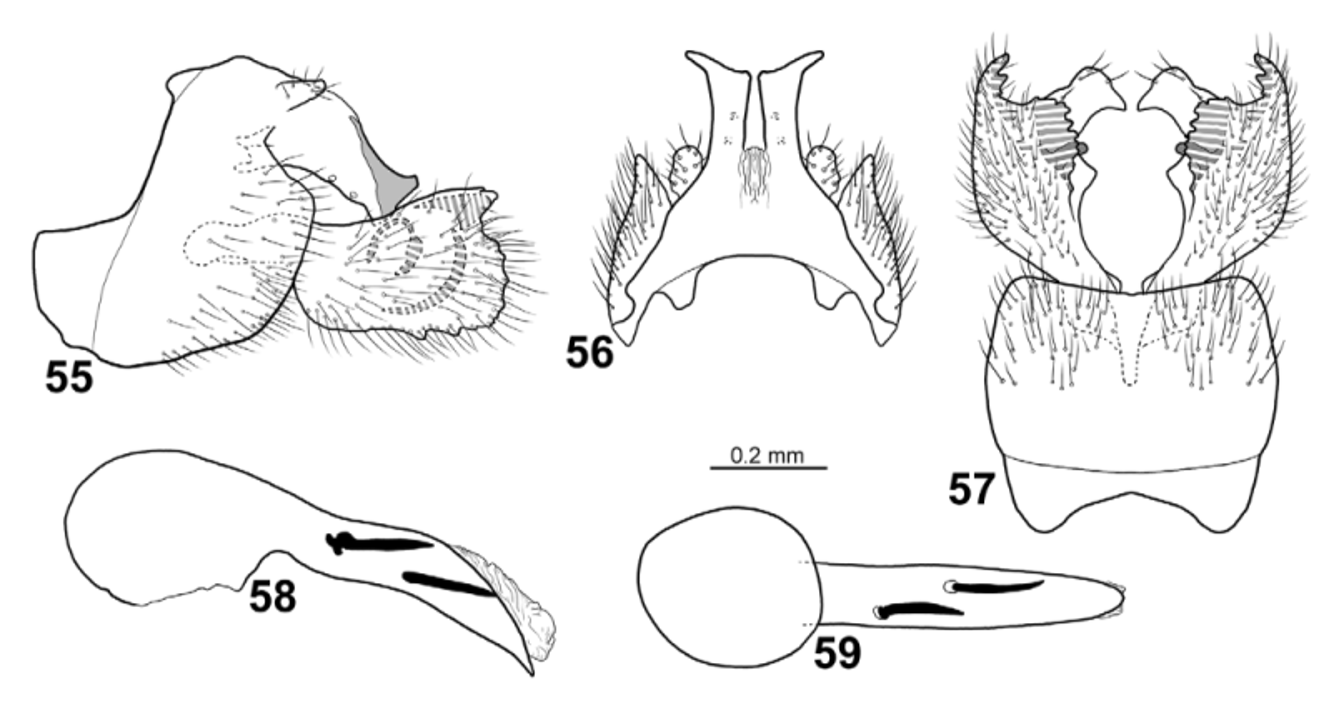
Figs. 10 View FIGURES 6 – 13 , 55–59 View FIGURES 55 – 59
Chimarra nathani has a large pale, hyaline spot on both wing pairs, like C. signata , C. schlingeri , C. braueri and C. vitiensis . It is easily distinguished from those species by the genitalia of the male. The gonopods each have a dorsal branch that appears as a small but distinct, triangular projection in lateral view, but in dorsal and ventral views this branch is seen to form a large, rectangular plate below the phallus.
Male. Head and thorax pale yellowish-brown. Area between ocelli yellowish-brown. Foreleg anterior claw as long as foreleg spur.
Wings ( Fig. 10 View FIGURES 6 – 13 ). Forewings 6.9 mm (n=1), brown; large pale, hyaline, nearly circular spot occupying central part of wing, including median cell and basal third of discoidal cell. Forewings broad, ratio of length to breadth 3.3; R1 nearly straight before crossvein r; radial sector strongly produced anterad immediately before discoidal cell; discoidal cell originating at mid-length of wing, nearly 3x longer than wide; median cell as long as discoidal cell; crossvein r fusing with basis of fork I; fork I originating before crossvein s at distance nearly equal to length of crossvein s; nygma located near base of fork II; fork III 1 /6th as long as wing; fork V about as long as fork II; Cu2 ending in wing margin close to A. Hind wings 5.6 mm (n=1), brown, with large, oval, pale hyaline spot centrally on anterior 1/2 of wings, occupying basal 1/2 of discoidal cell; broad, ratio of length to breadth 3.2; margin slightly incurved at arculus, where Cu1 and Cu2 fused with margin; fork III as long as discoidal cell and 1/9 as long as wing; fork V about as long as fork I; 1A+2A nearly as long as 1A.
Male genitalia ( Figs. 55–59 View FIGURES 55 – 59 ). Segment IX slightly longer than high; anterodorsal margins strongly concave in lateral view; ventral margins uniformly convex; each posterior margin produced posterad into narrow plate well below each cercus; ventral side of posterior 1/2 of segment IX densely covered by setae ( Fig. 57 View FIGURES 55 – 59 ). In dorsal view with pointed anterior lobes; anterodorsal margin with wide, shallow incision in dorsal view. In ventral view segment IX with concave anterior margin and very slightly concave posterior margin; posterior margin without central projection. Tergum X simple; slightly tapering till distal 3/ 4 in lateral view ( Fig. 55 View FIGURES 55 – 59 ); distal1/4 expanded dorsad into nearly triangular process; in dorsal view divided at mid-length into 2 parallel lateral branches ( Fig. 56 View FIGURES 55 – 59 ); each lateral branch nearly parallel-sided in dorsal and ventral view, apically nearly truncate, with strongly laterad-producing apicolateral corner; pair of sensillae located at mid-length of each lateral branch. Cerci slender in lateral view, located dorsally on segment IX and oriented slightly dorsad in lateral view and oriented posterad in dorsal view; covered by long setae. Gonopods shorter than segment IX, nearly quadrangular in lateral view; anterodorsal margin slightly concave; ventral margin nearly straight, undulating; dorsal branch forming strong, triangular process in lateral view, in dorsal and ventral view forming long, broad, nearly horizontal plates oriented postero mesad; ventral branch broad, posteriorly rounded in lateral view; in ventral view produced into nearly rectangular, darkly pigmented mesal plates; each mesal plate margin undulating. Mesal process situated centrally on gonopods, strongly sclerotized, slightly curved mesad. Phallic apparatus about as long as rest of genitalia; phallotheca, in lateral and ventral view, with anterior part 2x thicker than posterior part; apicoventral spine absent; phallotremal sclerite not observed; pair of nearly black, posterad directed, sub-equally large endothecal spines present, slightly longer than diameter of narrowest part of phallotheca.
Female. Unknown.
Holotype male: VITI LEVU: Naitasiri Prov., 3.3 km N Veisari, logging rd. to Waivudava, Malaise trap, 8–31.iii.2003, 18.0592°S, 178.367°E, 300 m, leg. M. Tokota’a [loc#20] [ FNIC].
Paratype: VITI LEVU: Naitasiri Prov., 4 km WSW Colo-i-Suva Vlg., Mt. Nakobalevu, Malaise trap, 24.iv–12.v.2004, 18.055°S, 178.424°E, 372 m, leg E. Schlinger & M. Tokota’a [loc#11] — 1 male [ NHRS, DNA voucher IL 9].
Etymology: Nathani , named after Nathan Banks who described several Chimarra from Fiji. Distribution: Viti Levu.

No known copyright restrictions apply. See Agosti, D., Egloff, W., 2009. Taxonomic information exchange and copyright: the Plazi approach. BMC Research Notes 2009, 2:53 for further explanation.
|
Kingdom |
|
|
Phylum |
|
|
Class |
|
|
Order |
|
|
Family |
|
|
Genus |