
Dalpada smaragdina ( Walker, 1868 )
|
publication ID |
https://doi.org/10.5281/zenodo.188737 |
|
DOI |
https://doi.org/10.5281/zenodo.6224214 |
|
persistent identifier |
https://treatment.plazi.org/id/9F64881E-FFF6-1017-FF24-FF0CFD53F21C |
|
treatment provided by |
Plazi |
|
scientific name |
Dalpada smaragdina ( Walker, 1868 ) |
| status |
|
Dalpada smaragdina ( Walker, 1868)
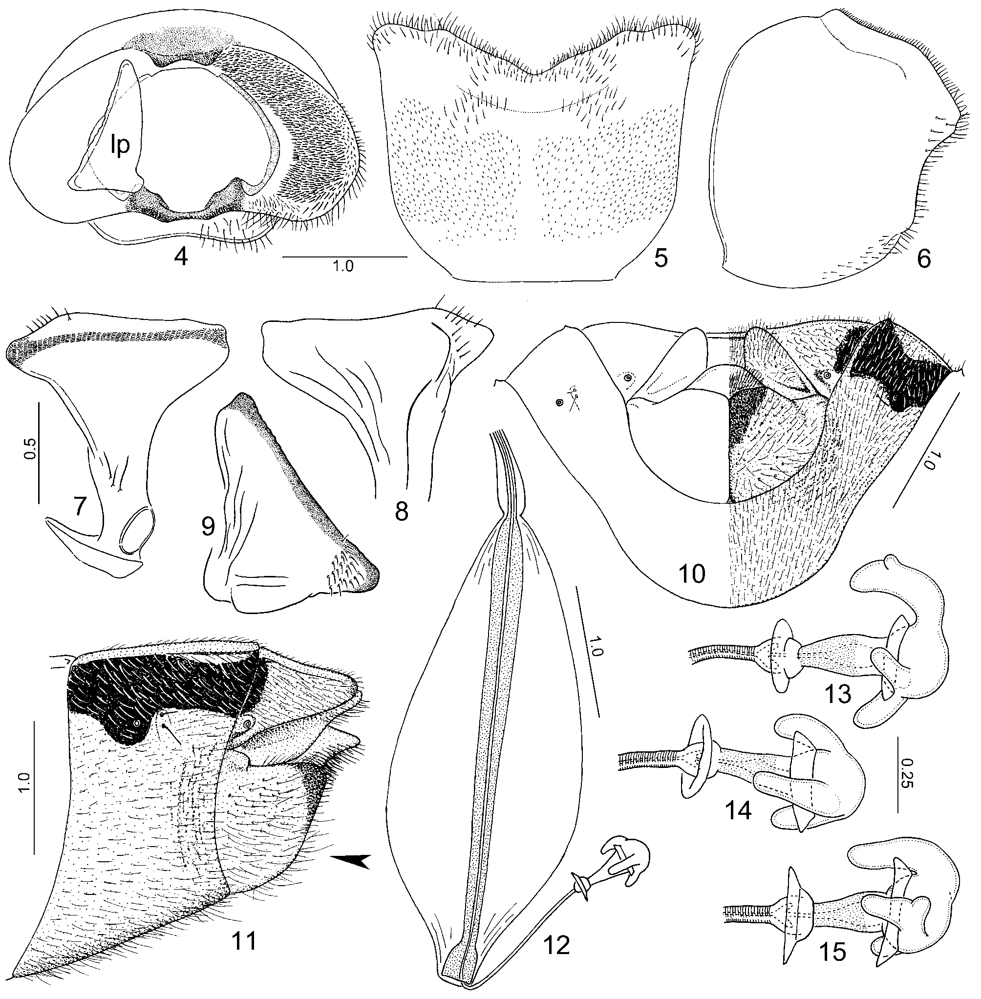
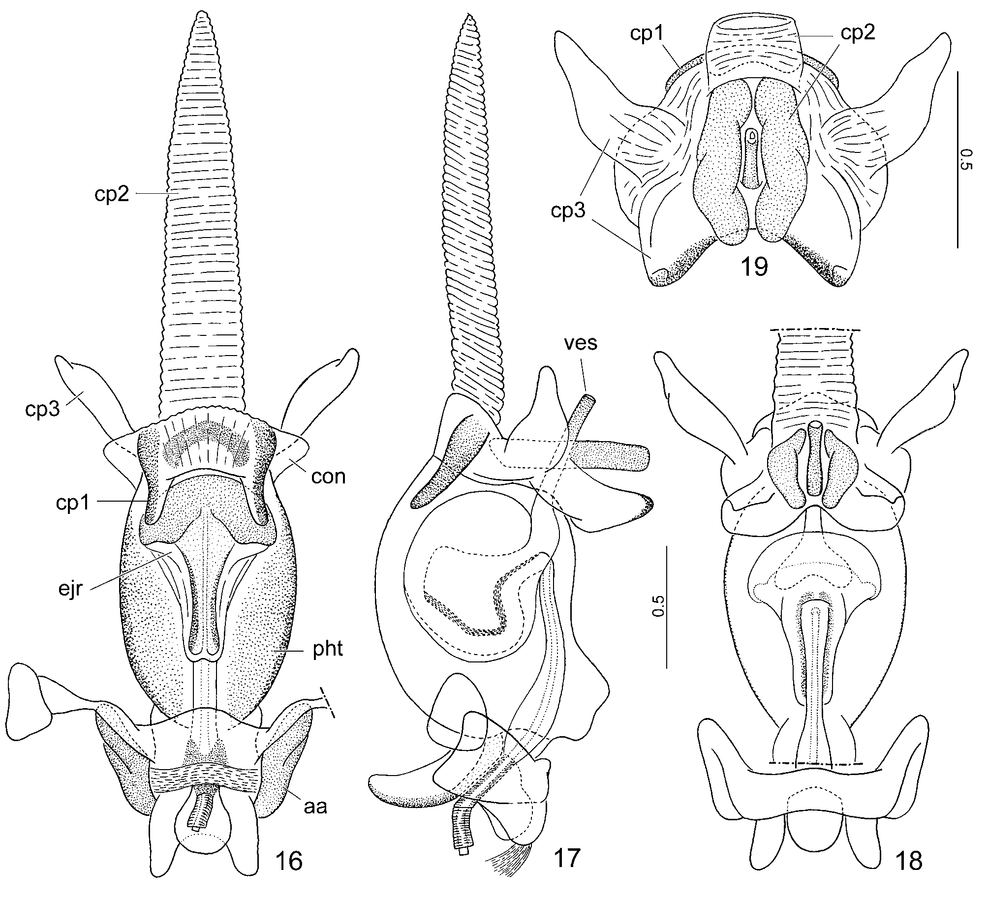
Figs. 4–19 View FIGURES 4 – 15 View FIGURES 16 – 19 .
Udana smaragdina Walker, 1868: 549 . Syntype (s) (3): „ Formosa ” [= Taiwan]; MVMA. = Amasenoides virescens Shiraki, 1913: 217. Syntype (s): „Taihoku” [= Taipei]; depository unknown. Synonymized by Esaki 1926: 147.
Main references. Shiraki 1913: 217 (as Amasenoides virescens, original description); Esaki 1926: 146 (synonymy, records from Taiwan); Yang 1962: 95 (redescription, colour habitus drawing); Hsiao & Zheng 1977: 118 (redescription, figure of head, pronotum, and scutellum, photo, listed from Taiwan); Rider 2006: 306 (catalogue).
Type material examined. SYNTYPE (3): „ TYPE ” [red square, printed]; „Udana \ smaragdina \ Formosa ” [Walker’s handwriting]; „ HOLOTYPE \ T-20529 \ Udana \ smaragdina \ Walker” [red square, printed + handwritten]; MVMA. Pinned; left antennal segments IIa–IV, right antenna, tarsus of left fore leg, tibia, and tarsus of right mid leg, tarsal segments II–III of right hind leg, right fore and left hind legs missing.
Other specimens examined. TAIWAN: Taichung County, Guguan, Taidian alley, 26. VI. 2004, leg. J.F. Tsai (1 3, NCHU). Male genitalia dissected and figured ( Figs. 4–9 View FIGURES 4 – 15 , 16–19 View FIGURES 16 – 19 ). „Takao” [= Kaohsiung], 1907, leg. H. Sauter (2 ♀, HNHM); „Mt. Hoozan” [= Fengshan], V. 1910, leg. H. Sauter (1 ♀, HNHM). Female genitalia dissected and figured ( Figs. 10–15 View FIGURES 4 – 15 ).
Redescription. Male genitalia. Pygophore ( Figs. 4–6 View FIGURES 4 – 15 ). Ventral rim deeply excised ( Fig. 5 View FIGURES 4 – 15 ), with an oblique depression below ventral rim ( Fig. 6 View FIGURES 4 – 15 ); genital opening large and rounded, with a pair of small ventrolateral excisions for reception of the parameres, with a rigid, scoop-like median sclerite (subgenital plate) ventrally; dorsal rim with median bifid projection; infolding of lateral rim flattened, densely setose, lateral and ventral rims with scattered hairs. Parameres ( Figs. 7–9 View FIGURES 4 – 15 ) symmetrical, apical part widely triangular, apical margin flattened. Phallus ( Figs. 16–19 View FIGURES 16 – 19 ). Phallotheca barrel-like, strongly sclerotized (sclerotization shown only in Fig. 16 View FIGURES 16 – 19 ), with a distinct ventral tubercle basally; conjunctiva with three sets of processes (cp1–cp3) as follows: cp1: a pair of dorsal, finger-like processes, directed anteriad in inflated condition; cp2: one single long, membaneous, flexible process dorsally, and a pair of rod-like, sclerotized processes ventrally, surrounding the vesica; cp3: a pair of bifurcate, wing-like, membraneous processes laterally, directed anteroventrad, and an elongate, membraneous process directed caudodorsad; ejaculatory reservoir large, occupying more than half of phallic cavity, distal part fan-shaped, its wall strongly sclerotized and pigmented; vesica rod-like, heavily sclerotized. Female genitalia. Posterior margin of abdominal sternite VII with a deep, rounded median excision, VIIIth gonocoxites situated within excision of sternite VII, strongly convex, their medial margin straight, posterior margin submedially convex, sublaterally concave; IXth paratergite impressed at base, rounded apically, its apex slightly surpassing posterior margin of tergite VIII; gonocoxite IX wide, transversal ( Figs. 10, 11 View FIGURES 4 – 15 ). Spermatheca ( Fig. 12 View FIGURES 4 – 15 ) with distimedial part (sclerotized rod) gradually narrowing towards apex, apical part abruptly widened, cup-like; distal part of spermathecal duct running outside of distimedial part straight, about 0.3 times as long as lenght of sclerotized rod; proximal and distal flanges well developed; distal flange joining seminal receptacle; seminal receptacle globose, with 3 projections of diverse shape, some of them also may bear small secondary projections ( Figs. 13–15 View FIGURES 4 – 15 ).
Discussion. Photographs of a male syntype and its labels were sent to us by K. Walker (MVMA) and we confirmed its identity. It is a well-known species easily recognized within Dalpada by its bright green dorsum and the head and pronotum not margined with orange or red. We examined several additional specimens from Taiwan and P.R. China. For facilitating recognition of the species, we present a brief redescription and illustrations of the male and female genitalia.
No known copyright restrictions apply. See Agosti, D., Egloff, W., 2009. Taxonomic information exchange and copyright: the Plazi approach. BMC Research Notes 2009, 2:53 for further explanation.
|
Kingdom |
|
|
Phylum |
|
|
Class |
|
|
Order |
|
|
Family |
|
|
Genus |
Dalpada smaragdina ( Walker, 1868 )
| Tsai, Jing-Fu & Rédei, Dávid 2009 |
smaragdina
| Esaki 1926: 147 |
| Shiraki 1913: 217 |
| Walker 1868: 549 |