
Protocalymene mcallisteri Ross, 1967
|
publication ID |
https://doi.org/ 10.11646/zootaxa.4859.1.1 |
|
publication LSID |
lsid:zoobank.org:pub:B7E3D096-CF3F-4915-BE47-1F256C0294C6 |
|
DOI |
https://doi.org/10.5281/zenodo.4537354 |
|
persistent identifier |
https://treatment.plazi.org/id/9F70020E-EB2E-AB3E-C1D9-26FCA0D734DC |
|
treatment provided by |
Plazi |
|
scientific name |
Protocalymene mcallisteri Ross, 1967 |
| status |
|
Protocalymene mcallisteri Ross, 1967 View in CoL
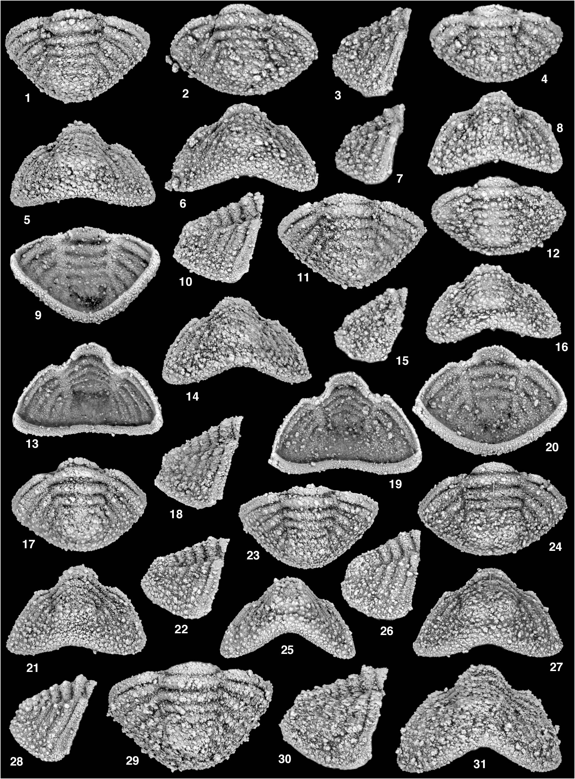
Plates 8–15 View PLATE 8 View PLATE 9 View PLATE 10 View PLATE 11 View PLATE 12 View PLATE 13 View PLATE 14 View PLATE 15
1967 Protocalymene mcallisteri Ross , p. D27, pl. 9, figs 1–28.
1970 Protocalymene mcallisteri Ross ; Ross, p. 92.
1977 Protocalymene macallisteri [sic] Ross; Kobayashi and Hamada, p. 125.
non 1983 Protocalymene mcallisteri Ross ; Andrawis et al. p. 66, fig. 1.
1990 Protocalymene mcallisteri Ross ; Geyer, p. 111.
1999 Protocalymene mcallisteri Ross ; Fortey and Droser, p. 197.
2002 Protocalymene mcallisteri Ross ; Turvey, p. 56.
2003 Protocalymene mcallisteri Ross ; Jell and Adrain, p. 431.
2005 Protocalymene mcallisteri Ross ; Turvey, text-fig. 1.
non 2012 Protocalymene mcallisteri Ross ; Basse, p. 35.
Material. Holotype, cranidium, USNM 145848 View Materials ( Ross, 1967, pl. 9, figs 5–7), and assigned specimens SUI 137444–137520 View Materials , Antelope Valley Formation (Dapingian), north of Pyramid Peak , west flank of Funeral Mountains, Death Valley National Park, California, USA.
Diagnosis. See genus diagnosis.
Description. Cranidial measurements were made on specimens from Pl. 8, figs 1–3, 10, 15, 21, Pl. 9, figs 1- 3, 11, 13, 19, 21, Pl. 10, figs 1–3, 10–12, 19, 20. Cranidium with strong dorsoventral relief, anterior and posterior fixigena strongly flexed downward from occipital lobe in lateral view, and anterior fixigena flexed upward toward glabella in anterior view; sagittal length 50.9% (46.5–55.2) width across posterior border; sagittal length 84.1% (79.2–89.1) width across δ; sagittal length 125.4% (115.2–140.9) width across anterior border furrow; sagittal length 106.1% (97.5–113.8) width (tr.) across γ; width across posterior border 247% (229.2–270.7) width across anterior border furrow; glabella subtrapezoidal in outline, sagittal length 83.4% (71.8–96.4) maximum width (tr.) across L1, moderately strong dorsal inflation, with apex sitting just below that of palpebral lobes in anterior view (e.g., Pl. 8, fig. 7), sculpture of prominent larger tubercles present, especially on median portion of glabella, with background sculpture of coarse granules; L1 clearly developed, bulbous, displaced laterally giving glabella overall bell shape, inner margin not isolated from glabella, deep “C”-shaped furrow composed of S1, axial furrow, and distal portion of LO define external margin of L1; S1 deep, short, directed posteromedially; L2 less well developed than L1, but still prominent, set off anteriorly by extremely short, deep S2; axial furrow narrow and shallow from intersection with posterior cranidial margin to base of L1, deep and laterally deflected around L1, shallower and broad opposite L2 (on most specimens), faint continuation of axial furrow from fossulae anteriorly to intersection with anterior border furrow clearly separates preglabellar field from anterior fixigena, furrows are anteriorly divergent; distance between intersection of axial furrows with posterior cranidial margin 96.9% (91.3–104.0) maximum glabellar width; fossulae deep and pit-like on dorsal surface, ventrally expressed as small raised region; eye ridge poorly developed, oriented obliquely with proximal termination creating an indentation in lateral margin of glabella, defined anteriorly by shallow furrow running from fossulae to γ; preglabellar fur- row incised along entire course, medial portion slightly deeper on some specimens (e.g., Pl. 8, fig. 3), anteriorly arched; preglabellar field relatively long, with sagittal length 12.7% (8.2–16.7) cranidial length, lengthens (exsag.) slightly abaxially, sculpture of densely spaced granules; anterior border furrow moderately short, shallow, lateral portions opposite anterior fixigenae deeper than medial portion opposite anterior glabellar margin, gently anteriorly arched; anterior border in dorsal view longest medially, with sagittal length 14.1% (10.7–17.6) cranidial length (sag.), shorter abaxially with distal portion tapered to a point at intersection with anterior facial suture, anterior margin more strongly anteriorly arched than posterior margin; dorsal inflation weak; anterior border in anterior view strongly dorsally arched (e.g., Pl. 8, fig. 7), thickens nearly equal along entire course, covered with densely spaced granules of medium and small size; lateral profile of preglabellar area forms moderate ventrally sloping surface, slight break in slope at anterior border furrow, with anterior border slightly independently inflated; articulating furrow for rostral plate present ventrally beneath anterior border, broad, forming distinct groove, with posterior and anterior margins defined by distinct inflated rims, anteriorly arched as anterior border; fixigena covered by small to medium densely spaced granules, with few isolated small tubercles (especially on interocular fixigena); interocular fixigena relatively broad; palpebral lobe with length between γ and ε 51.8% (45.0–58.5) glabellar length (sag.), gentle independent inflation, δ situated opposite L2, γ situated opposite point between S2 and fossulae, and ε opposite anterior portion of L1; lobe set off from fixigena by narrow shallow furrow; posterior projection extends far beyond lateral extent of palpebral lobe in dorsal view, strongly downturned from horizontal plane (e.g., Pl. 8, figs 14, 17); narrow inflated ridge continuing from posterior portion of palpebral lobe around anterior margin of posterior fixigena, progressively narrower and weaker toward intersection with posterior border furrow, deep narrow furrow bounds inner margin of ridge opposite point just behind ε; posterior fixigena tapering (exsag.) to a point abaxially, portion distal to fulcrum directed posterolaterally; posterior border furrow distinct, shallow adjacent to axial furrow, lengthening (exsag.) toward fulcrum, deepest at fulcrum, abruptly shallow just before intersection with posterior facial suture so that furrow terminates before reaching intersection; posterior border short (exsag.) adjacent to LO, directed subparallel to transverse axis, gently lengthening abaxially to fulcrum, then significantly longer (exsag.) from fulcrum abaxially, portion distal to fulcrum directed posterolaterally, confluent with genal spine; doublure beneath posterior border subtriangular in outline; genal spine long and stout, directed subparallel to sagittal axis; SO relatively short (sag., exsag.), clearly incised, slightly shallower medially, deepest opposite posterior margin of L1, gently anteriorly arched, merging smoothly around L1 into axial furrow; LO generally lenticular in outline, sagittal length 18.6% (16.7–22.0) cranidial length (sag.), anterior margin anteriorly arched, posterior margin posteriorly arched, with median portion nearly subparallel to transverse axis in some specimens (e.g., Pl. 8, figs 2, 15), median tubercle prominent and elongated, in lateral view tubercle merges smoothly with anterior margin of LO so that an uninterrupted slope is formed, tubercle angled upwards and back in lateral view, in some specimens tubercle appears to be hooked backwards (e.g., Pl. 8, fig. 22); a few small isolated tubercles present along posterior margin of median portion of LO, some specimens exhibit two distinct tubercles flanking larger median tubercle (e.g., Pl. 8, figs 15, 21, Pl. 9, fig. 2), others possess three or four small tubercles situated in an arc along posterior margin (e.g., Pl. 8, fig. 2); LO and median tubercle visible in anterior view sitting just above glabellar apex; doublure present beneath LO broad, terminates before reaching ventral expression of SO, surface largely smooth, with a few scattered very fine granules mostly situated near posterior margin.
Librigena (Pl. 12, figs 10, 11, 13, 14, 16–18, 20–28) with maximum width 57.1% (52.6–63.3) exsagittal length (excluding anterior projection); length of anterior portion of facial suture 36.5% (30.6–42.3) exsagittal length of main body, length of posterior facial suture 44.8% (42.3–50.6) exsagittal length of main body;
visual surface of eye large; eye socle clearly developed, short, with width less than half that of visual surface, clearly set off from visual surface by narrow distinct furrow;furrow separating eye socle from field broader with width almost equal to that of eye socle, marks distinct change in slope between eye socle and field; field large, subtrapezoidal in outline, moderately inflated sitting above border in lateral view, with sculpture of densely spaced small granules, overlain by small less densely spaced tubercles, sculpture subdued in band adjacent to border furrow; lateral border furrow shallow and nearly effaced at intersection with posterior facial suture, becoming deeper anteriorly, abruptly shallow at intersection with anterior facial suture; lateral border broad in external view, narrowest anteriorly, progressively broader posteriorly from about midlength to posterior facial suture, in lateral view border tall, (e.g., Pl. 13, fig. 26) and gently flexed upward medially, sculpture similar to that on field, with fewer prominent isolated small tubercles, sculpture present on entire external surface; anterior projection short, hook-like; anterior facial suture nearly straight to very gently arcuate across field, strong change in course at intersection with lateral border; posterior projection longer than anterior; posterior facial suture nearly straight to very gently convex across proximal portion of field, becoming concave across distal portion of field and posterior projection, with maximum point of curvature across from field just adjacent to lateral border furrow; broad doublure present beneath lateral border and anterior and posterior projections, portion beneath posterior projection forming small flange that is visible in external view as small subtriangular point (e.g., Pl. 12, fig. 14); doublure with sculpture of very fine granules present along external margin, becoming progressively effaced toward inner margin, narrow inflated ridge-like rim developed along external margin (e.g., Pl. 12, fig. 17, Pl. 13, fig. 24).
Hypostome elongate with maximum width achieved across anterior wings; width (tr.) across shoulder 82.7% (80.5–83.9) that across anterior wings; anterior margin forming gently anteriorly bowed curve, cut strongly posterolaterally at anterolateral corner forming obtuse angle; anterior border flattened, broad, strongly flexed ventrally (e.g. Pl. 13, figs 6, 7, 12, 13), with narrow rim developed along anterior margin; anterior border furrow forming, broad depression medially, lateral portions shallower; anterior wing large, posterolateral corner forming nearly right angle in ventral view, flexed dorsally (e.g., Pl. 13, fig. 9); pit of wing process deep ventrally, clearly visible dorsally (Pl. 13, figs 4, 14); hypostome strongly constricted (tr.) just below anterior wing and opposite anterior portion of middle body before shoulder, with lateral margin strongly expanded (tr.) outward around shoul- der, then turning sharply posteromedially toward posterior margin; middle body elongate, with length 138.5% (135.4–142.3) maximum width, anterior margin sloping gently toward anterior border furrow, with only moderate dorsoventral convexity; anterior lobe of middle body with length (sag.) approximately just over twice that of posterior lobe; middle furrow moderately incised, directed posteromedially, deepest abaxially, shallowing medially eventually becoming effaced so that anterior portion of middle body is very faintly isolated; lateral border clearly set off from middle body by distinct lateral border furrow; lateral border furrow with portion anterior to middle furrow deepest, slit-like, directed almost parallel to sagittal axis; lateral border furrow shallower just posterior to intersection with middle furrow, slightly deeper again opposite shoulder; posterolateral corners developed into two distinct posteriorly directed pointed projections (Pl. 13, fig. 3); posterior margin strongly arched medially, forming deep “V” with apex directed anteriorly; posterior border furrow shallow, clearly setting off posterior margin of middle body from posterior border; broad doublure present beneath lateral and posterior borders (Pl. 13, fig. 4), narrow inflated rim developed along inner margin, which forms a broadly rounded “U”.
Rostral plate wide (tr.) (Pl. 13, figs 15, 16, 18, 19), relatively short in lateral profile (Pl. 13, fig. 17); border sector wider (tr.) than doublural sector, anterior and posterior margins moderately arched so that border sector forms curved band (Pl. 13, fig. 19), of similar length (sag., exsag.) along majority of band with distal portions opposite connective sutures pinched out; distinct and abrupt change in slope between border and doublural sectors; doublural sector forms curved band similar to border sector, but shorter (sag., exsag.) and slightly concave, similarly shorter opposite connective sutures, but with distal tips more rounded; connective sutures cut strongly obliquely across border and doublural sectors, forming deep “V” with apex pointing medially; scattered coarse granules cover external surface of border sector, especially on distal portion opposite connective suture; external surface of doublural sector more smooth.
Thoracic segment (Pl. 13, figs 21–23, 25, 28) with overall strong dorsal convexity (Pl. 13, figs 25, 28), axis strongly convex, pleural region between axis and fulcrum nearly parallel to horizontal plane, and portion distal to fulcrum strongly downturned from horizontal; fulcrum set relatively close to axis; distinct articulating halfring, with sagittal length 72.2% that of axial ring, anterior margin gently anterior arched, posterior margin almost exactly transverse, distal tips forming distinct points; ring furrow incised along entire course with medial portion shallow, distal portion deep and slit-like, medial portion transverse with distal tips directed more anterolaterally; axial ring of similar length (sag. and exsag.) along entire course, with lateral portions directed more anterolaterally and medial portion very gently anteriorly arched; axial furrow shallow; width (tr.) of pleurae in dorsal view 71.5% (68.8–75.0) that of axis (tr.); pleural furrow deeply incised from axial furrow abaxially, abruptly terminated before reaching margin (Pl. 13, fig. 23); anterior and posterior pleural bands of similar length (exsag.), both shortest adjacent to axis, lengthening slightly abaxially; posterior pleural band extended distally into short articulating facet, with distal tip forming distinct, small posterolaterally directed spike, anterior corner of facet developed into short articulating process; semilunate strip of doublure present beneath articulating half-ring (Pl. 13, fig. 22); doublure also present below pleural facet (Pl. 13, fig. 28), continuing along anterior margin of pleurae and terminating at fulcrum (Pl. 13, fig. 22); sculpture of closely spaced granules covers external surface of segment, with additional larger granules present along posterior portion of posterior pleural band.
Pygidial measurements made on the most complete specimens of Plates 14 View PLATE 14 and 15 View PLATE 15 . Pygidium with strong dorso-ventral convexity, sagittal length (excluding articulating half-ring) 55.3% (49.0–63.3) maximum width (tr.); axis with strong independent inflation, composed of articulating half-ring, four clearly defined axial rings, terminated by broadly rounded and inflated terminal piece; maximum axial width (tr.) 37.0% (33.5–40.8) maximum pygidial width; axis gently tapered (tr) posteriorly; posterior margin of axis faintly discernible in dorsal view, more clearly defined in lateral and posterior view by change in slope between terminal piece and pleural region; dorsal surface of axis gently sloping downward toward posterior margin in lateral profile, with articulating half-ring and first axial ring situated highest, axial rings distinct with clear independent inflation; articulating half-ring with sagittal length similar to that of first axial ring, anterior margin gently anteriorly arched, posterior margin nearly transverse (e.g., Pl. 14, fig. 13) to gently anteriorly arched (e.g., Pl. 14, fig. 28), lateral margins pinch out to form distinct points before reaching lateral extent of first axial ring, sculpture of fine granules, prominent paired tubercles absent; first axial ring with sagittal length 14.4% (9.8–17.4) that of pygidium (excluding articulating half-ring), length generally similar across ring, except for slightly shorter distal tips, which appear to merge abaxially into pleural band across a shallow axial furrow on some specimens (e.g., Pl. 14, fig. 13), sculpture of densely spaced granules, overlain by scattered distinct small tubercles (on some specimens, e.g., Pl. 14, fig. 1), with distinct pair of prominent tubercles adjacent to sagittal line (e.g., Pl. 14, fig. 11); second axial ring similar to first, but shorter (sag., exsag.), width (tr.) nearly equal to that of first axial ring, sculpture also similar; third and fourth rings generally similar to first two, progressively smaller; articulating furrow distinct along entire course, nearly transverse to gently anteriorly arched, clearly setting off articulating half-ring from first axial ring; second and third ring furrows deepest and longest (exsag.) abaxially, shallower and shorter (sag.) medially, directed almost exactly transverse medially; subsequent ring furrows similar to previous, but progressively shallower and more effaced; anterior pygidial margin gently sloping posterolaterally from axial furrow to fulcrum, with slight change in course at fulcrum so remainder of margin directed slightly more posteriorly; first pleural furrow clearly incised, portion between axis and fulcrum deep, straight, directed posterolaterally, portion abaxial to fulcrum progressively shallower towards pygidial margin, generally directed posterolaterally with gentle anterolateral arch, terminated before reaching pygidial margin; first interpleural furrow weakly incised (e.g. Pl. 15, fig. 1), difficult to discern on many specimens; subsequent pleural and interpleural furrows progressively effaced; prominent isolated tubercle present on posterior pleural bands at fulcrum (e.g., Pl. 14, figs 11, 12, 15); fulcrum set very close to axis, becoming progressively closer to axis posteriorly; pleural region distal to fulcrum strongly downturned from horizontal plane; sculpture of densely spaced granules covers pleural region, overlain by larger isolated tubercles around and adjacent to pygidial margin; lateral and posterior pygidial margin broadly rounded in dorsal view, in posterior view margin moderately arched upward medially; border not clearly differentiated, in posterior view appears to possess very slight independent inflation, sculpture more dense and finer compared to that of pleural region; doublure short (e.g., Pl. 15, fig. 13), strongly turned upward to ventral surface, covered with sculpture of densely spaced granules, in ventral view inner margin describes broadly rounded and slightly “V” shaped curve.
Ontogeny. The glabella on the smallest recovered specimens is elongate and rectangular (e.g., Pl. 12, figs 2, 3) and strongly dorsally inflated. It becomes shorter, broader, and more trapezoidal in outline throughout ontogeny and also less strongly inflated. The genal spine becomes longer relative to the length (sag.) of the cranidium. In the smallest specimens it is about 25–30% as long as the cranidium, whereas in larger specimens (e.g., Pl. 8, fig. 21) it is more than 50% as long. The median tubercle on LO occupies almost entire length (sag.) of LO and is slightly posteriorly situated. It becomes reduced in size and more anteromedially situated. The tubercle also exhibits extreme dorsal relief (see lateral and anterior views) in smaller specimens and becomes less tall in larger specimens. L1 is small and poorly developed, becoming larger and more clearly defined by more prominent S1. L2 and S2 are not clearly expressed on smallest cranidia, but are progressively more well developed and clearly visible on larger specimens. The preglabellar field lengthens throughout ontogeny. The anterior margin of the anterior border in dorsal view is subparallel to the transverse axis, but becomes more anteriorly arched so that on the largest specimen the anterior margin is strongly arched. A similar change occurs in the course of the anterior border furrow. The anterior facial suture between γ and the intersection with the anterior border furrow is nearly straight and directed anteromedially. Throughout ontogeny the posterior portion of the suture becomes oriented more subparallel to sagittal axis with a strong anteromedial bend just before intersecting the anterior border furrow. There is also a change in the sculpture from being more coarsely tuberculate to more subdued, densely spaced granules with scattered tubercles. The pygidium becomes more elongate relative to width, with a more posteriorly tapered margin. The sculpture overall is reduced in coarsness, and the prominent tubercles on the axial rings reduce to mainly the paired axial tubercles. The axis becomes more strongly vaulted (sag., tr.) and each ring becomes slightly more independently inflated and set off by longer inter-ring furrows. Anterior bands of second and sometimes third pleurae develop stronger independent inflation. The lateral and posterior borders become slightly more independently inflated.
Discussion. Andrawis et al. (1983, p. 66) assigned a single cranidium from a well core in western Egypt to P. mcallisteri . As pointed out by Geyer (1990, p. 111), the specimen appears to represent a species of the Cambrian Series 2 ellipsocephalid Kingaspidoides Hupé, 1953b .
Although Protocalymene is not monotypic, we have not attempted to separate the genus diagnosis from that of the only well known species, P. mcallisteri . This is because the other two occurrences are extremely poorly known (a total of four specimens).
Protocalymene sp. of Ross (1970, p. 92, pl. 18, figs 2–5) is known from a cranidium, a librigena, and a pygidium. The quality of the material and illustrations hampers comparison, but it could conceivably be related to P. mcallisteri . However, it has a strongly inflated anterior border which directly abuts the front of the glabella, versus a long preglabellar field in P. mcallisteri . The librigenal lateral border appears deeper than that of P. mcallisteri . The pygidium has six well defined axial rings, whereas those of P. mcallisteri have only four. The Ikes Canyon species is obviously calymenoidean, but more material would be required to confirm its genus assignment.
Loch and Ethington (2017, fig. 13.6) illustrated a single partial pygidium as “ Protocalymene sp.” Their specimen has fine tuberculate sculpture on its pleural areas, which matches the morphology in P. mcallisteri , but obviously much more material would be necessary to meaningfully evaluate the occurrence.


No known copyright restrictions apply. See Agosti, D., Egloff, W., 2009. Taxonomic information exchange and copyright: the Plazi approach. BMC Research Notes 2009, 2:53 for further explanation.