
Atlanticalymene bardensis, Adrain & Karim, 2020
|
publication ID |
https://doi.org/ 10.11646/zootaxa.4859.1.1 |
|
publication LSID |
lsid:zoobank.org:pub:B7E3D096-CF3F-4915-BE47-1F256C0294C6 |
|
DOI |
https://doi.org/10.5281/zenodo.4537332 |
|
persistent identifier |
https://treatment.plazi.org/id/AA4495DF-D58D-4930-A486-3A36472CB7A2 |
|
taxon LSID |
lsid:zoobank.org:act:AA4495DF-D58D-4930-A486-3A36472CB7A2 |
|
treatment provided by |
Plazi |
|
scientific name |
Atlanticalymene bardensis |
| status |
sp. nov. |
Atlanticalymene bardensis n. sp.
Plates 1–7 View PLATE 1 View PLATE 2 View PLATE 3 View PLATE 4 View PLATE 5 View PLATE 6 View PLATE 7
1965 Ischyrophyma ? sp. ind.; Whittington, p. 340, pl. 19, figs 16, 19, 20.
1965 Komaspidid? pygidium; Whittington, p. 374, pl. 39, figs 8, 9.
1965 aff. Calymenidius sp. ind.; Whittington, p. 419, pl. 59, figs 10, 12–15.
1967 Protocalymene sp.; Ross, p. D27.
1970 Protocalymene sp.; Ross, p. 92.
1977 Protocalymene sp.; Siveter, p. 388.
1990 Protocalymene sp.; Fortey, p. 568.
1997 Sthenarocalymene sp.; Adrain and Fortey, p. 105.
1999 “aff. Calymenidius sp. ind.” of Whittington; Fortey and Droser, p. 199.
2002 Protocalymene sp.; Turvey, p. 56.
2011 Calymenia sp.; Shaw and Bolton, p. 417.
2014 Sthenarocalymene sp.; Zhou et al., p. 115.
2016 Sthenarocalymene sp.; Zhou et al., p. 322.
Material. Holotype, cranidium, GSC 135298 View Materials (Pl. 2, figs 10, 13–15), and assigned specimens GSC 135289–135297 View Materials , 135299–135358 View Materials , from horizons TCM 13, TCM 14, and TCM 18, Table Cove Formation (Darriwilian), coast opposite Marechal Island , near Main Brook , Hare Bay , Great Northern Peninsula , western Newfoundland. Assigned specimens GSC 18345, 18346 View Materials , 18369 View Materials , 18569 View Materials , Table Cove Formation (Darriwilian), Table Cove , west coast of Great Northern Peninsula , western Newfoundland, Canada.
Etymology. From Bard Island Tickle, a local name for the type locality.
Diagnosis. Interocular fixigena very broad; librigena subtriangular in outline, posterior facial suture hardly bowed compared with other calymenine taxa; eye small; rostal plate with gently ventrally curved ventral sector, longer exsagittally than sagittally in plan view, smooth and posteriorly concave doublural sector; hypostome with large anterior wings, lacking rhynchos, and with posteromedian spine in addition to the conventional paired posterolateral spines, posterior border long (sag.; exsag.); thoracic segments with anterior and posterior pleural bands subequal in length (exsag.), tuberculate sculpture on both but more prominent on posterior band; pygidium with four distinct axial rings, fourth not fully defined by ring furrow posteriorly in some specimens; pleural furrow of only the first segment well impressed distal to fulcrum; posterior of axis slightly inflated with a pair of subdued lateral swellings in large specimens; pleural region with faint interpleural furrows and dense fine tuberculate sculpture.
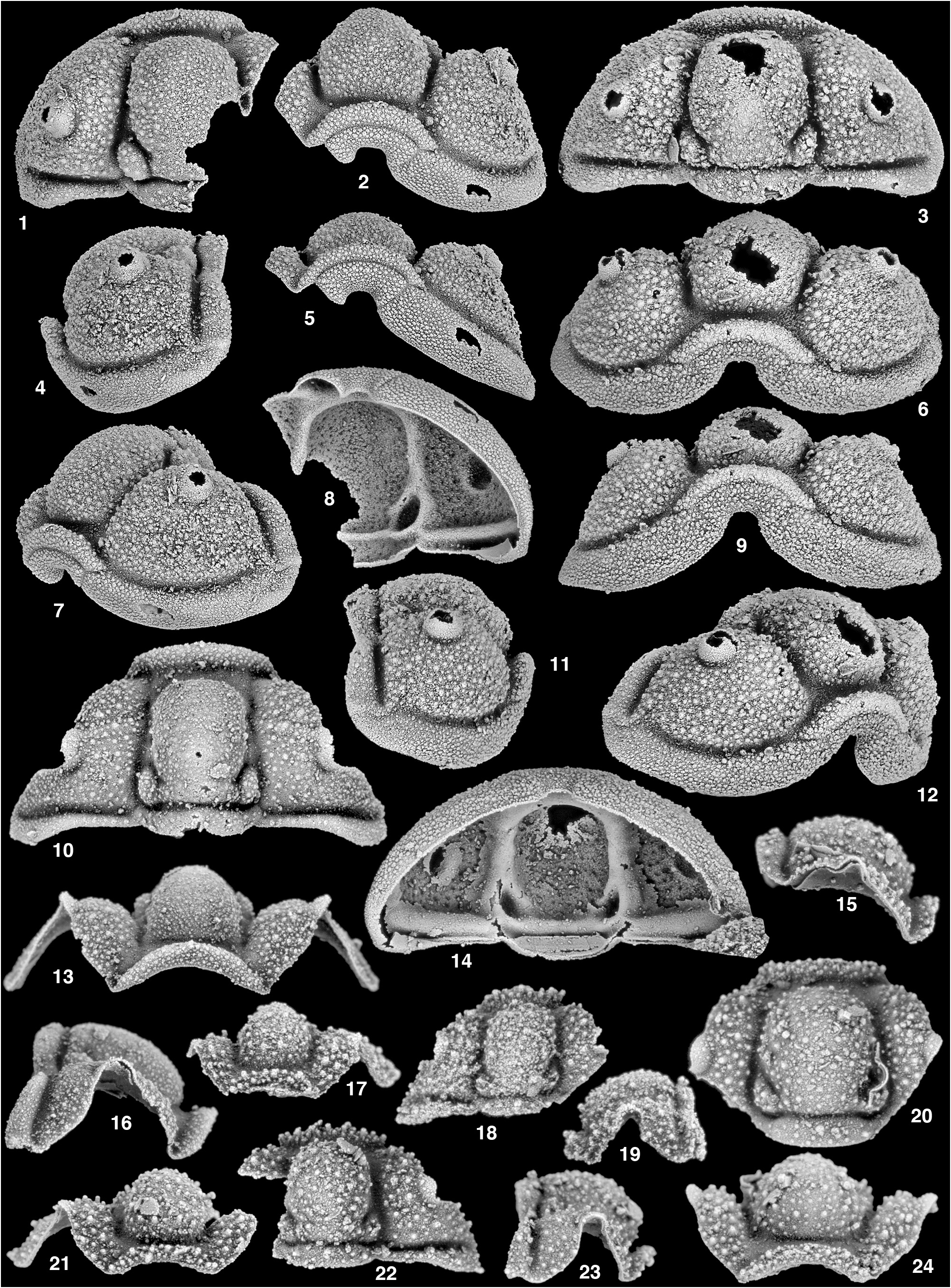
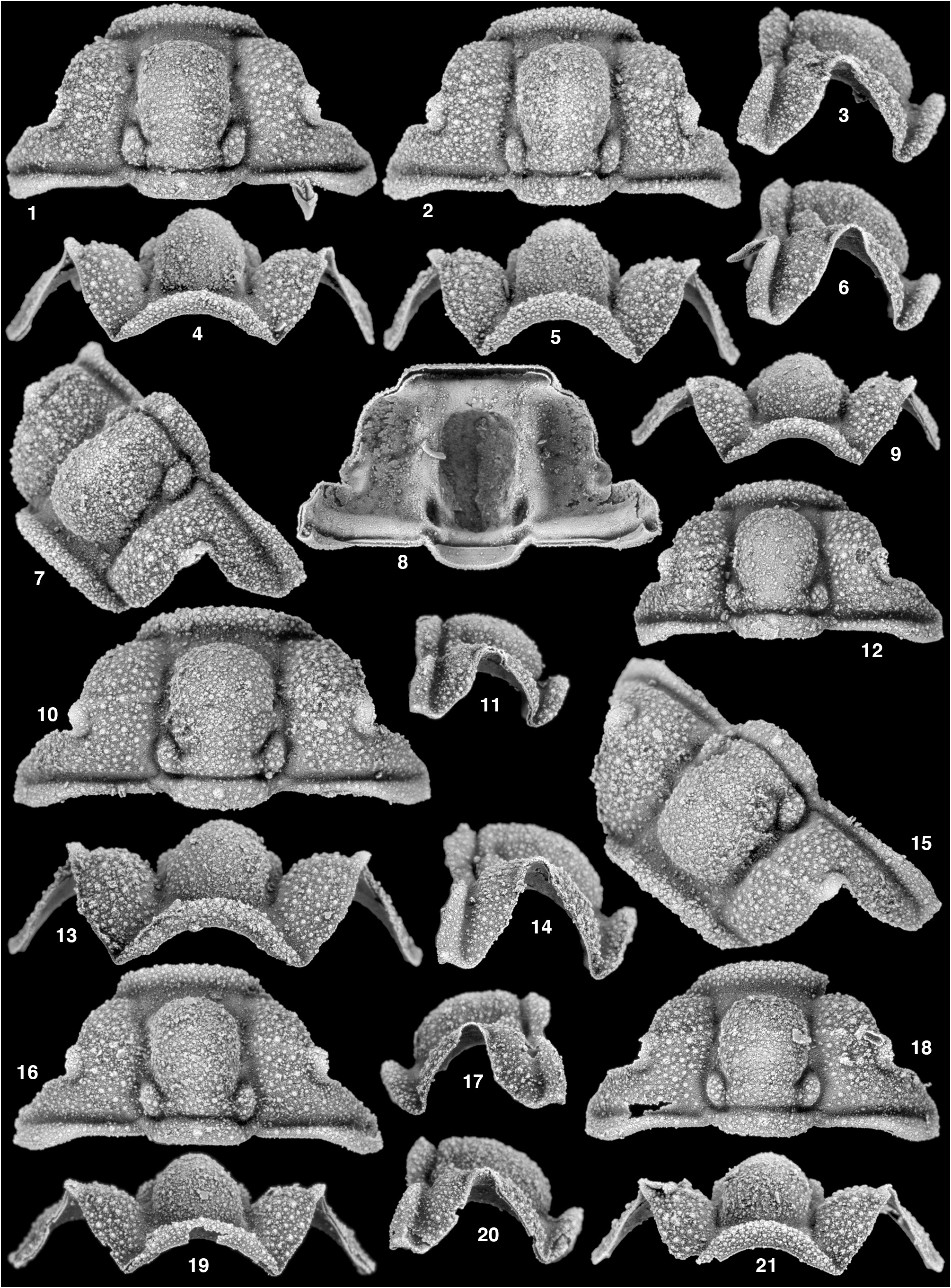
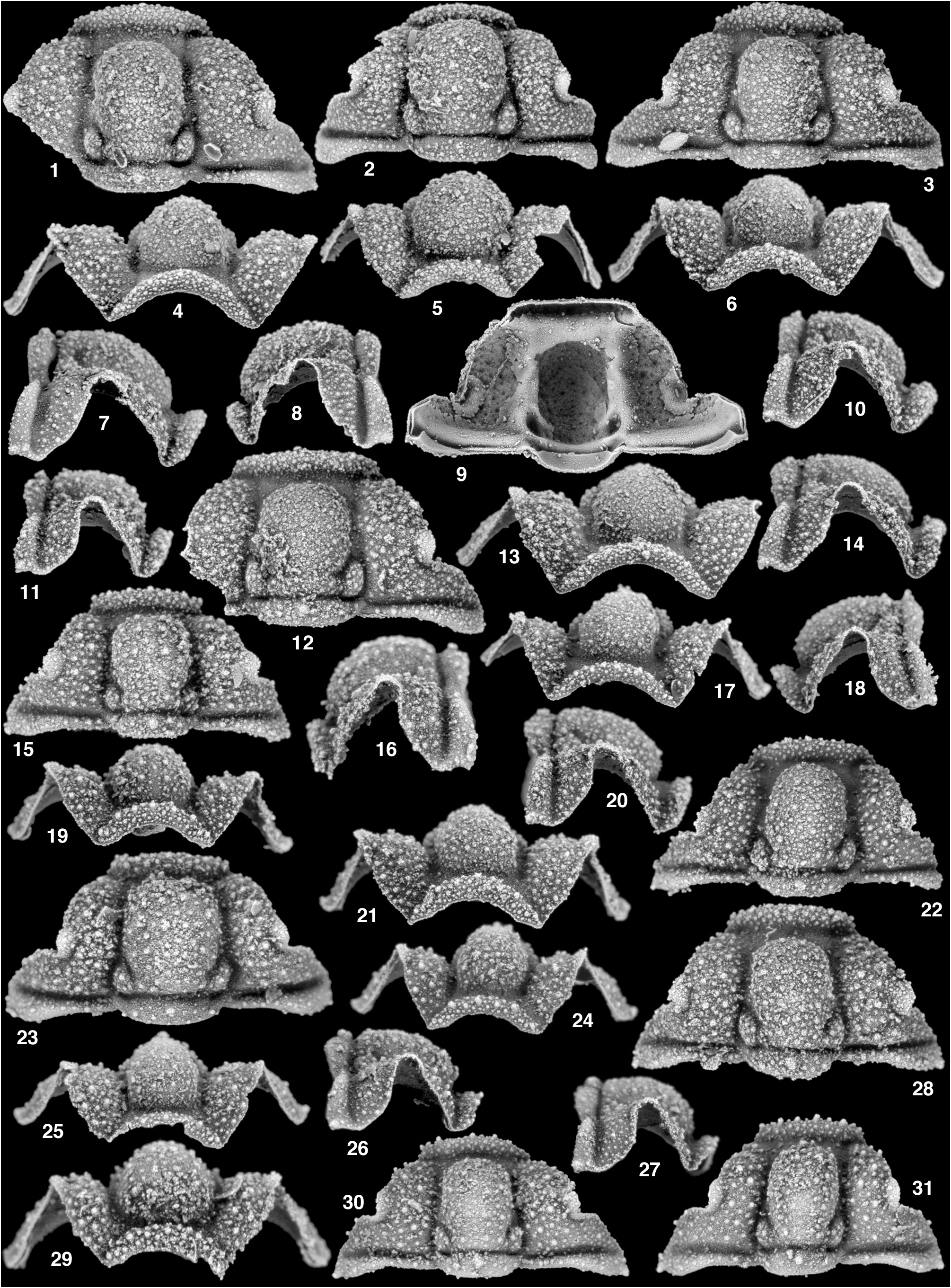
Description. Cranidial measurements are based on the large, intact specimens of Pl. 1, fig. 10, Pl. 2, figs 1, 2, 10, 12, 16, 18, and Pl. 3, figs 1–3, 12. Where part of the cranidial morphology was preserved on only one side, the distance to the sagittal line from this side was doubled. Cranidium strongly vaulted, covered with densely spaced tubercles that are sometimes flattened; maximum cranidial sagittal length 50.6% (47.5–55.3) maximum width across posterior border; width across anterior border furrow 42.1% (39.1–45.4) maximum width across posterior border; distance across γ 66.5% (61.8–70.9) maximum cranidial width; anterior border with length (sag.) 75.9% (62.7–95.5) LO sagittal length, slightly shorter (exsag.) abaxially, forming strongly bowed arch in anterior profile; anterior border flexed upward and back toward anterior tip of glabella, so that it partially obscures border furrow in anterior view, but it does not abut anterior margin of glabella in transverse profile; anterior border with sculpture of small and medium sized, densely spaced somewhat flattened tubercles, tubercles become smaller toward rostral suture; anterior border furrow confluent with preglabellar furrow, long (sag.), with length (sag.) nearly equal to that of anterior border in dorsal view on some specimens, dorsally concave, much deeper and shorter (exsag.) opposite frontal area; anterior sections of facial sutures gently laterally convex opposite anterior border, laterally concave opposite anterior border furrow, and strongly, but unevenly, laterally bowed opposite frontal areas with bow strongest opposite anterolateral corners of glabella; frontal areas strongly inflated, sloped strongly downward and mostly facing anteromedially with exsagittal portion facing slightly obliquely, anteriormost portion nearly vertical to slightly recurved under cranidium; palpebral lobe small, lobe strongly laterally bowed, comma shaped, with portion posterior to δ slightly longer than portion anterior to δ; distance across γ 131.5% (124.3–142.7) cranidial sagittal length; distinct furrow clearly differentiating palpebral lobe from fixigena, furrow is shallow anteriorly, but deep along posterolateral corner of lobe; eye ridge faint, but discernible on dorsal surface of most specimens, clearly visible ventrally (Pl. 2, fig. 8), running obliquely from anterior portion of palpebral lobe to point just behind fossula; interocular fixigena very broad, with moderately strong dorsal inflation, sloping toward glabella; posterior fixigena with inflation similar to frontal area, strongly sloping downward from horizontal plane; fixigena covered with sculpture of medium and small sized scattered tubercles, tubercles become smaller and less dense toward genal angle; posterior section of facial suture nearly straight, slightly posterolaterally directed to nearly transverse; posterior border furrow relatively long (exsag.) and deep, nearly transverse, deeper and forming a sharp contact posteriorly with posterior border, shallower and forming a more gradational contact anteriorly with posterior fixigena, distal and proximal tips shallower; posterior border dorsally inflated, shortest (exsag.) proximally, lengthened distally from fulcrum, then slightly shortened distally from point opposite distal margin of posterior facial suture; border with fine background tubercles and a few larger tubercles arranged linearly along portion of border between fulcrum and axis; posterior margin of posterior border transversely straight to fulcrum, directed more posterolaterally from fulcrum to point opposite distal margin of posterior facial suture, directed anterolaterally from this point distally; ventral margin of posterior border between fulcrum and axis with narrow transverse articulating groove, additional thin subtriangular strip of doublure present beneath posterior border from fulcrum distally; genal angle developed into very short posterolaterally directed projection; glabella subtrapezoidal in dorsal view, with maximum width across L1 98.8% (87.9–104.1) sagittal length excluding LO, sagittal length (excluding LO) 64.8% (61.9–71.7) that of cranidium, strongly convex and dorsally inflated, with apex sitting above fixigena and situated opposite posterior fixigena in lateral view, anterior portion of glabella faces anteriorly and is nearly recurved under margin; axial furrows clearly impressed along majority of course, shallow and nearly effaced opposite LO, deeper and laterally bowed around L1, furrow broad in front of L1 forming a subtriangular depression along the lateral glabellar margin, tapers anteriorly, but broadens again at intersection with anterior border and preglabellar furrows, width across posterior contact of furrows with posterior margin 58.8% (53.6–63.7) cranidial sagittal length, laterally confluent with posterior bor- der furrow; S1 deep, completely isolating L1 on some specimens (e.g., Pl. 2, fig. 2), but not quite on others (e.g., Pl. 2, fig. 10); S2 poorly developed, barely visible on some specimens; L1 bulbous, subtriangular, with strong dorsal inflation; L2 and L3 not prominent and without independent inflation; sculpture of mostly small densely spaced tubercles covers glabella, with a scattering of larger tubercles superimposed; LO long, sagittal length 36.9% (35.0–40.7) that of cranidium, longer sagittally than exsagittally, anterior and posterior margins nearly transverse medially, with distal tips directed anterolaterally, sculpture of small scattered tubercles, slightly less densely spaced than on glabella, prominent median node, situated slightly anterior to midline; SO deep and short (sag.) medially, with distal tips deeper and deflected around base of L1, nearly transverse medially, anterior and posterior edges with sharp contact with rear of main glabella and LO respectively, joining axial furrow in smooth curve without obvious disruption in course; doublure of broad, semicircular articulating surface underlying LO, anterior margin terminating just before ventral expression of SO, mostly smooth with very faint transverse lines; fossulae expressed dorsally on most specimens.
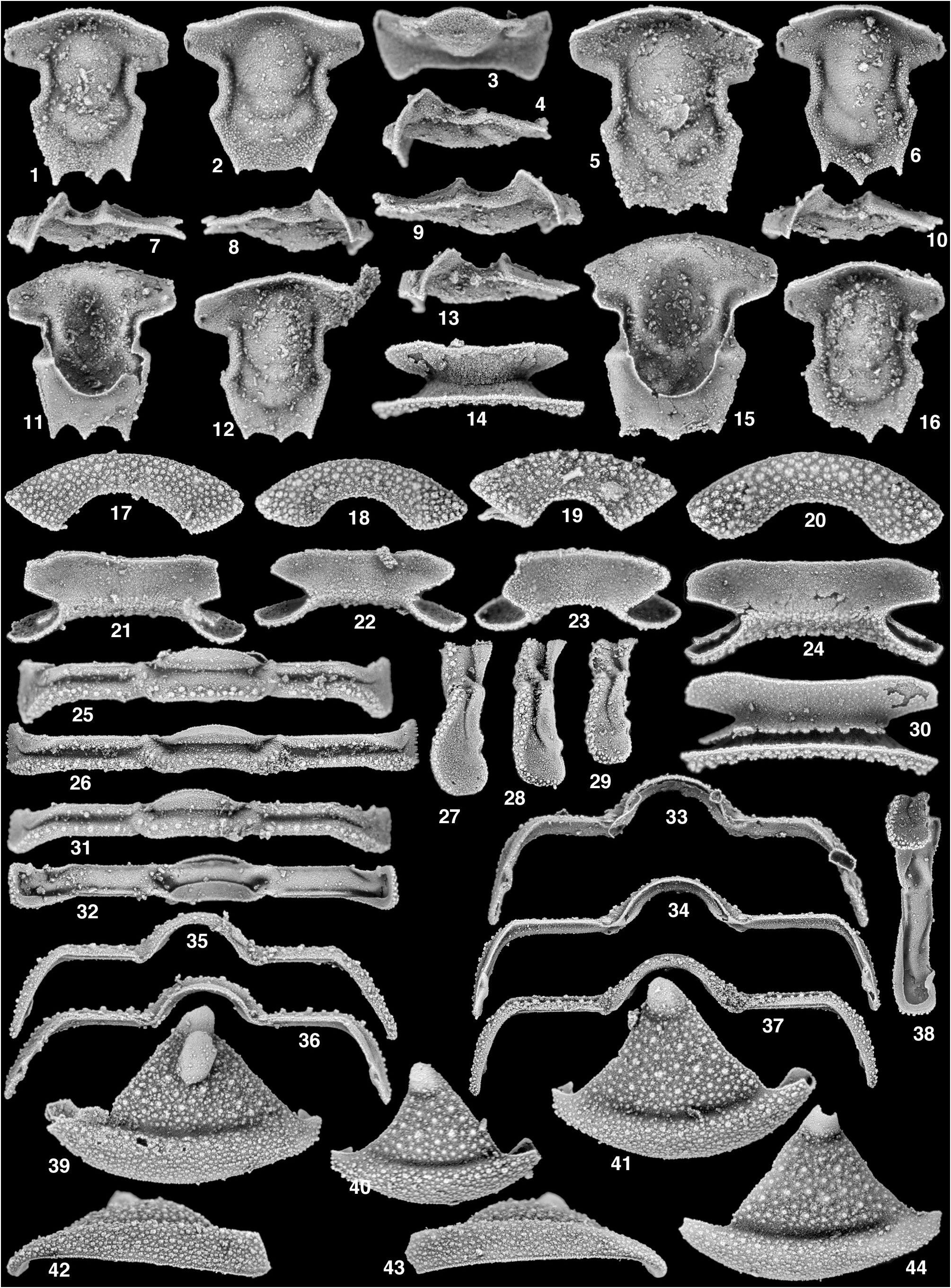
Librigenal measurements were made on the larger specimens of Pl. 4, figs 28–30, 34, 36, Pl. 5, figs 41, 44. Librigena subtriangular, with main body (excluding anterior projection) with maximum width 74.0% (66.7–76.9) exsagittal length; anterior section of facial suture with length 62.0% (55.1–68.3) exsagittal length of main body and posterior section of facial suture with length 53.5% (48.6–59.2) exsagittal length of main body; anterior section gently convex to nearly straight opposite field, with curve slightly more strong opposite abaxial half of field, curve strongly bent across lateral border; posterior section with gentle concave bow, curve continues smoothly across lateral border in some specimens, but in others curve is more strongly bent at lateral border; visual surface small, short, bulbous; eye separated from field by shallow furrow, deeper posteriorly, furrow covered by sculpture of small tubercles; field with background sculpture of very fine granules overlain by more prominent sculpture of densely spaced small and medium sized tubercles; lateral border furrow broad and relatively deep, distal tips slightly shallower on most specimens, background sculpture of fine granules covers furrow, but larger tubercles are mostly absent; lateral border with margin strongly but not evenly arcuate, curve strongest just anterior to midlength; border broad, strongly inflated with margin flattened forming a nearly vertical wall in ventrolateral profile (Pl. 5, figs 42, 43), with sculpture of very densely spaced small flattened tubercles, some slightly larger tubercles are present along the internal margin adjacent to lateral border furrow, ventrolateral margin with prominent raised line bounding margin; anterior projection moderately short, downturned from horizontal plane (Pl. 5, fig. 42), sculpture from border continues without obvious disruption onto anterior projection; doublure broad and slightly narrower than lateral border and anterior projection, narrower posteriorly, strongly upturned toward ventral surface, largely smooth, lacking coarsely tuberculate sculpture present on dorsal surface; inner margin of doublure describes continuous arc, with sharp angular change posteriorly.
Rostral plate measurements were made on the four specimens of Plate 5 View PLATE 5 . Rostral plate with width (tr.) at juncture of connective suture and inner arc 52.1% (44.5–60.8) width at rostral suture; length (sag.) of border sector 19.0% (16.7–22.6) maximum width at juncture of connective and rostral sutures, border sector lengthened (exsag.) abaxially; border sector strongly bowed, inner arc tighter than arc along rostral suture, with sculpture of densely spaced, flattened, medium and small sized tubercles; intersection of border and doublural sectors describing smooth arc; doublural sector smooth, lacking obvious tuberculate sculpture, posteriorly concave.
Hypostomal measurements were made on the six specimens of Plate 5 View PLATE 5 . Hypostome with anterior border strongly ventrally flexed, forming a short wall along the anterior margin; anterior border furrow deep and incised medially along anterior margin of middle body, otherwise shallow; anterior margin of hypostome describing smooth arc, with medial portion more strongly arched than portion opposite anterior wing; lateral margin somewhat sinuous to nearly straight, but overall directed nearly parallel to sagittal axis; minimum hypostomal width (tr.) across middle body 57.5% (53.8–60.0) maximum hypostomal width across anterior wing; width across shoulder 69.6% (63.8–71.3) width across anterior wing; anterior wing large, extended far beyond lateral extent of shoulder, with subrectangular outline and small pit at posterolateral corner; lateral margin of shoulder directed gently posteromedially toward posterior margin; posterior wing developed into short dorsally directed process; lateral border furrow deep and incised, deeper opposite anterior portion of middle body; lateral border relatively wide (tr.), strongly inflated; posterior bor- der furrow shallow, slightly deeper medially on some specimens, but clearly outlining posterior margin of middle body; posterior border long (sag., exsag.), flattened, extended into three relatively short posteriorly directed spines, held nearly parallel to horizontal plane; portion of posterior margin between spines strongly arched; middle body with moderate inflation, anterior lobe more strongly inflated than posterior lobe, lacking rhynchos; middle furrow generally shallow, but deeper anteriorly and nearly effaced medially, directed strongly posteromedially, forming a “V” or a “U” shape overall, separating middle body into ovate anterior lobe and roughly U-shaped posterior lobe; posterior lobe unevenly arcuate; posterior furrow variably impressed, describing more shallow U-shape than middle furrow (Pl. 5, figs 1, 5, 6, 12, 16); broad doublure present beneath posterior border, continues along hypostomal margin beneath posterior and lateral portion of shoulder; sculpture of fine granules covers entire ventral surface of hypostome; maculae indistinct.
Thorax known from three segments with the following morphology: axial ring with maximum width (tr.) 35.8% (34.4–38.3) maximum width of segment; axial ring with a band of medium and small sized tubercles, band located posteriorly on larger segments; narrow and deep ring furrow, with distal tips longer (exsag.); articulating half ring moderately large, lenticular to crescentic in outline, covered by small densely spaced granules; axial furrows very shallow to effaced; pleural furrow deep and long (exsag.) between axis and fulcrum, slightly shorter from fulcrum abaxially to facet, furrow continues a short distance onto facet, but does not reach margin; anterior and posterior pleural bands of nearly similar length (exsag.), with posterior band slightly longer (exsag.) on larger segment (Pl. 5, fig. 25); band of prominent tubercles present on axial ring continues across posterior pleural band as a single row of tubercles, tubercles continue around lateral margin of pleurae (Pl. 5, figs 27–29); anterior band with less prominent row of small tubercles along posterior margin; anterior and posterior bands with background sculpture of fine densely spaced granules; very narrow, transverse furrow set anterior to anterior band between axis and fulcrum, with the anterior edge of the articulating flange forming a very narrow transverse tongue which articulates with a narrow ventral transverse groove beneath the posterior edge of the pleurae of the next segment anteriorly (or the posterior border of the cranidium); anterolateral tip of posterior pleural band developed into broad facet, subtriangular in plan view with anterolateral corner developed into short point, rounded and lobate in lateral profile; very small axial process, that appears to be a continuation of the abaxial tip of the axial ring, set at the anterior end of the axial furrow articulates with a small pit in the posterior end of the next segment anteriorly (or the posterior of the cranidium); ventrally, doublure forms a broad articulating surface beneath the axial ring, small portion of doublure is also present along posterior margin of the posterior pleural band from the fulcrum distally; fulcrum set far distally, with portion of pleurae distal to fulcrum in transverse profile just slightly longer than portion between fulcrum and axis; portion of pleura distal to fulcrum turned down from horizontal plane between 55-65 degrees, portion of pleurae between fulcrum and axis nearly parallel to horizontal plane to just slightly inclined toward axis.
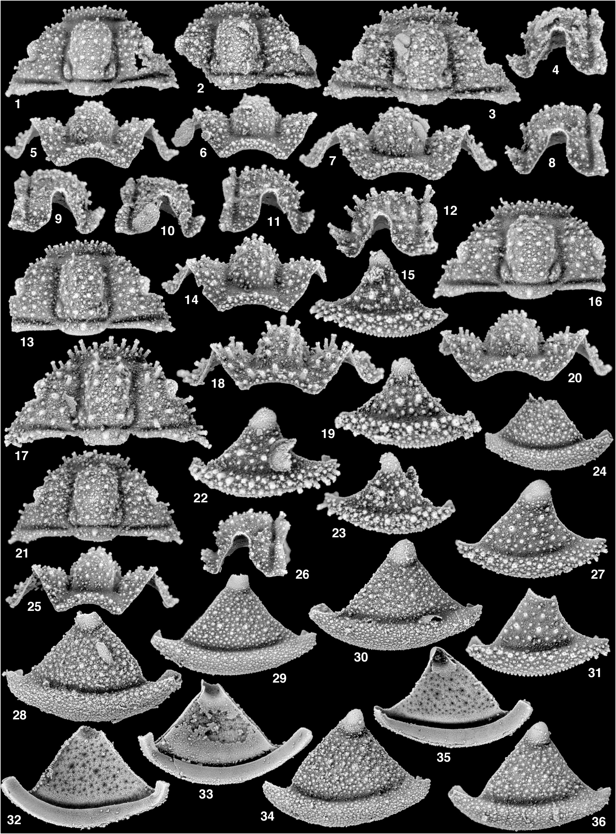
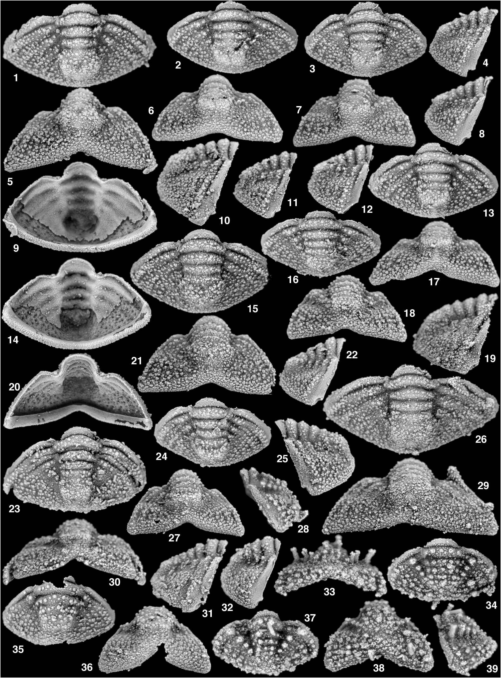
Pygidial measurements were made on the largest and most complete specimens of Pl. 6, figs 1–3, 13, 15, 16, 26. Distance to sagittal line was doubled where one side of the pygidium was broken. Pygidium with maximum width across anterior margin of fourth segment 178.5% (163.4–194.2) sagittal length; axis of four segments and terminal piece, with width across first segment 32.9% (28.1–36.4) maximum pygidial width, width across fourth segment 77.9% (70.0–93.9) width across first segment, length of axis excluding articulating half ring 78.7% (74.3–82.0) total sagittal length of pygidium; articulating half ring semilunate, slightly smaller than first axial segment, sagittal length 11.5% (10.7–12.4) total pygidial length, anterior margin subsemicircular, ring held almost horizontal to sagittal profile of axis, weakly dorsally convex in sagittal profile, strongly bowed in transverse profile (along with rest of axis), with sculpture of very small tubercles; articulating furrow deep, short, and moderately strongly anteriorly bowed; first ring furrow as deep as first, very slightly more transverse; second ring furrow very shallow medially but with distal tips deep, nearly transverse to describing gently anteriorly concave arc; third ring furrow shallow, mostly effaced medially, describing more strongly anteriorly concave arc; first axial ring longer sagittally than exsagittally, with sculpture of about five to six tubercles oriented linearly (tr.) just anterior to mid-length of segment, background sculpture of fine closely spaced granules; posterior rings progressively smaller, but with essentially the same morphology; axis terminated by broad, lightly inflated terminal piece, poorly developed into two lateral swellings in larger specimens (Pl. 6, figs 1–3), densely tuberculate; axis distinctly terminated by change in slope and shallow furrow; axial furrow shallow opposite first axial segment, deepest opposite second and third segments, remaining deep opposite anterior portion of fourth segment, but shallower opposite posterior portion of fourth segment and terminal piece; first segment with maximum exsagittal pleural length 23.6% (21.6–24.7) sagittal axial length, anterior margin almost exactly transversely straight to fulcrum, which is set extremely close to axis, margin directed sharply posterolaterally distal to fulcrum; anterior pleural band set off from margin by very faint transverse accessory furrow (Pl. 6, fig. 10), well inflated, with sculpture of small tubercles arranged linearly along posterior margin of band, background sculpture of fine granules, band of similar length (exsag.) along entire course; posterior pleural band of similar length (exsag.) as anterior band, but slightly longer distally, with mixture of medium and smaller sized tubercles scattered across entire band, slightly more densely spaced distally, finer background tubercles less prominent; pleural furrow deep from axis, past fulcrum, to point just before lateral margin, short (exsag.), terminated before pygidial margin; interpleural furrow much shallower than pleural furrow, deepest between axis and fulcrum, shallower from fulcrum abaxially, extending to pygidial margin; subsequent pleural and interpleural furrows progressively shallower, but remaining deep and discernible adjacent to axial furrow; subsequent pleural segments progressively less clearly defined, overall shorter (exsag.) adjacent to axis, with anterior bands much shorter than posterior bands and nearly pinched out adjacent to axis; pleural region covered by sculpture of prominent medium and a few small sized tubercles, which are distinct and closely spaced adaxially, but become more densely spaced and somewhat flattened abaxially, faint row of single isolated tubercles present on first four segments at fulcrum, background sculpture of fine granules present; border covered by very densely spaced, flattened medium sized tubercles, which merge without disruption from pleural region onto border; ventral aspect of border covered by sculpture of fine granules, that are slightly larger than those composing background sculpture on dorsal surface of pygidium; pygidial margin bowed upward medially in posterior profile; doublure strongly upturned against ventral surface of pygidium in ventral profile (Pl. 6, figs 9, 14), in anterior profile (Pl. 6, fig. 20) doublure clearly visible, apparently smooth and lacking tuberculate sculpture present elsewhere on pygidium.
Ontogeny. Ontogenetic stages of calymenids are relatively poorly known, with descriptions by Chatterton et al. (1990) the main exception. Small specimens of A. bardensis compare closely with those of Silurian species of “ Calymene sensu lato ” illustrated by Chatterton et al. (1990, fig. 9).
The cranidium undergoes several changes in morphology during ontogeny in addition to changes in size. Small- er cranidia (Pl. 4, fig. 17) are covered in densely spaced small spines, especially on the anterior portion of the cranidium and along the facial suture, which become greatly reduced throughout ontogeny into prominent tubercles (Pl. 1, fig. 20). The glabella features sets of paired spines. L1 becomes more clearly outlined and bulbous throughout ontogeny and the glabella become slightly wider with respect to length.
The overall shape of smallest librigenae is similar to that of larger librigenae, but again there is an obvious change in the morphology of the sculpture throughout ontogeny from longer spines/tall tubercles into densely spaced and more flattened tubercles. This is most apparent on the lateral border and field. The smaller librigenae possess a very faint background sculpture of fine granules with clearly isolated, tall tubercles/spines on the field. This sculpture changes throughout ontogeny into the overall more prominent background sculpture of fine granules with more subdued isolated tubercles on the larger cheeks. The same change is seen on the lateral border. The field also become larger with respect to the lateral border throughout ontogeny.
The smallest pygidium (Pl. 6, fig. 37) possesses prominent tall tubercle/spine pairs on each axial segment, whereas the next smallest pygidium (Pl. 6, fig. 34) has the tall tubercles/spines reduced into a band of prominent tubercles along each axial segment. These tubercles continue to become more reduced and subdued throughout ontogeny. A similar patten of reduction is seen in the line of tubercles present on the pleural region adjacent to the axis. The smallest pygidium displays a prominent row of spines that runs along the fulcrum of pleural region, with the largest sitting anteriorly, and that become progressively smaller and shorter posteriorly. The spines are reduced, but still clearly visible and distinct on the next smallest pygidium, and throughout ontogeny the row of spines is further reduced to a band of small tubercles that are progressively harder to distinguish on larger specimens. The posterior margin of the smallest pygidia is broadly rounded and lacking a point medially. This curve becomes more strong throughout ontogeny and with larger pygidia having more of a broadly rounded point medially. The pygidium also becomes more strongly vaulted with the portion distal to the fulcrum lengthened throughout ontogeny (cf. Pl. 6, fig. 33, Pl. 6, figs 27, 38).
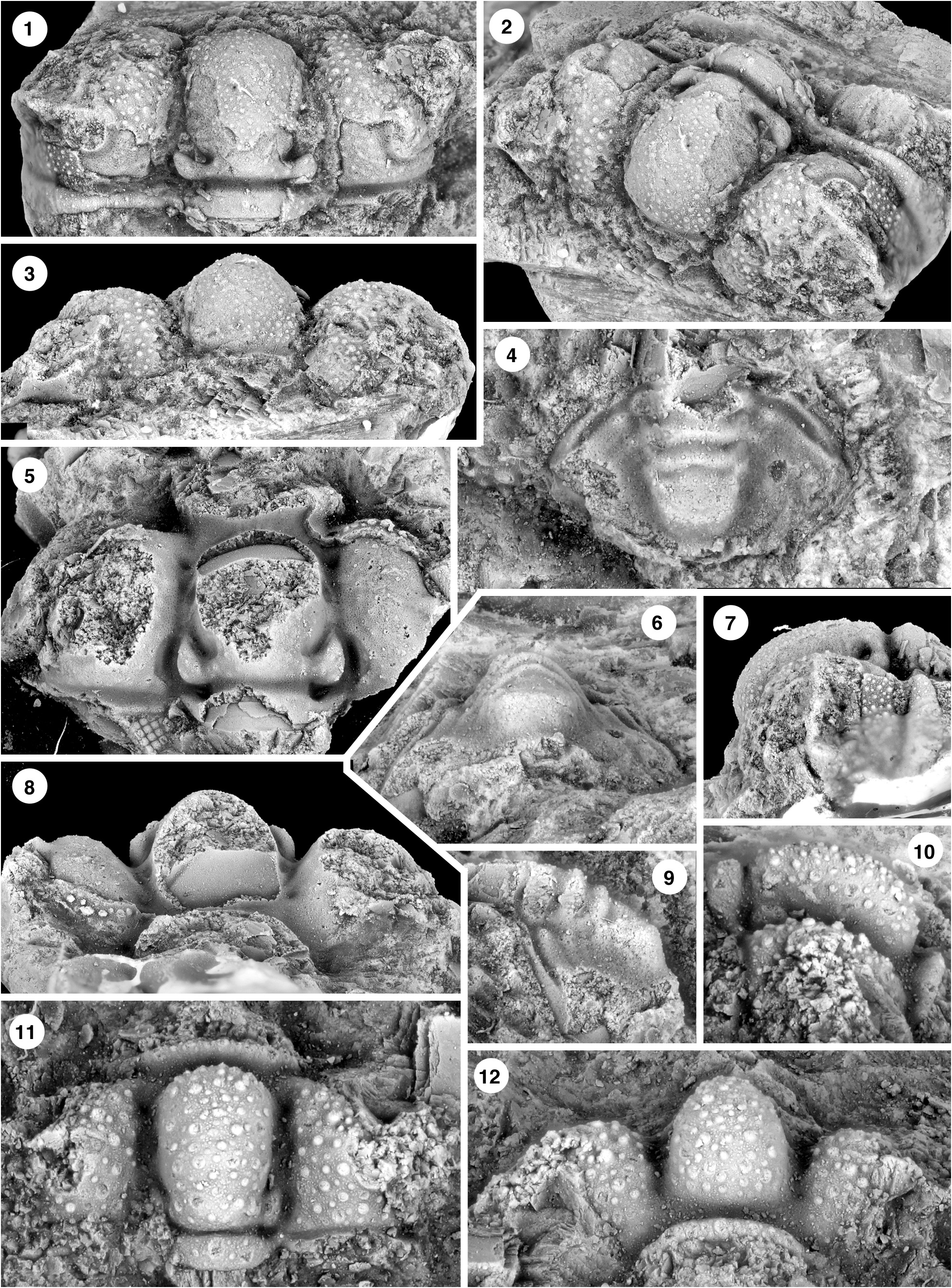
Discussion. Two cranidia assigned by Whittington (1965, p. 419, pl. 59, figs 10, 12-15) to “aff. Calymenidius sp. ind.” have been cited repeatedly in the literature (figured herein as Pl. 7, figs 5, 8, 10–12). With description of A. bardensis on the basis of abundant material, it becomes evident that these specimens belong to the species and that Whittington also illustrated other material of the species in his monograph, misassigned to other taxa. Another cranidium ( Whittington, 1965, p. 340, pl. 19, figs 16, 19, 20; reillustrated as Pl. 7, figs 1–3, 7 herein) was assigned to “ Ischyrophyma ? sp. ind.” This reflects his earlier assignment of a cephalon and thoracic segments from the older Shallow Bay Formation (Whittington, 1963, p. 50, pl. 6, figs 13–15) to the same taxon (see Atlanticalymene n. sp. A below). Finally, an exfoliated pygidium ( Whittington, 1965, p. 374, pl. 39, figs 8, 9; Pl. 7, figs 4, 6, 9, herein) was illustrated as “Komaspidid? pygidium.”
| GSC |
Geological Survey of Canada |
No known copyright restrictions apply. See Agosti, D., Egloff, W., 2009. Taxonomic information exchange and copyright: the Plazi approach. BMC Research Notes 2009, 2:53 for further explanation.