
Phasmotaenia salomonense, Hennemann, Frank H. & Conle, Oskar V., 2009
|
publication ID |
https://doi.org/ 10.5281/zenodo.185796 |
|
DOI |
https://doi.org/10.5281/zenodo.6224557 |
|
persistent identifier |
https://treatment.plazi.org/id/9F7B87E1-437F-FF91-E2AE-659DFE16FD79 |
|
treatment provided by |
Plazi |
|
scientific name |
Phasmotaenia salomonense |
| status |
sp. nov. |
Phasmotaenia salomonense View in CoL n. sp.
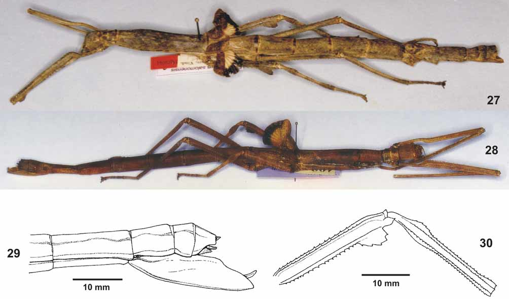
[ Figs. 27–30 View FIGURES 27 – 30 , 57 View FIGURES 48 – 58 , 59 View FIGURE 59 , 60 View FIGURE 60 ]
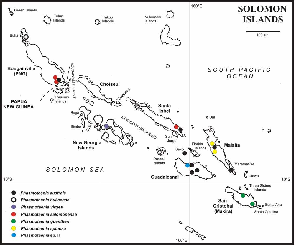
HT, Ƥ + 1egg (ex ovipositor): Salomonen, Santa Isabel Isl., Albatros, ex. Mus. Caesar (NHMW). PT, Ƥ: Solomon Islands, Bougainville, Crown Prince Range, 4000 ft., 1965 C. RA. Exploration; No. F3224.179 (MMUE).
PT, Ƥ + 1 egg (ex ovipositor): Kieta, Bougainville, 193., 1937-9; australis K. Gthr. det. (SMTD). PT, Ƥ: Bougainville, Solomon Is, II.1928, Ac. 28250, Am. Mus. Nat. Hist. Dept. Invert. Zool. (ANSP).
Diagnosis: The prominent sub-apical ventral lobes of the mesofemora, dorsally lobed probasitarsus, dark orange and broadly black marginated anal fan of the alae, and plain dark brown capsule indicate close relation to P. spinosa n. sp. from Malaita Island. However, ƤƤ of P. s a l o m o n e n s e n. sp. at once differ from this species by the lack of spines on the mesonotum and metapleurae, lack of posterolateral lobes on abdominal tergites III–VII and much shorter subgenital plate ( Fig. 29 View FIGURES 27 – 30 ).
Etymology: Named after its distribution, the Solomon Islands.
Description: Ƥ ( Fig. 27–28 View FIGURES 27 – 30 ): Medium-sized (body length 138.6–151.0 mm, incl. subgenital plate 142.2–157.0 mm), moderately elongate species for the genus with a gently broadened mesothorax, developed tegmina (6.9–7.4 mm) and alae (10.8–12.0 mm), the latter with a bright orange and broadly black marginated anal region, very short subgenital plate and sub-basally elevated mesotibiae.
Colouration: General colouration of body and legs mid to dark brown. The HT is greyish brown and has the entire body furnished with irregular yellowish brown speckles, with abdominal tergite VI paler than remaining and in anterior portion with two triangular dark brown markings. The PT’s in SMTD and ANSP are of a rather plain reddish brown colour. The PT in MMUE is reddish mid brown with bold blackish markings on the mesothorax and along the lateral surfaces of the abdominal tergites, the dorsal portion of the abdomen being mostly pale cream. Granules of the mesonotum and mesopleurae dark orange. Tegmina and costal region of alae plain brown, in HT with irregular darker brown mottling. Anal region of alae orange (presumed bright red when alive) with a broad black outer margin. The PT in MMUE has the black outer margin with several of the transverse veins transparent. Meso- and metatibiae with three ± distinct dark brown transverse bands (indistinct in the SMTD Ƥ). Antennae mid to dark brown, eyes reddish brown.
Head: Sub-globose, oval in dorsal aspect, broadest at the eyes and about 1.3x longer than wide; vertex gently rounded. Eyes circular, rather small and projecting hemispherically; their length contained a little more than 3x in that of cheeks. Between the eyes with a shallow transverse bulge which at both ends terminates in a small hump. Antennae almost reaching to posterior of mesonotum. Scapus dorsoventrally compressed, about 1.8x longer than wide and decidedly narrowed towards the base. Pedicellus cylindrical and less than ½ the length of scapus. Third antennomere a little longer than pedicellus, IV almost 2x longer than III. Following antennomeres first increasing then decreasing in length.
Thorax: Pronotum slightly shorter and narrower than head, about 1.3x longer than wide, indistinctly widened towards the posterior and very gently narrowed medially. Transverse median depression distinct but very short and not reaching the lateral margins of the segment. Complete segment with an impressed longitudinal median line. Mesothorax a little more than 5x the length of pronotum, constricted at the anterior, gently swollen pre-medially and almost parallel-sided in the posterior half. Mesonotum with a fine longitudinal median carina and sparsely set with a few small, rounded granules. Mesopleurae with a longitudinal row of small spiniform tubercles, the metapleurae with a row granules. Mesosternum with two roughly parallel rows of small granules, metasternum smooth. Tegmina roughly reaching to posterior margin of metanotum, broadened in the apical half and distinctly constricted towards the base; central hump indistinct. Alae slightly longer than tegmina and reaching about ¾ the way along median segment.
Abdomen: Median segment 1.5x longer than metanotum, about 2x longer than wide and rectangular; unarmed. All segments unarmed, except for a transverse, scale-like swelling on posterior margin of tergite V and VI (lacking in the PT in SMTD). Tergites VI–IX may have two irregular and indistinct longitudinal carinae. Segment II longer than median segment, II–VI about equal in length and width, almost 2x longer than wide and parallel-sided. VII slightly shorter and narrower than previous and about 2.5x longer than wide. Praeopercular organ formed by a small, scale-like, medial swelling at the posterior margin of sternite VII. Tergite VIII about half the length of VII, slightly longer than wide and widened towards the posterior. IX shorter than VIII, transverse. Anal segment a little longer than IX, lateral margin with a shallow concave excavation, the posterolateral angles acute and the posterior margin with a small, median indention; dorsal surface with a faint longitudinal median carina. Supraanal plate roundly triangular and slightly projecting over posterior margin of anal segment. Cerci round in cross-section, slender, slightly up-curving and tapered towards the tip; finely bristled. Lower gonapophyses elongate, filiform and roughly reaching to apex of subgenital plate; apex slightly thickened and up-curving. Subgenital plate strongly keeled and projecting over the apex of abdomen by less than the length of tergites IX and X combined; apex rounded ( Fig. 29 View FIGURES 27 – 30 ).
Legs: All of moderate length for the genus, with most carinae dentate. Profemora longer than mesonotum, metatibiae shorter than mesonotum, metafemora about ½ the way along abdominal tergite IV and metatibiae reaching about half way along tergite VI. Posteroventral and anteroventral carinae of profemora acutely serrate. The posteroventral carina in the HT with a rounded, dentate sub-apical lobe. Medioventral carina distinct and armed with a variable number of small spines. Protibiae with all carinae slightly lamellate but unarmed; merely a few small serrations on anteroventral carina. Meso- and metafemora with all carinae dentate, the two outer ventral carinae of the mesofemora with a prominent dentate sub-apical lobe. Medioventral carina faint and armed with a few small spines which decrease in size towards apex of femur. Mesotibiae with the dorsal and medioventral carinae roundly elevated sub-basally ( Fig. 30 View FIGURES 27 – 30 ), all carinae dentate. Metatibiae with all carinae dentate but slender. Probasitarsus as long as the remaining tarsomeres combined, except claw with the dorsal carina ± strongly rounded and raised; all carinae unarmed. Meso- and metabasitarsi slightly longer than the following three tarsomeres combined, ventral carinae minutely dentate.
Variation: Already the few known specimens show ƤƤ of this species to exhibit considerable variation concerning to various morphological features. The Ƥ in MMUE has a distinct scale-like posteromedian tubercle on tergites V and VI, which are much less distinct in the HT and lacking in the Ƥ in SMTD. The HT has a dentate sub-apical lobe on the posteroventral carina of the profemora, which is not present in any of the other three known paratypic ƤƤ. The dorsal lobe of the probasitarsus is very prominent and large in the MMUE ƤƤ, considerably smaller in the HT and rather shallow in SMTD Ƥ. For the variability of the colouration see description above.
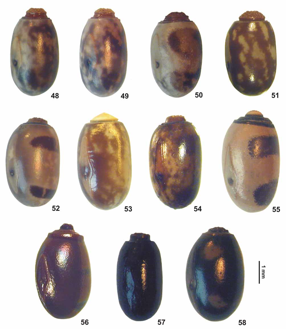
Eggs ( Fig. 57 View FIGURES 48 – 58 ): Two eggs (paratypes) could be extracted from the ovipositors of the HT in NHMW and PT in SMTD. They show slight variation regarding to the size and the egg taken from the HT has the posterior portion of the micropylar plate slightly relatively shorter than in the example from Bougainville.
Rather small, ovoid, capsule almost 2x longer than wide and slightly oval in cross-section; the lateral surfaces in the median portion almost parallel-sided. Capsule surface very slightly uneven and strongly shiny. Micropylar plate elongate and slightly less than 2/3 the length of capsule, anterior 2/3 slender and gradually widening towards a roundly elevated sub-median portion, the posterior portion first constricted and then gradually tapered. Both ends of plate rather pointed. Outer margin bluntly raised. Micropylar cup small, rounded and placed in posterior half of plate. Operculum almost circular and in the centre with a rather prominent knob-like pseudo-capitulum, which has the dorsal surface with several deep and irregularly shaped impressions. General colouration of capsule and micropylar plate plain blackish brown to black, the pseudocapitulum dark brown with the dorsal portion reddish mid brown.
Measurements [mm]: length (incl. operculum) 3.6–3.8, length 3.1–3.3, width 1.9–2.0, height 2.1–2.3, length of micropylar plate 2.4–2.6.
Comments: Zompro (2003: 33) erroneously listed the PT Ƥ from Kieta, Bougainville in SMTD as a PT of Phasmotaenionema australe Günther, 1933 , although Günther (1933) did not mention a PT from this locality nor a specimen in SMTD.
There are five 3 and an additional Ƥ in ANSP which were collected from the same locality as the Ƥ in the same collection, here designated a PT. Unfortunately, all of these specimens are in rather poor condition and were only briefly examined, so no formal description of the 3 can be presented herein.
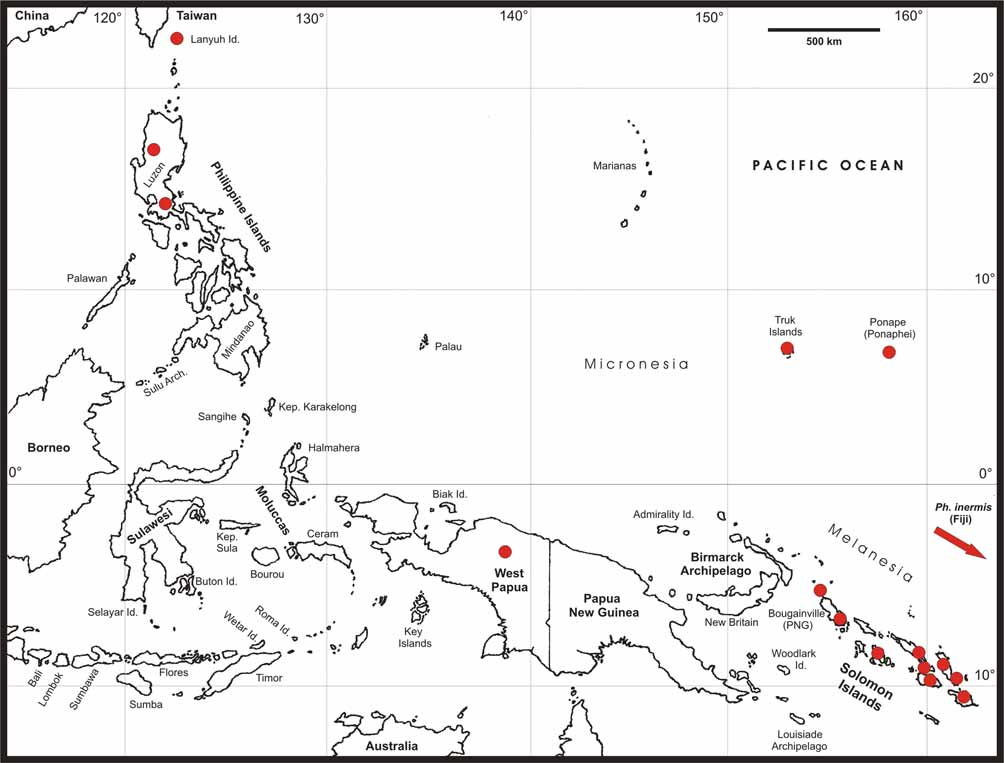
Distribution ( Fig. 60 View FIGURE 60 ): Solomon Islands. Santa Isbel Island (Albatros) & Bougainville Island (Kieta & Crown Prince Range).
No known copyright restrictions apply. See Agosti, D., Egloff, W., 2009. Taxonomic information exchange and copyright: the Plazi approach. BMC Research Notes 2009, 2:53 for further explanation.