
Elysia ellenae Ortea, Espinosa & Caballer in Ortea, Espinosa, Buske & Caballer, 2013
|
publication ID |
https://doi.org/ 10.11646/zootaxa.4148.1.1 |
|
publication LSID |
lsid:zoobank.org:pub:91353147-FDA8-45CC-A8F1-1DE801C835A6 |
|
DOI |
https://doi.org/10.5281/zenodo.5664223 |
|
persistent identifier |
https://treatment.plazi.org/id/A04A7E6D-9C11-FFE1-46C9-FAC0FE7D1CF1 |
|
treatment provided by |
Plazi |
|
scientific name |
Elysia ellenae Ortea, Espinosa & Caballer in Ortea, Espinosa, Buske & Caballer, 2013 |
| status |
|
Elysia ellenae Ortea, Espinosa & Caballer in Ortea, Espinosa, Buske & Caballer, 2013 View in CoL
( Figs. 54–55 View FIGURE 54 View FIGURE 55 , 56 View FIGURE 56 C, 57)
Elysia ellenae Ortea, Espinosa & Caballer in Ortea, Espinosa, Buske & Caballer 2013: 185 View in CoL –188, pl. 11 (Type locality: South of Port Louis and west from Petit Canal, Guadeloupe); Krug et al. 2015: 990, fig. 3B. Thuridilla View in CoL sp. — Redfern 2013: 288, fig. 797.
Elysia View in CoL sp. 4—Turner et al. 2012: 54.
Type material. Elysia ellenae— holotype (MNHN IM-26975), paratype (MNHN IM-26976).
Material examined. Bahamas: Stocking Island , Exumas , Bahamas, 15 January 2009, 1 specimen ( CPIC 00071 ), Goulding Point, New Providence , 13 July 2010, 1 specimen ( LACM 178663 View Materials ). Photographs of four additional specimens from Cayman Islands (courtesy of Everett Turner) examined.
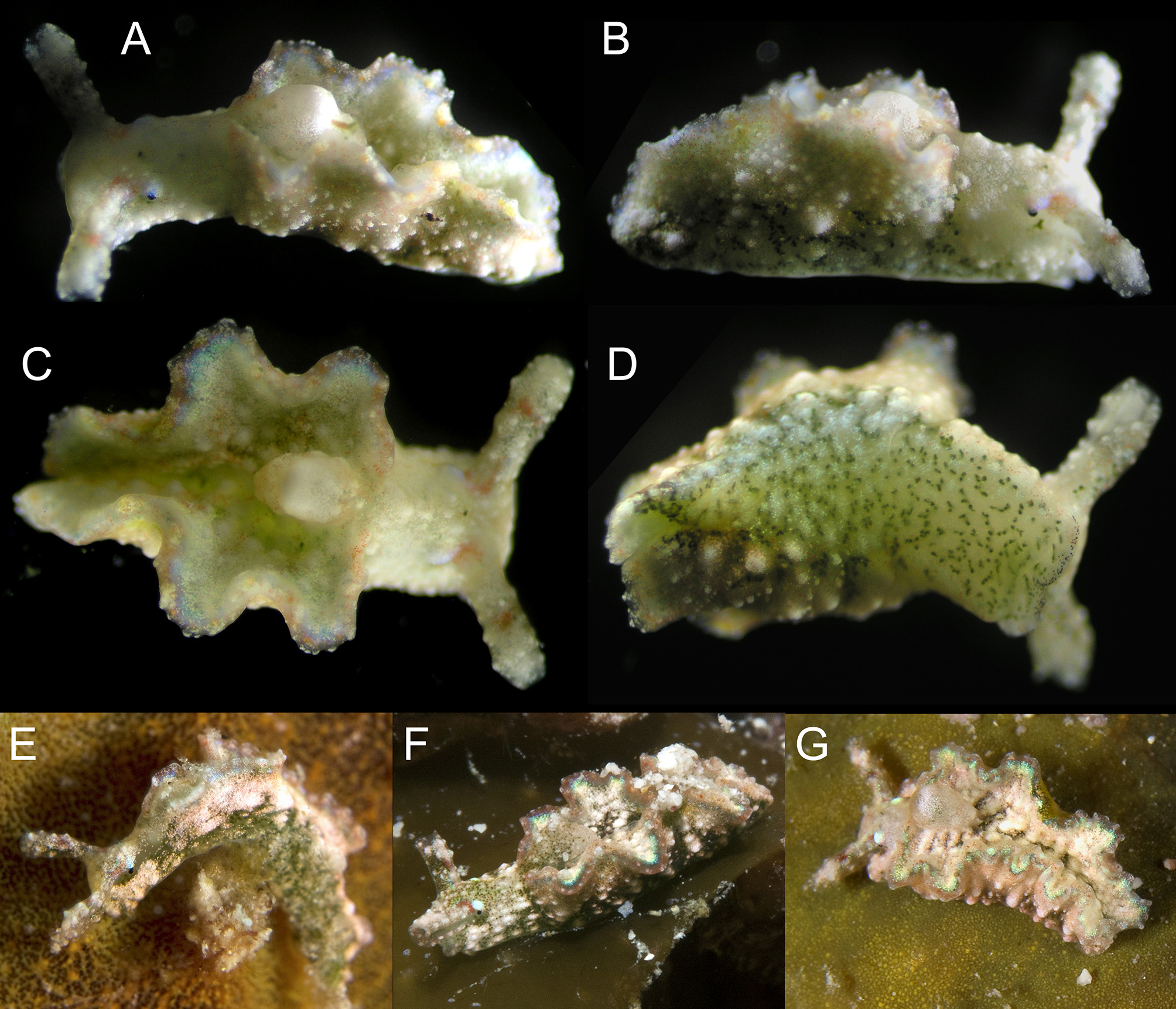
Live animal. According to Ortea et al. (2013) “this species moves crawling, not jumping, not swimming. When at rest, it flattens and expands the parapodia, taking the appearance of a heart-shaped leaf.”
External anatomy. Head white. Light blue patch or streak above each eye, like an eyebrow, extending part way up rhinophores on some specimens ( Fig. 54 View FIGURE 54 A–C, E–F). Black row of dots forming “moustache” above upper lip of mouth ( Fig. 54 View FIGURE 54 D). Rhinophores short relative to length of animal, and wider at tip than base. Rhinophores papillose; overall color white, with green digestive diverticula scattered across anterior surface. Green digestive diverticula scattered across anterior face of rolled rhinophores. Rose-red or brick-red spot at midpoint of rhinophores; red patch or curved line located at base of rhinophores, just anterior to each eye ( Fig. 54 View FIGURE 54 C).
Foot white with sparse diverticula visible, and covered with scattered patches of light blue iridescence ( Fig. 54 View FIGURE 54 D). Transverse groove extends halfway across left side of foot but does not fully delimit head from foot. No distinct tail; foot narrowing to a point, where ends of parapodia fuse.
Parapodia notably thick, separated at anterior edge by gap. Parapodia form three siphonal openings on living animal, with slightly undulating margins ( Fig 54 View FIGURE 54 A, C, F–G). Overall color white. Outer face of each parapodium has two distinct halves: bottom half penetrated by green diverticula, and spotted with large white papillae, irregularly spaced; top half lacks visible diverticula, dotted with small white papillae except for smooth band just below parapodial margin. Entire outer parapodial face uniformly dotted with minute red spots. Parapodial margin with dark red band and row of regularly spaced yellow spots; marginal edge thickened, with regularly spaced white papillae. Faint submarginal band of iridescent blue may be present on outer parapodial surface ( Fig. 54 View FIGURE 54 C, G). Inner face of parapodium dotted with white papillae and dark green diverticula. Inner submarginal band of iridescent light blue, widest inside siphonal openings.
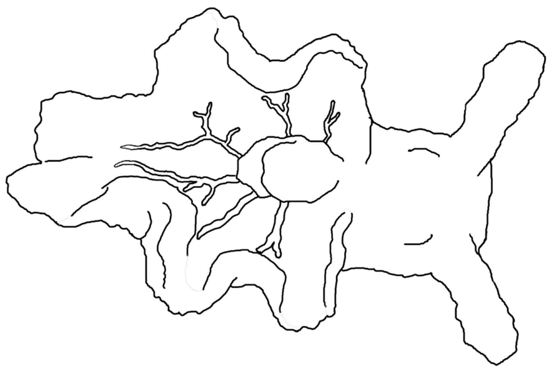
Pericardium large, white, round; completely filling anterior siphonal opening ( Fig. 54 View FIGURE 54 C). Pericardium swelling to height of anterior parapodial edge, clearly visible as dorsal bulge on living animal. Renopericardial extension running about one-third of body length, more grey in color than pericardium. In a specimen used for DNA analysis, three dorsal vessels radiating from right side of renopericardial complex, and two vessels from left side, outlined in red spots ( Figs. 54 View FIGURE 54 C, 55). Vessels unbranched or forking once terminally, except elongated posterior vessels having two lateral side branches reaching up to parapodial margin, respectively at midpoint and posterior end of second siphonal fold of parapodium.
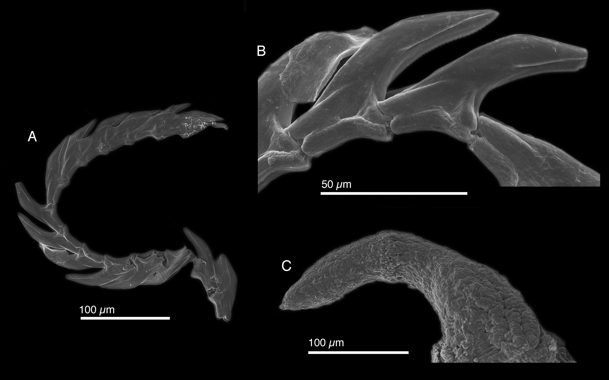
Internal anatomy. Radula with 11–14 teeth (CPIC 0 0 0 71, LACM 178663), 5–6 teeth in ascending limb and 6–8 in descending limb ( Fig. 57 View FIGURE 57 A). Leading tooth elongate and robust with cusp bearing a finely denticulate keel and smooth lateral edges on each side ( Fig. 57 View FIGURE 57 B). Housing depression for interlocking teeth “V”-shaped and extending ½ of tooth lengh. Base of tooth ½ to ⅓ total tooth length. Ascus containing three slightly smaller discarded teeth, according to Ortea et al. (2013)
Penis curved and elongate with rigid musculature that did not deform after drying ( Fig. 54 View FIGURE 54 C), bearing a resistant, hollow tip ( Fig. 57 View FIGURE 57 C) visible by SEM, but not light microscopy. Deferent duct long, thin, and convoluted.
Reproduction and development. No data available.
Host ecology. Elysia ellenae was not found associated with any algal host in either the Bahamas or Cayman Islands, and no food source has been reported.
Phylogenetic relationships. In phylogenetic analyses, E. ellenae was recovered as sister to Elysia crispata , while the eastern Pacific E. diomedea was sister to ( E. ellenae + E. crispata ) ( Fig. 4 View FIGURE 4 ). The morphological similarity of E. ellenae to E. crispata is consistent with their phylogenetic affinity. The clade comprising E. diomedea , E. crispata and E. ellenae is a derived member of subclade 2. The nested position of the clade containing E. ellenae suggests an ancestral lineage colonized the Caribbean from the northwestern Atlantic, after which closure of the Isthmus of Panama isolated the ancestor of E. diomedea in the eastern Pacific from the last common ancestor of E. ellenae and E. crispata , which subsequently diverged in the Caribbean.
Range. Bahamas: Abaco (Redfern 2013), New Providence (present study), Stocking Island (present study), Exumas (present study), Cayman Islands (present study); Guadeloupe (Ortea et al. 2013).
Remarks. Where visible, major features described from the Bahamas specimen were also present on photographs of additional specimens from the Cayman Islands and type material depicted from Guadeloupe, including the distinctive red and blue patches on the head, and bright iridescent blue marginal band. Morphologically, E, ellenae resembles its sister taxon E. crispata but has a much larger and more rounded pericardium and thicker parapodia. Elysia crispata has more undulating parapodia, and lacks the distinct siphonal openings of E. ellenae .
The probable presence of an apical penial stylet in E. ellenae but not in E. crispata suggests that divergence in reproductive armature may have been involved in the speciation process for these sister taxa, which have sympatric ranges. Confusion about this structure qualifying as a true stylet has to do with the penis apex bearing a resistant tip, but no obvious barb, spike, or scoop. However, as Gascoigne (1974) defined a penial stylet as simply “a hollow, cuticular extension of the vas deferens,” we conclude that E. ellenae possesses a stylet.
Patchy coverage of the foot by thick digestive diverticula also distinguishes E. ellenae from both the typical morph of E. crispata , which lacks diverticula in the foot, and the ‘ clarki ’ morph, in which the foot has an overall green appearance due to uniform coverage by small diverticula. In E. crispata , the foot is also more blunt-ended rather than tapering to a point as in E. ellenae .
Interestingly, although the algal host of Elysia ellenae is unknown, feeding in this species causes marked wear on its radular teeth ( Fig. 57 View FIGURE 57 B). It is clear that such wear is the outcome of feeding because the leading tooth has a blunt tip, but teeth in the ascending radular row do not. Evidence of extreme tooth wear was not observed in any other Caribbean elysiid.


| LACM |
Natural History Museum of Los Angeles County |
No known copyright restrictions apply. See Agosti, D., Egloff, W., 2009. Taxonomic information exchange and copyright: the Plazi approach. BMC Research Notes 2009, 2:53 for further explanation.
|
Kingdom |
|
|
Phylum |
|
|
Class |
|
|
Order |
|
|
Family |
|
|
Genus |
Elysia ellenae Ortea, Espinosa & Caballer in Ortea, Espinosa, Buske & Caballer, 2013
| Krug, Patrick J., Vendetti, Jann E. & Valdés, Ángel 2016 |
Elysia ellenae
| Ortea, Espinosa & Caballer in Ortea, Espinosa, Buske & Caballer 2013: 185 |