
Elysia hamanni, Krug, Patrick J., Vendetti, Jann E. & Valdés, Ángel, 2016
|
publication ID |
https://doi.org/ 10.11646/zootaxa.4148.1.1 |
|
publication LSID |
lsid:zoobank.org:pub:91353147-FDA8-45CC-A8F1-1DE801C835A6 |
|
DOI |
https://doi.org/10.5281/zenodo.5664231 |
|
persistent identifier |
https://treatment.plazi.org/id/A04A7E6D-9C20-FFF1-46C9-FB20FBAA1EA0 |
|
treatment provided by |
Plazi |
|
scientific name |
Elysia hamanni |
| status |
sp. nov. |
Elysia hamanni View in CoL new species
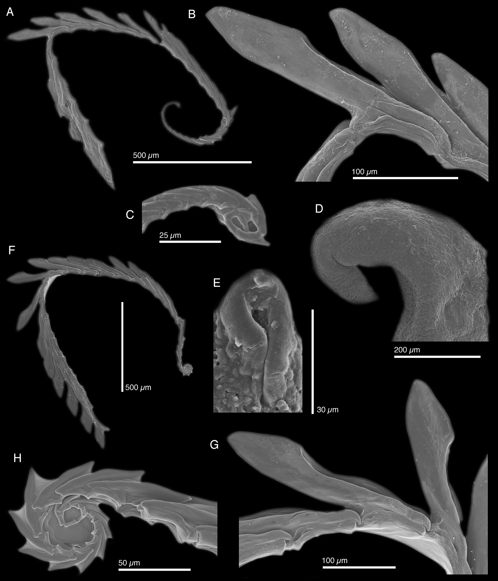
( Figs. 56 View FIGURE 56 H, 67–69)
Elysia sp. 5 — Valdé s et al. 2006: 76–77.
Type material. Banana River , Florida, USA, May 1988, (Holotype LACM 3310 View Materials ) collected by Jeff Hamann ; True Blue Point , True Blue Bay, Grenada, December, 1987, (Paratype LACM 3311 View Materials [8 in lot]), collected by Jeff Hamann .
Type locality. Banana River , Florida, USA
Material examined. Banana River , Florida, USA, May 1988, 5 specimens (Holotype LACM 3310 View Materials , LACM 178668 View Materials [1 in lot], LACM 178669 View Materials [3 in lot]) ; Grenada: September, 1981, 3 specimens ( LACM 178671 View Materials ), True Blue Point, True Blue Bay, December , 1987, 8 specimens (Paratype LACM 3311 View Materials ) ; Isla Mujeres, Mexico, December, 1993, 8 specimens ( LACM 178670 View Materials [5 in lot], LACM 178673 View Materials [3 in lot]); Ranguana Caye , Belize, 31 March 1992, 1 specimen ( LACM 178667 View Materials ) ; Port Antonio , Jamaica, August 1990, 5 specimens ( LACM 178672 View Materials ) .
Live animal. Resting slugs held their parapodia apart, forming a series of irregular openings ( Fig. 67 View FIGURE 67 A–B). The animals leave characteristic circular feeding marks on their algal host, Caulerpa prolifera . When stressed the animals release a milky white substance.
External anatomy. Holotype (LACM 3310) measuring 21 mm long, 10 mm wide at widest point with parapodia flattened; paratype (LACM 3311) 17 mm long, 6 mm wide. Other examined specimens: LACM 178672, 19 mm long × 10 mm wide; LACM 178667, 18 mm long.
Background coloration pale green with a pinkish tinge on external side of parapodia. Epidermis of head and body covered by small dark purple to black spots. Body elongate when parapodia contracted over dorsum. Head pale green with pinkish pigment dorsally, and elongate lighter area on each side running from eyes into base of rhinophores. Rhinophores elongate, rolled, with flat tips. Body covered with numerous conical papillae of various sizes, larger papillae occur mainly on external sides of parapodia. Parapodia margins lighter, with faint, thin, dark grey line, also covered with papillae.
Pericardium small and oval, pale green, dotted with few white spots. Renopericardial extension runs almost entire length of body, pale green. Extension narrows immediate upon exiting pericardium and expands again. Numerous very thin dorsal vessels, opaque white. One vessel extending out of pericardium and running along dorsal surface of renal extension ( Fig. 68 View FIGURE 68 ). Two dorsal vessels emerging from pericardium on each side, numerous remaining vessels emerging from renopericardial extension at regular intervals along whole body length. Vessels symmetrical, with most paired with a vessel on opposing side of body. Most vessels bifurcating about halfway up inner parapodial surface margin and then forking again near parapodial margin; vessels anastomosing into complex network ( Fig. 68 View FIGURE 68 ).
Internal anatomy. Radula with 28 teeth (LACM 3311), 6 teeth in ascending limb and 22 in descending limb ( Fig. 69 View FIGURE 69 A). Leading tooth wide and robust with a parallelogram-like shape and lacking denticles. Housing depression for interlocking teeth extending ⅔ the tooth length ( Fig. 69 View FIGURE 69 B). Base of tooth approximately ⅓ of total tooth length. Ascus with 16 teeth, becoming progressively smaller in size, in a spiral or whorl ( Fig. 69 View FIGURE 69 C).
Penis broad, and relatively large with rigid musculature resistant to desiccation ( Fig. 56 View FIGURE 56 H), tapering distally into a conical apex lacking armature ( Fig. 69 View FIGURE 69 D–E). Deferent duct long, narrow, and highly convoluted.
Reproduction and development. Egg masses were photographed in the field along with the specimens. The egg strand formed a tight elysiid spiral, with one egg per capsule. Within the strand, capsules alternated on either side of a continuous ribbon of bright orange ECY ( Fig. 67 View FIGURE 67 C). The egg ribbon was flat. Development mode could not be determined from the photographs.
Host ecology. All specimens were found on the alga Caulerpa prolifera , on which they leave a characteristic round feeding scar ( Fig. 67 View FIGURE 67 A–B).
Phylogenetic relationships. No specimens of E. hamanni n. sp. were available for molecular work, so the phylogenetic relationships of this species are unknown. Based on its diet, radula, and elongated renopericardial complex, we hypothesize this species belongs to subclade 4, the E. tomentosa complex.
Range. Ranguana Caye, Belize (present study), Banana River, Florida, USA (present study), Grenada (Valdés et al. 2006), Jamaica (present study), Isla Mujeres, Mexico (present study).
Etymology. Named in honor of Jeff Hamann, who over several years compiled a comprehensive collection of sea slugs from the Caribbean, including numerous new species such as this one.
Remarks. Elysia hamanni n. sp. is clearly different from other species of Elysia described to date in the Caribbean region. Morphologically, the most similar species may be E. subornata , which also has a body covered with conical papillae, similar dorsal vessels, and wide parapodia with a marginal line. Although the dorsal vessel pattern is similar in these two species, E. hamanni n. sp. has comparatively more vessels. Both species also feed on Caulerpa . However, the radular morphology of these two species is somewhat different: E. subornata has narrower, more elongate radular teeth and the ascus is highly disorganized. The radular teeth of Elysia hamanni n. sp. are proportionally shorter and wider and the ascus forms a highly organized spiral or whorl. Radular morphology of E. hamanni n. sp. is also similar to E. pawliki n. sp. and E. zemi n. sp., although teeth are more pointed and blade-like in E. hamanni n. sp. The tightly spiraled ascus is a notable similarity between E. hamanni n. sp. ( Fig. 69 View FIGURE 69 C) and E. zemi n. sp. ( Fig. 63 View FIGURE 63 H).
An unusual feature shared between E. hamanni n. sp. and the Indo-Pacific species Elysia (= Pattyclaya ) brycei is a single dorsal vessel extending out from the pericardium and running along the dorsal surface of the elongated renal gland (fig. 20 in Jensen & Wells 1990). As both species feed on Caulerpa , it is possible this may be a synapomorphy, and that “ Pattyclaya ” spp. are derived members of subclade 4 with lateral lammellae. Molecular data are needed to resolve the phylogenetic placement of both E. hamanni n. sp. and Pattyclaya spp. to assess homology versus convergence in characters such as the ascus and dorsal vessel patterns.


| LACM |
Natural History Museum of Los Angeles County |
No known copyright restrictions apply. See Agosti, D., Egloff, W., 2009. Taxonomic information exchange and copyright: the Plazi approach. BMC Research Notes 2009, 2:53 for further explanation.