
Elysia zemi, Krug, Patrick J., Vendetti, Jann E. & Valdés, Ángel, 2016
|
publication ID |
https://doi.org/ 10.11646/zootaxa.4148.1.1 |
|
publication LSID |
lsid:zoobank.org:pub:91353147-FDA8-45CC-A8F1-1DE801C835A6 |
|
DOI |
https://doi.org/10.5281/zenodo.5664227 |
|
persistent identifier |
https://treatment.plazi.org/id/A04A7E6D-9C28-FFE8-46C9-FE33FB481FA4 |
|
treatment provided by |
Plazi |
|
scientific name |
Elysia zemi |
| status |
sp. nov. |
Elysia zemi View in CoL new species
( Figs. 56 View FIGURE 56 F, 61–63)
Type material. Martinique, 5 March 2014, (Holotype LACM 3305 View Materials ), collected by Yan Buske ; Cul de sac du Marin, Martinique, 1987, (Paratype LACM 3306 View Materials ), collected by Jeff Hamann ; Martinique, 14 March 2013, (Paratype LACM 3307 View Materials ), collected by Yan Buske .
Type locality. Martinique.
Material examined. Martinique, 5 March 2014, 1 specimen, 12 mm long × 10 mm wide (Holotype LACM 3305 View Materials ) , 14 March 2013, 1 specimen, 16 mm long × 14 mm wide (Paratype LACM 3307), 2 February 2014, 1 specimen (isolate Ezem_ 14Mar02); Cul de sac du Marin, 1987, 1 specimen, 12 mm long (Paratype LACM 3306, LACM 178664–65); Petit Nevis Island, Saint Vincent and the Grenadines, January 1987, 1 specimen ( LACM 178666 View Materials ) . Underwater photographs of live specimens from the Cayman Islands were also examined, courtesy of Evertt Turner.
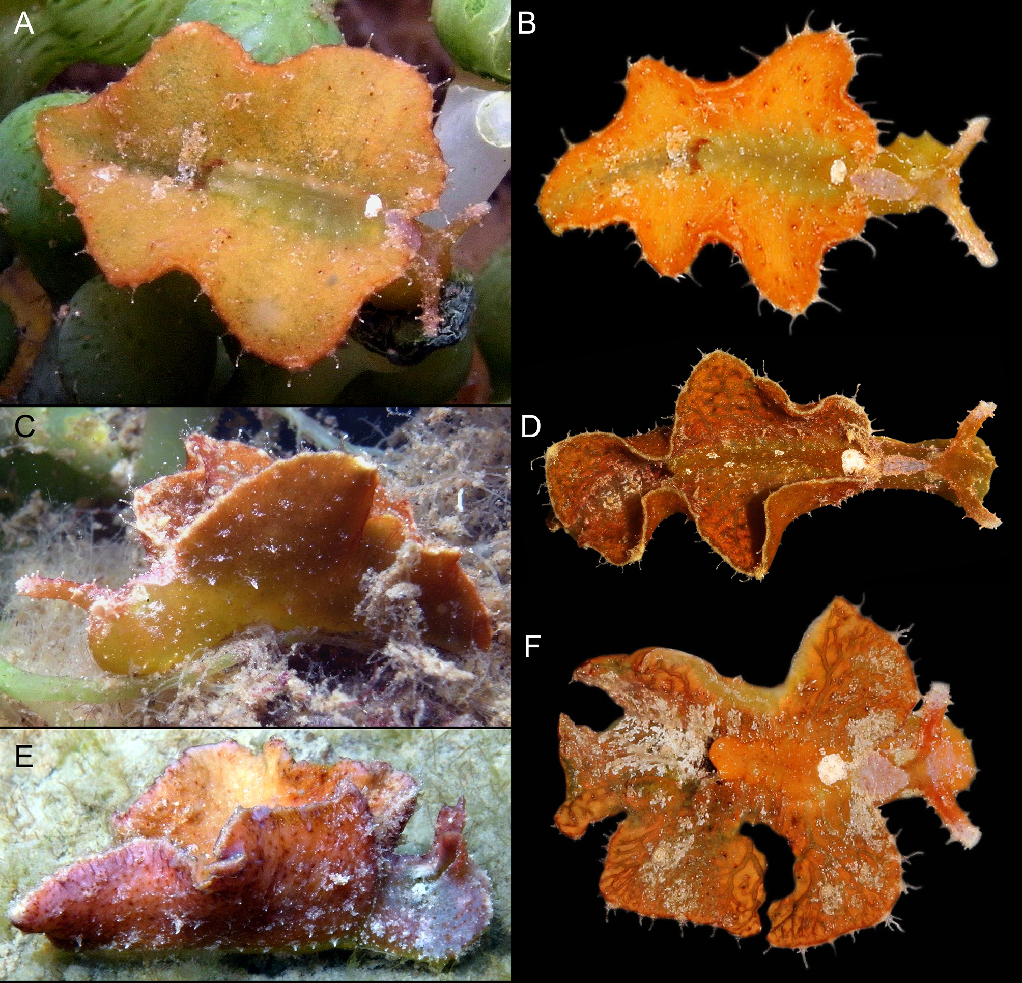
Live animal. Specimens were collected subtidally in association with the alga Caulerpa racemosa . Parapodia were often held open by the resting animal, exposing the dorsum ( Fig. 61 View FIGURE 61 A–B).
External anatomy. Overall coloration mottled orange-brown overlaying dark green ( Fig. 61 View FIGURE 61 A–C), ranging to dark red ( Fig. 61 View FIGURE 61 D, F), with pinkish-rose or whitish patches on some specimens ( Fig. 61 View FIGURE 61 E). Body shape dominated by large parapodia with two pairs of laterally extended side-flaps with rounded margins. When parapodia close over dorsum, flaps create three siphonal openings — a small anterior opening over the pericardium, and two prominent openings at the middle and posterior end of the body ( Fig. 61 View FIGURE 61 A–B, D, F). These large siphonal openings may be same size, or posterior opening smaller. Elongated middle flaps giving cruciform appearance to live animal when held open. Outer surface of body, head and rhinophores heavily dotted with elongated, hair-like, branching papillae, grey-white in color, often splitting into 2–4 branches with the central branch the longest ( Fig. 61 View FIGURE 61 A–B, D, F). Papillae imparting a hairy appearance to live animal. Posterior of body narrows to blunt end or slightly pointed tip, but no extended tail.
Head rounded in front. Top of head bearing species-diagnostic feature, a triangular or acorn-shaped patch of light grey-purple pigment, with anterior point placed medially along head and directly between eyes; posterior end narrowing slightly just before pericardium. Second patch of light grey-purple situated between rhinophores, sometimes extending anteriorly onto face, or posteriorly toward top of head. Paired white patches on upper sides of head, one above each eye, running alongside the region between the two purple patches (usually of background color) ( Fig. 61 View FIGURE 61 B–C, E–F). Irregular pigment patches like lichen may occur on sides or top of head, ranging from white to pink to grey ( Fig. 61 View FIGURE 61 E). Eyes tiny, located in patch of background coloration posterior to base of rhinophores, sometimes directly under a white pigment patch located on either side of the central purple patch atop head. Rhinophores short relative to body length, rolled, tips blunt-ended or rounded; may be held at 90° angle to head on crawling specimens, giving hammerhead shark-like appearance ( Fig. 61 View FIGURE 61 A). Rhinophores matching background body color but with light purple or grey patches at tips, and bearing rows of long white papillae.
Exterior surface of parapodia uniform in color, but dotted with elongate, white papillae of varying lengths, mostly unbranched, densely covering sides of some specimens but sparse on others. Parapodial margin thin and tan, with submarginal band of brown or dark orange. Row of elongated, thin papillae running along margin, white to clear, sometimes with swollen white tips ( Fig. 61 View FIGURE 61 A–B, D, F). Tiny, scattered, dark brown glandular inclusions dotting exterior and interior of parapodia. Interior of parapodia with scattered white flecks across whole surface.
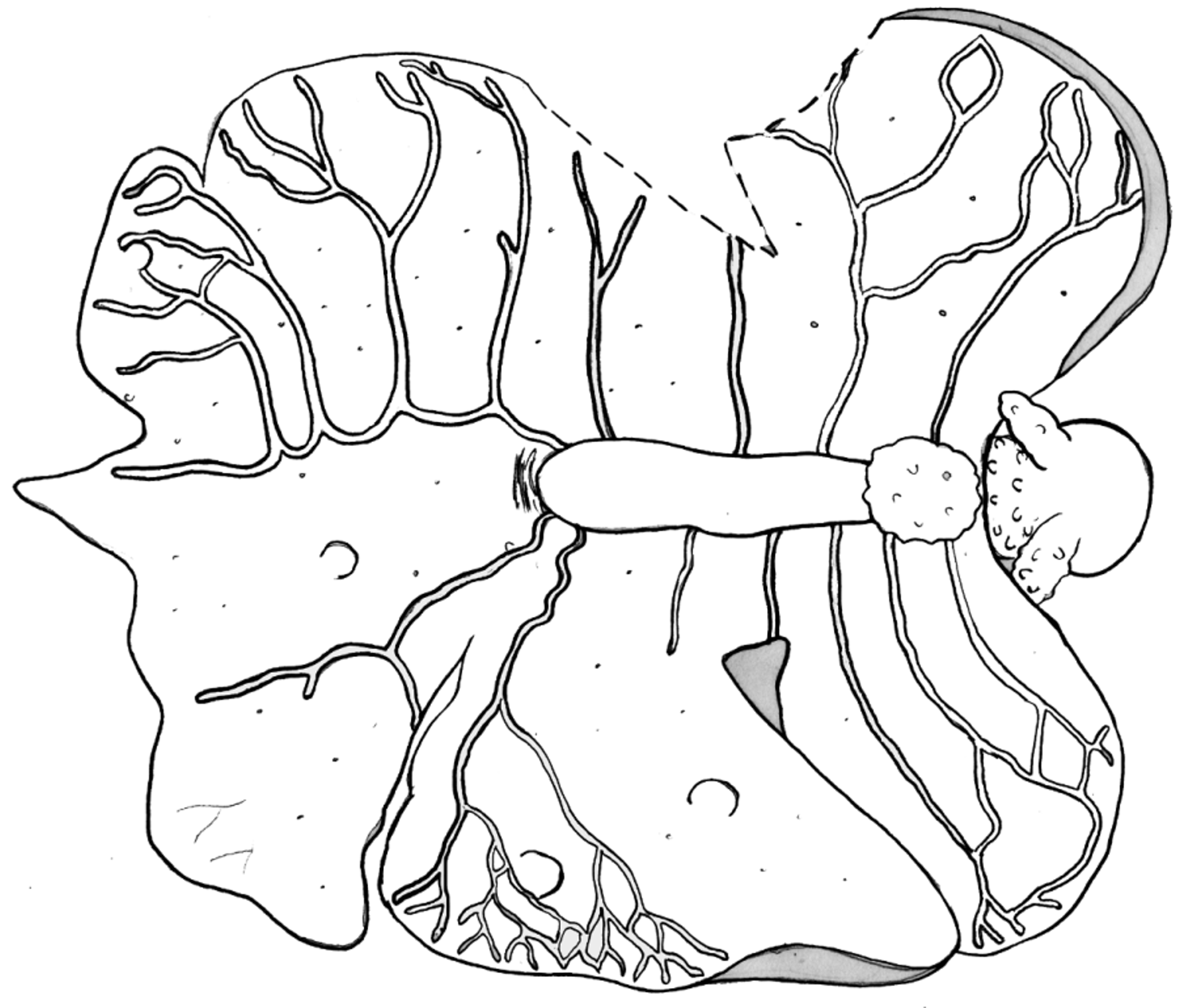
Pericardium with prominent and distinctive white round patch on top ( Fig. 61 View FIGURE 61 A–B, D, F). Thick renopericardial extension same color as dorsum, difficult to see, running halfway down body to start of second parapodial side-flap. Irregular splotch of dark pigment (brown to black) forming band on dorsum immediately posterior to end of renopericardial extension, followed by larger irregular white patches of pigment.
Dorsal vessels asymmetric, with five to seven vessels per side emerging from renopericardial complex ( Fig. 62 View FIGURE 62 ). Vessels branch into anastomosing network covering most inner parapodial surface. Elongated posterior vessel running to tail and sending off numerous lateral side branches which run to parapodial margin. Vessels clear on some specimens ( Fig. 61 View FIGURE 61 A), but notably lighter in color ( Fig. 61 View FIGURE 61 B) or much darker than dorsal surface ( Fig. 61 View FIGURE 61 D, F) on other specimens.
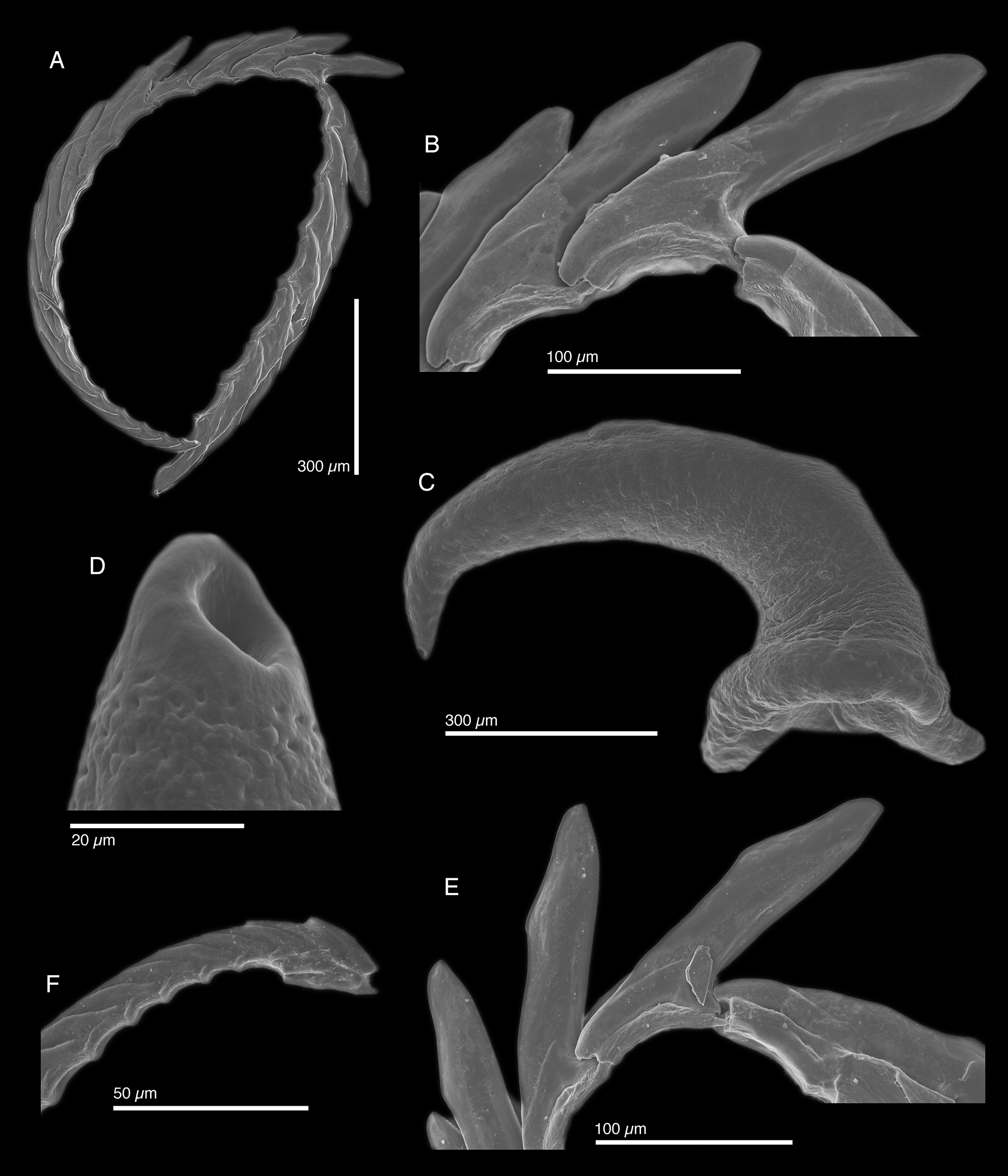
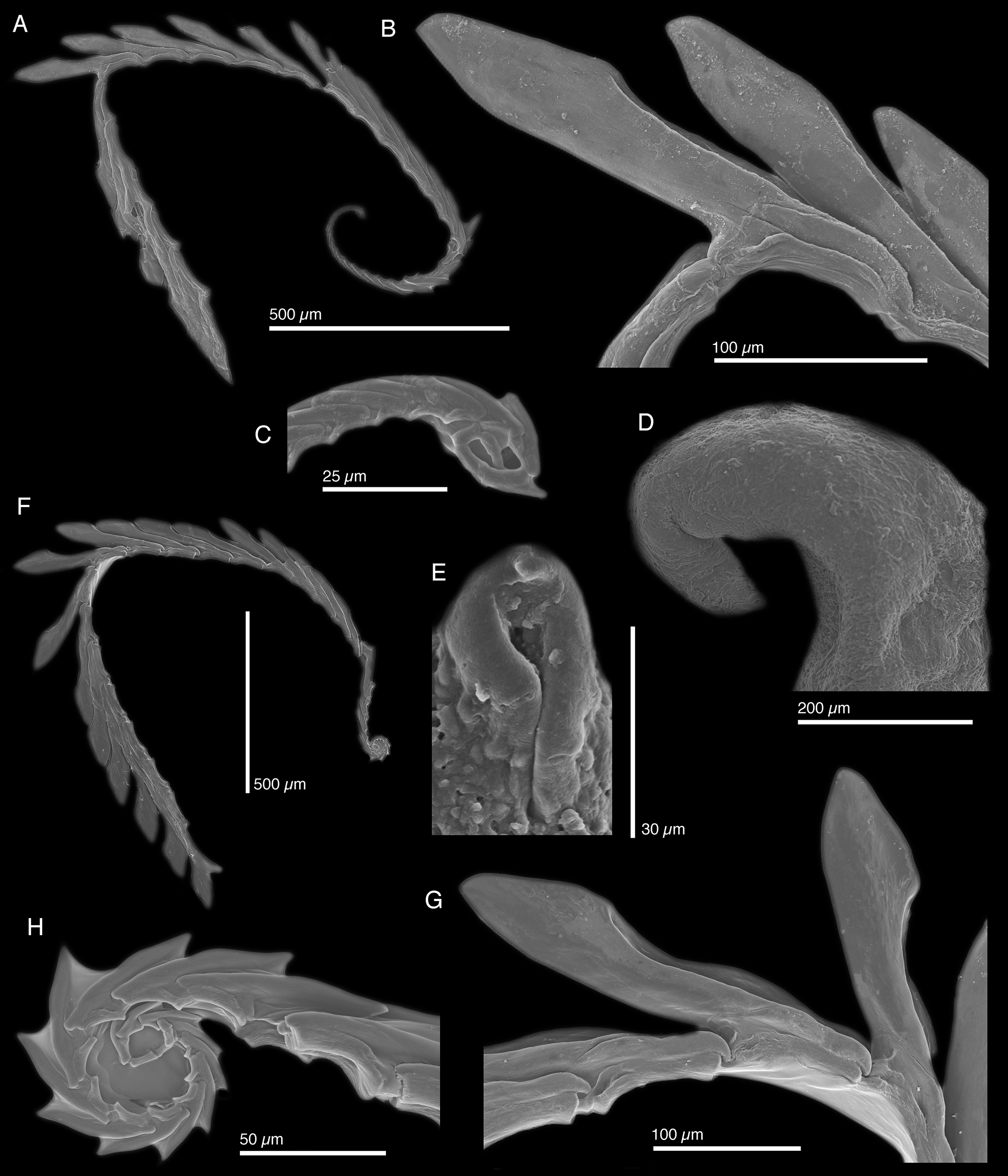
Internal anatomy. Radula with ~24 teeth (LACM 3307, LACM 178664, LACM 178666), 5–7 teeth in ascending limb and ~ 14–17 in descending limb ( Fig. 63 View FIGURE 63 A, F). Leading tooth wide and flat, with fine, blunt denticles on slightly convex cusp, with pointed tip on club-shaped apex ( Fig. 63 View FIGURE 63 B, G). Housing depression for interlocking teeth extending approximately ⅔ of the tooth length. Base of tooth approximately ⅓ total tooth length. Ascus of small teeth in a variable spiral or whorl ( Fig. 63 View FIGURE 63 C, H).
Penis elongate and curved ( Fig. 56 View FIGURE 56 F) with rigid musculature that did not deform after drying, tapering into a conical apex bearing a resistant, hollow tip ( Fig. 63 View FIGURE 63 D–E). Penial stylet is not a scoop or barb; hardened penial tip visible by SEM, but not light microscopy. Deferent duct long, thin, and highly convoluted.
Reproduction and development. No data available.
Host ecology. Specimens of E. zemi n. sp. were associated with the alga Caulerpa racemosa .
Phylogenetic relationships. Elysia zemi n. sp. belongs to subclade 4, the E. tomentosa complex, which includes related Caribbean species E. subornata , E. pratensis and E. pawliki n. sp. ( Fig. 4 View FIGURE 4 ). No sister species was recovered with significant support.
Range. Cayman Islands (present study), Martinique (present study), Saint Vincent and the Grenadines (present study), Venezuela (Valdés et al. 2006).
Etymology. Named after zemí , an embodiment of natural forces and ancestral spirits of the Taíno , an indigenous people of the Lesser Antilles. The distinctive triangular purple patch on the head of this species resembles the shape of the three-pointed sculptural zemí carved by the Taíno .
Remarks. A large elysiid, E. zemi n. sp. appears to be rare in most of the Caribbean, but locally common in Martinique and the Cayman Islands. Morphologically, the most similar Caribbean species is E. pawliki n. sp., while the most similar species from the eastern Atlantic is E. manriquei Ortea & Moro 2009 , from the Canary Islands off of West Africa. The purple patch on top of the head, white pericardium, brown marking just posterior to the renopericardial extension, and elongated hair-like papillae all differentiate E. zemi n. sp. from E. pawliki n. sp.
In addition to its east Atlantic type locality, E. manriquei differs in having symmetrical and non-anastomosing dorsal vessels, large black spots dotting the entire body and head, and more vertically exaggerated siphonal openings. Molecular data clearly differentiate E. zemi n. sp. from all other members of subclade 4.
The radular teeth of E. zemi n. sp. are most similar to E. pawliki n. sp. in overall morphology, but can be clearly distinguished. Radulae of both species have minute denticles, possess a sharp tooth tip on a rounded apex, and a bend of the radula at ⅔ the length of the tooth. The teeth of E. zemi n. sp. are distinct in having convex tooth cusps, concave “V”-shaped depressions, and wider, more club-like tooth apices. The ascus of E. zemi n. sp. also forms a spiral that varies in size and number of teeth ( Fig. 63 View FIGURE 63 C, H), not featured in E. pawliki n. sp. ( Fig. 60 View FIGURE 60 F). The tooth of E. manriquei has a flat, serrated cutting edge, and smooth top edge that curves towards a pointed tip, like a disposable plastic knife.
Like in E. pawliki n. sp., the resistant penial tip of E. zemi n. sp. is not scoop or barb-like. Instead, the stylet of E. zemi n. sp. appears as a curved cuticle with a “seam” observable under SEM ( Fig. 63 View FIGURE 63 E).


| LACM |
Natural History Museum of Los Angeles County |
No known copyright restrictions apply. See Agosti, D., Egloff, W., 2009. Taxonomic information exchange and copyright: the Plazi approach. BMC Research Notes 2009, 2:53 for further explanation.