
Cilliba sellnicki (Hirschmann & Zirngiebl-Nicol)
|
publication ID |
https://doi.org/ 10.5281/zenodo.184196 |
|
DOI |
https://doi.org/10.5281/zenodo.5664155 |
|
persistent identifier |
https://treatment.plazi.org/id/A07887BC-163F-FF9F-FF7A-83E41163FAE8 |
|
treatment provided by |
Plazi |
|
scientific name |
Cilliba sellnicki (Hirschmann & Zirngiebl-Nicol) |
| status |
|
Cilliba sellnicki (Hirschmann & Zirngiebl-Nicol)
Uropoda (Cilliba) sellnicki Hirschmann & Zirngiebl-Nicol, 1964: 19 , Fig. 15 View FIGURES 11 – 16 ; Hirschmann & Zirngiebl-Nicol, 1965: 4; 1969a: 30; Gwiazdowicz & Biernacik, 2000: 205; Hirschmann, 1979a: 21; 1979b: 79; 1993: 363; Wiśniewski, 1993a: 256; 1993b: 421; Wiśniewski & Hirschmann, 1993: 193.
Cilliba sellnicki .— Athias-Binche, 1979: 570; Bloszyk, 1992: 324.
Uropoda (Cilliba) sopronensis Wiśniewski & Hirschmann, 1990: 157 ; Wiśniewski 1993a: 265; 1993b: 421; Wiśniewski & Hirschmann, 1990: 157; Wiśniewski & Hirschmann, 1993: 193; Bloszyk et al., 2004: 1506 (synonymy by Bloszyk, 1999).
Uropoda sopronensis .— Mašán, 2001: 287.
Uropoda soproniensis (sic).— Kontschán, 2003a: 55; 2003b: 119.
Material examined. 5 ΨΨ, 3 ɗɗ, apparently syntypes, slides numbered 28a/1, 28a/3, 1392, no other data ( BSCZ).
Other material examined: Croatia: 1 Ψ, Postojne (1 sample, 1963); 2 ΨΨ, 1 ɗ, Plitrièka Jezera (1 sample, 1963). Germany: 6 ΨΨ, 10 ɗɗ, Schwaingangaer Valley (1 sample, 1945). Iran: 16 ΨΨ, 20 ɗɗ, Shahsavar, 30°51'09" N, 61°35'14" E (1 sample, 1972); 8 ΨΨ, 11 ɗɗ, Chalus, 37°00'00" N, 52°00'00" E (1 sample, 1972). Poland: 1 ɗ, Czartowe Pole Res., FA 48 (1 sample, 1971); 2 ΨΨ, Jasło, EA 30 (1 sample, 1977); 1 Ψ, Komañcza, EV 76 (1 sample, 1971); 45 ΨΨ, 59 ɗɗ, Orle Mts., EV 98 (3 samples, 1957–60); 4 ɗɗ, 2 DN, Bieszczady, FV 16 (1 sample, 1977); 1 Ψ, 1 ɗ, Bieszczady, FV 23 (1 sample, 1968); 1 Ψ, 1 ɗ, 1 PN, Western Bieszczady, FV 24 (2 samples, 1959, 1976); 2 ɗɗ, 3 DN, Bieszczady (2 samples, 2001). Romania: 3 ΨΨ, 3 ɗɗ, Bezirak Lasi (1 sample); 9 ΨΨ, 7 ɗɗ, Bucegi (1 sample). Russia: 2 ΨΨ, 5 ɗɗ (no other information).
Turkey: 9 ΨΨ, 9 ɗɗ, Dûzce, 36°10'00" N, 36°10'00" E (2 samples, 1977). (in AMU).
Redescription. Female. Strongly sclerotised, brown.
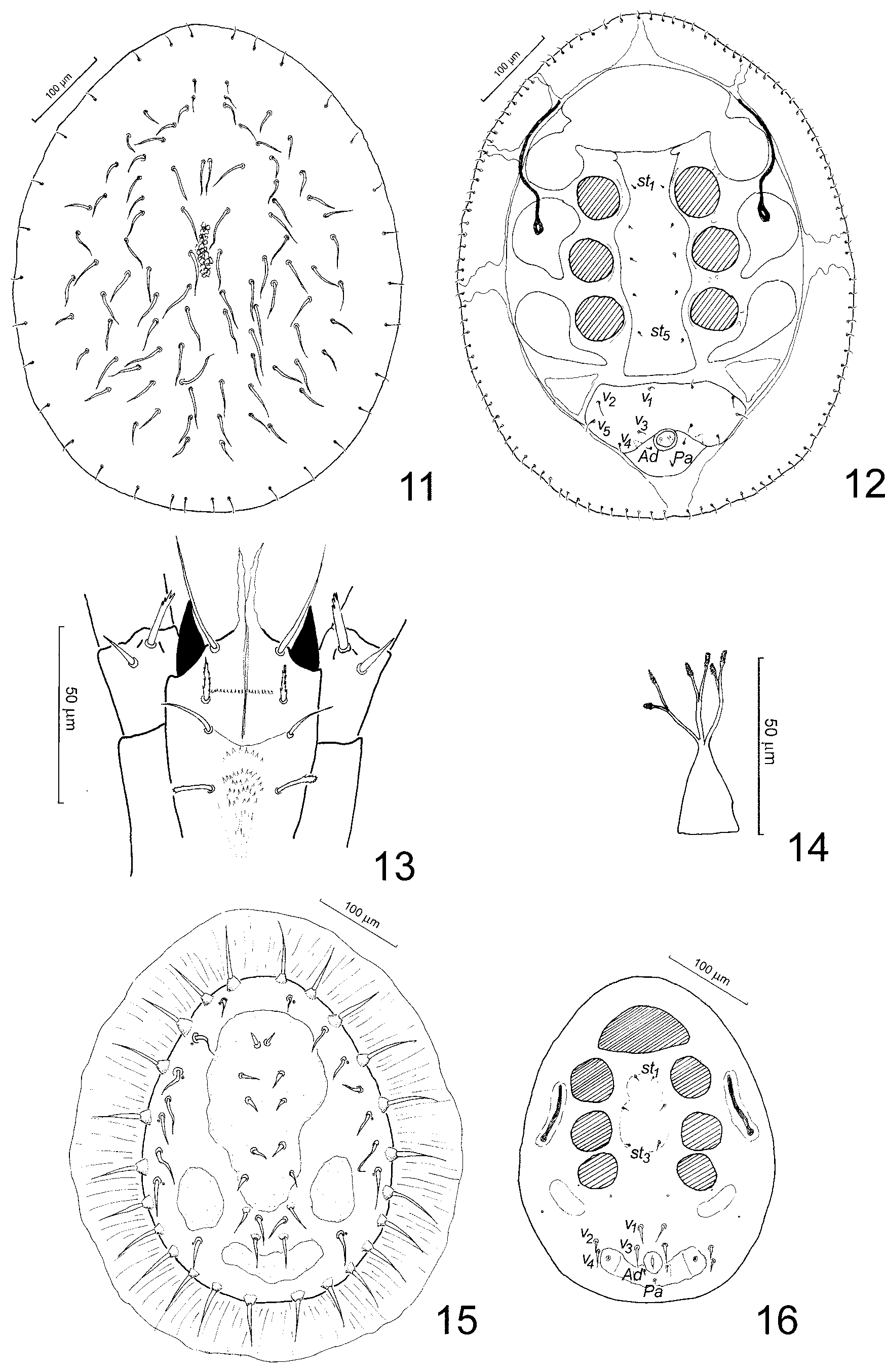
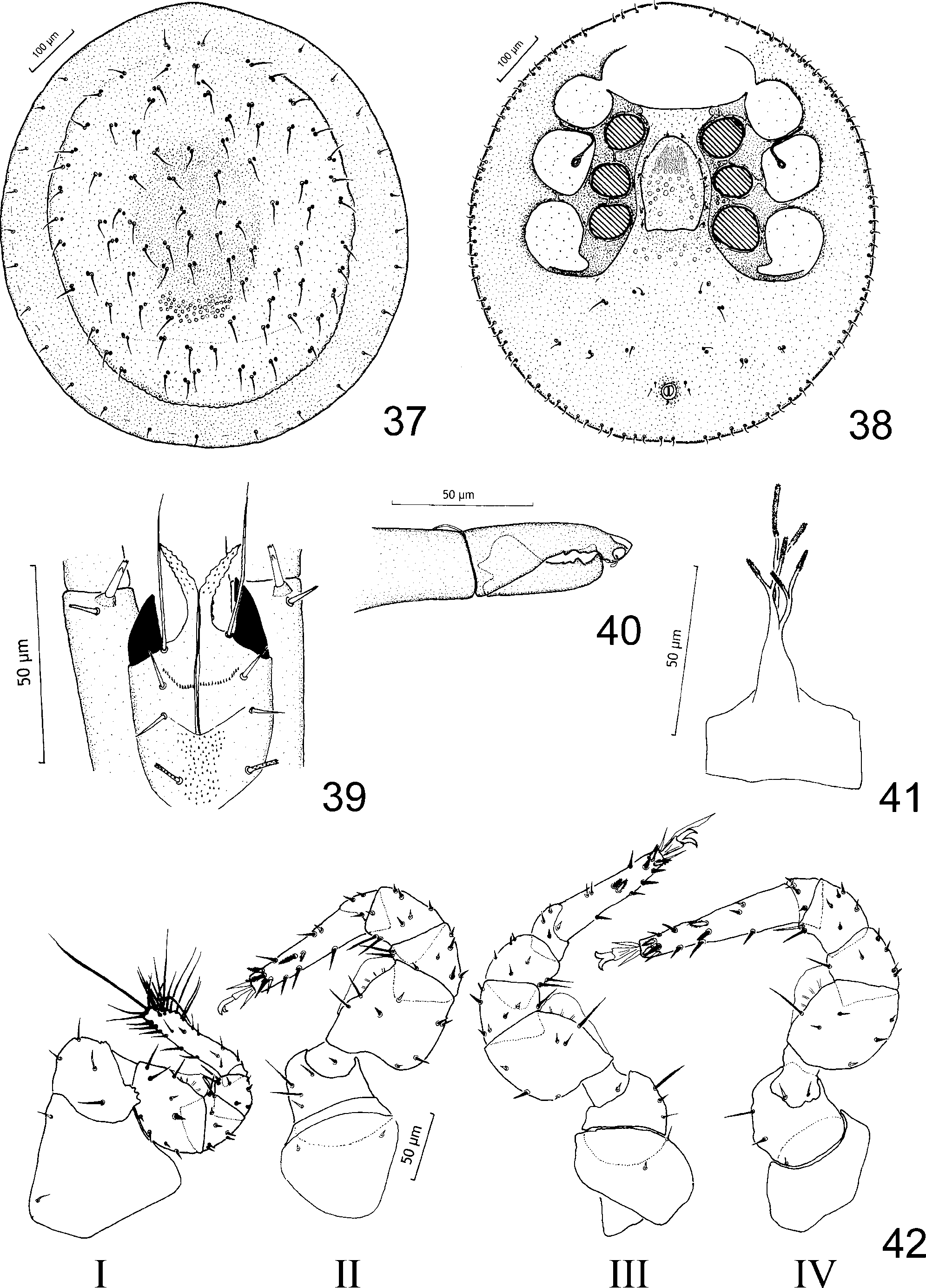
Dorsal idiosoma ( Fig. 37 View FIGURES 37 – 42 ). Length 641–773 μm, width 613–745 ìm (n = 36). Dorsal shield subcircular, cuticle smooth, with characteristic ornamentation of scattered circular pits in the posterior half. Marginal shield smooth, with numerous lyrifissures (id), anteriorly fused with dorsal shield. Dorsal setae numerous, simple (ca 35 µm long). Submarginal setae simple (ca 15 µm long), marginal setae numerous, very short, hook-like.
Ve n t r a l idiosoma ( Fig. 38 View FIGURES 37 – 42 ). Surface smooth, except for small pits on endopodal shields and anterior part of opisthosoma between coxae IV. Sternal setae st1 located close to anterior edge of epigynium, st2–st5 lateral to epigynium. Epigynium tongue-shaped (length 144–181 μm, width 91–125 μm), with distinct central ornamentation. Ventral setae (v1–v5) simple, v1 = ½ length v2; setae v4 and v5 located on shield, not on tubercles. Adanal setae Ad1 very short, Ad2 = 2 x Ad1. Unpaired seta Pa = Ad1. Anal opening small, oval. Peritremes V-shaped, length ca 123 μm. Tritosternum 6-branched, with broad base, laciniae with finely serrated tips ( Fig. 41 View FIGURES 37 – 42 ).
Gnathosoma . Epistome narrow, laterally serrated, anterior processes finely serrated. Hypostomal setae h1 very long (ca 40 μm), simple; h2 short (ca 9 μm), robust, serrated; h3 short (ca 8 μm), simple; h4 short (ca 7 μm), robust and finely serrated ( Fig. 39 View FIGURES 37 – 42 ). Hypostomal surface covered with fine denticles of various sizes; denticles grouped in single row at the level of setae h2 and distributed irregularly between setae h3–h4. Ventral setae pv1–pv2 on palp trochanters robust, pv1 ca 11 μm long, pv2 ca 7 μm long. Chelicerae of average length, fixed digit with globular sensillus and small denticles on the internal surface, movable digit shorter than fixed digit, with single denticle on internal surface ( Fig. 40 View FIGURES 37 – 42 ).
Legs ( Fig. 42 View FIGURES 37 – 42 ). Trochanter I with seta pl elongated, seta pl on femur I short, robust. Tarsi II–IV subdivided into basitarsus and telotarsus by complete peripodomeric fissure. On tarsus II proximal and distal ad and pl setae serrated. Tarsus III with proximal setae ad and pd and distal dorsal setae serrated. On tarsus IV proximal seta d and distal setae ad and pd serrated.
Male. Strongly sclerotised, brown.
Dorsal idiosoma. Length 641–783 μm, width 632–764 μm (n = 39). Ornamentation and chaetotaxy as for female.
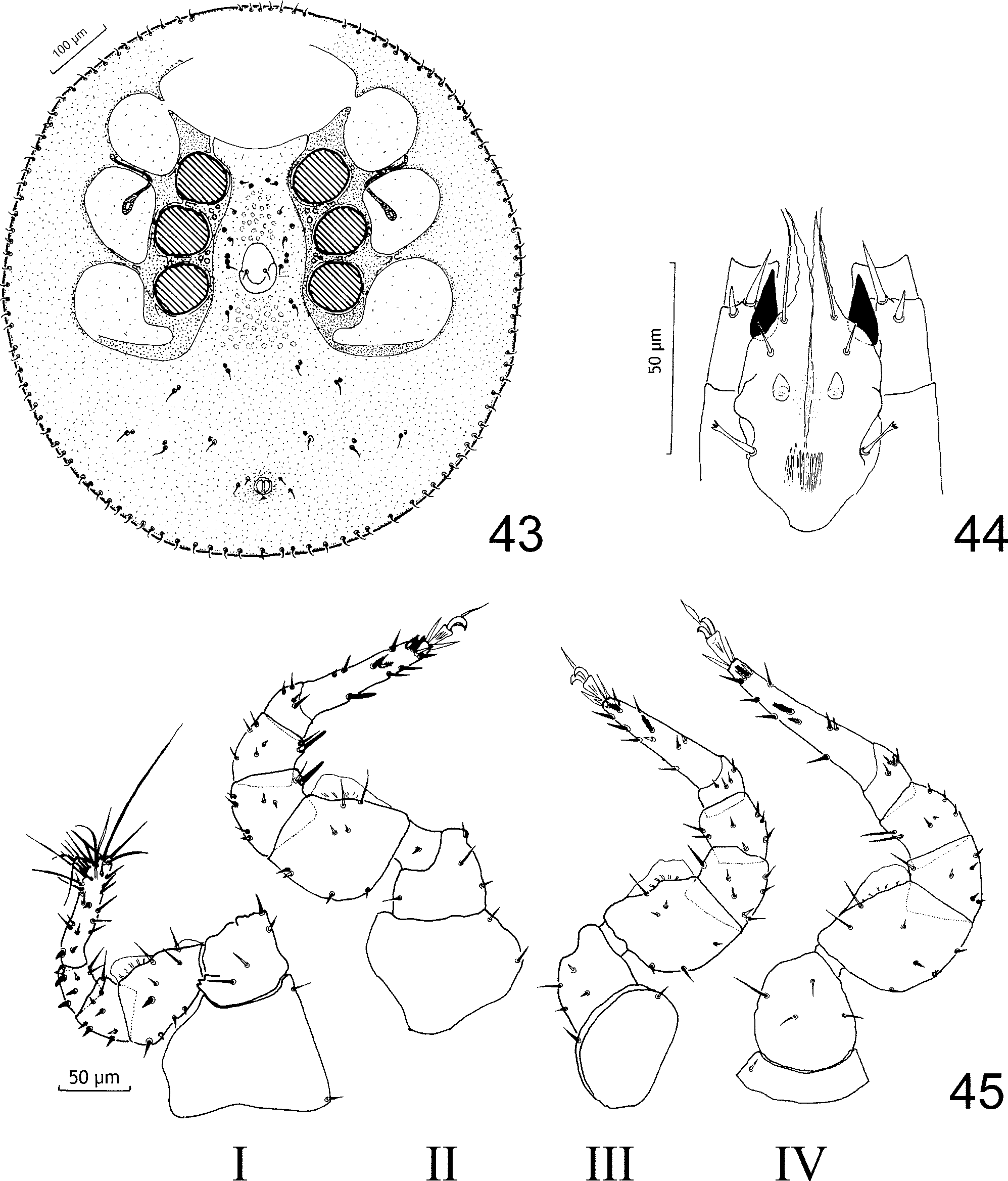
Ve n t r a l idiosoma ( Fig. 43 View FIGURES 43 – 45 ). Sternal region at level of coxae II–III and anterior part of opisthosoma ornamented with small circular pits. Operculum oval (60–79 x 45 –57 μm) with a pair of short eugenital setae, located at a level between coxae III and IV. Sternal setae st1–st2 very short, st3–st5 longer. Two pairs of glands located near setae st3. Ornamentation and chaetotaxy of opisthosoma and peritremes as for female.
Gnathosoma . Epistome as for female. Hypostomal setae h1 ca 30 μm, simple; h2 ca 8 μm, simple; h3 short (4 µm), blunt, rounded; h4 ca 13 μm, curved, serrated distally, located far from hypostomal axis ( Fig. 44 View FIGURES 43 – 45 ). Hypostomal groove smooth. Ventral setae pv1–pv2 on palp trochanters robust, pv1 ca 16 µm long, pv2 ca 7 µm.
Legs ( Fig. 45 View FIGURES 43 – 45 ). Structure and chaetotaxy as for female, except setae ad on trochanter I located on small tubercles, seta pl on femur robust and short, dorsal setae of genu and tibia robust, short. Tarsus II–IV subdivided into two segments. On genu and tibia II setae av robust, massive; on tarsus II setae al and pl and distal dorsal setae ad and pd serrated. Proximal and distal seta pd on tarsus III serrated. On tarsus IV setae pd and proximal seta ad serrated.
Deutonymph. Partly sclerotised, colour yellowish to light brown.
Dorsal idiosoma ( Fig. 46 View FIGURES 46 – 51 ). Length ca 634 µm, width ca 593 µm. Dorsal shield subcircular, smooth, marginal shield absent. Dorsal setae numerous, simple (ca 42 μm); some of them accompanied by circular pores. Between the setae ca 6 pairs of dorsal lyrifissures (id). Submarginal setae simple (ca 18 μm). Marginal setae very numerous, short, hook-like.
Ve n t r a l idiosoma ( Fig. 47 View FIGURES 46 – 51 ). Sternal shield amphora-shaped, with narrow base, finely ornamented in central and posterior part, extending behind coxae IV (ca 335 μm). Sternal setae (st1–st5) simple. Two pairs of lyrifissures: iv1 near camerostome, iv2 behind setae st5. Ventral shield wide, smooth. Ventral setae short, v1 = 1/3 length v2. One pair of lyrifissures (iv3) located near setae v2. Anal shield triangular, anterior and postero-lateral margins convex, anal opening covered by a small valve and a pair of very short setae Ad1. Setae Ad2 and Pa on shield surrounding the anus, Ad2 = Pa. Peritreme ca 190 μm long, with characteristic curve at level of coxae II, without poststigmatic extension, prestigmatic section reaching camerostome, stigmata at level of coxae III.
Gnathosoma . Epistome narrow and serrated, distally bifid. Structure of hypostome and tritosternum similar to female.
Protonymph. Weakly sclerotised, colour white to yellowish.
Dorsal idiosoma ( Fig. 48 View FIGURES 46 – 51 ). Length ca 464 µm, width ca 361 µm. Podonotal shield pear-shaped, large, smooth. Mesopodal shields small (ca 71 μm), elongate; mesonotal shields irregular in shape (diameter ca 88 μm); pygidial shield crescent-shaped (ca 149 μm in width); all shields smooth. Setae j3–j6 and z5 simple, short (ca 21 μm). Setae Z1, J1 and J2 longer, inserted on pleura between podonotal and pygidial shield. Setae j2, z2, z3, s3–6, S1, Z2 short (ca 33 µm), positioned submarginally; J4 longer (ca 60 μm), inserted on small protuberances. Setae j1, s2, r3–r5, R1, R3, S3, S4, Z3, Z4 and J5 long (ca 80 µm), massive, inserted on protuberances along margin of idiosoma, apparently supporting a soft, membranous fringe surrounding the idiosoma (ca 120 μm in width). Several idiosomal setae with associated circular pores.
Ve n t r a l idiosoma ( Fig. 49 View FIGURES 46 – 51 ) weakly sclerotised, smooth. Sternal shield poorly defined, with three pairs of simple sternal setae ca 14 µm long. Metapodal shields elongate (ca 85 µm), smooth. Ventral setae (v1, v3–v5) simple, short (ca 23 µm); v1 and v3 anterior to ventri-anal shield, v4 and v5 lateral to ventri-anal shield. Three pairs of glands inserted in soft ventral pleura near metapodal shields (gv1, gv2 and gl6). Ventri-anal shield oval, smooth, with anal opening, a pair of setae Ad and an unpaired seta Pa; Ad = 2 x Pa. Peritreme short, simple, without poststigmatic section, prestigmatic section straight, ca 64 μm in length; stigmata at level of coxae III. Tritosternum typical for the genus ( Fig. 51 View FIGURES 46 – 51 ).
Gnathosoma . Structure of hypostome as for female except setae h3 long, serrated ( Fig. 50 View FIGURES 46 – 51 ). Ventral setae pv1 and pv2 on palp trochanters serrated.
Larva. Unknown.
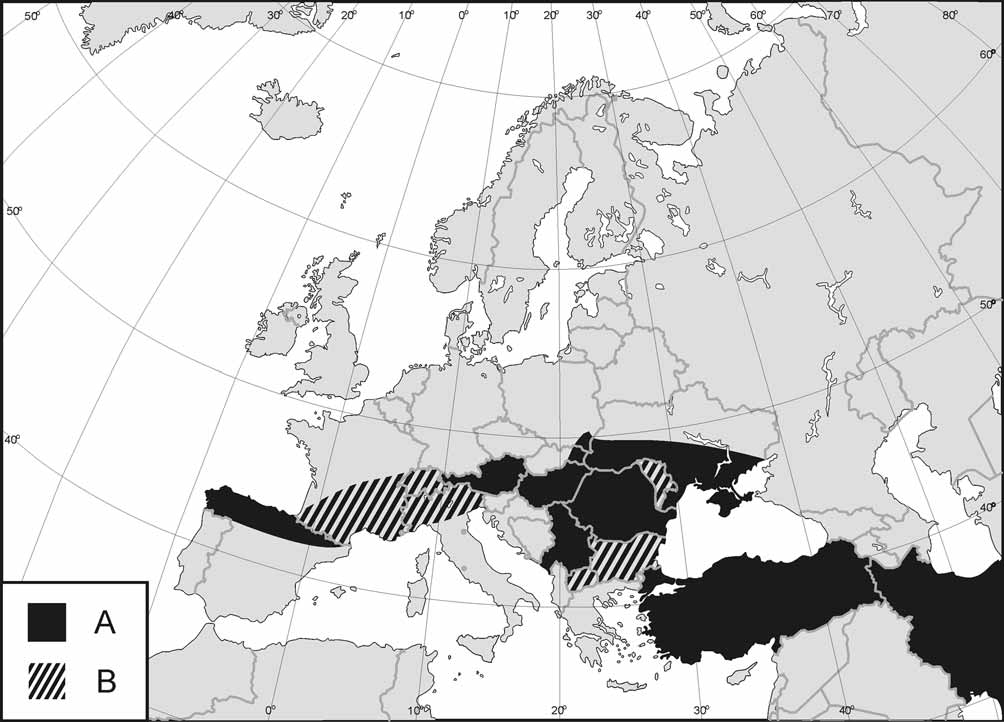
Ecology. This species is widely distributed in Europe and the near East ( Fig. 52 View FIGURE 52 ). It has been recorded from Austria, Croatia, Hungary, Iran, Poland, Slovakia, Slovenia, Spain, Turkey and Ukraine. It shows significant geographic variability, and some authors regard populations from Slovakia and Hungary as a separate species, C. sopronensis . In Poland C. sellnicki is the rarest member of the genus ( Fig. 67 View FIGURE 67 E). It inhabits oakhornbeam forests, beech-fir forests, Carpathian beech wood, mixed deciduous forests and fir forests. Several specimens were also found in rotting wood. The optimal altitude for this species is below 600 m a.s.l.


No known copyright restrictions apply. See Agosti, D., Egloff, W., 2009. Taxonomic information exchange and copyright: the Plazi approach. BMC Research Notes 2009, 2:53 for further explanation.
|
Kingdom |
|
|
Phylum |
|
|
Class |
|
|
Order |
|
|
Family |
|
|
Genus |
Cilliba sellnicki (Hirschmann & Zirngiebl-Nicol)
| Stachowiak, Marcin, Halliday, Bruce & Bloszyk, Jerzy 2008 |
Uropoda soproniensis
| Kontschan 2003: 55 |
Uropoda sopronensis
| Masan 2001: 287 |
Uropoda (Cilliba) sopronensis Wiśniewski & Hirschmann, 1990 : 157
| Bloszyk 2004: 1506 |
| Wisniewski 1993: 265 |
| Wisniewski 1993: 193 |
| Wisniewski 1990: 157 |
| Wisniewski 1990: 157 |
Cilliba sellnicki
| Bloszyk 1992: 324 |
| Athias-Binche 1979: 570 |
Uropoda (Cilliba) sellnicki
| Gwiazdowicz 2000: 205 |
| Wisniewski 1993: 256 |
| Wisniewski 1993: 193 |
| Hirschmann 1979: 21 |
| Hirschmann 1965: 4 |
| Hirschmann 1964: 19 |