
Macvicaria adomeae, Aken'Ova & Cribb & Bray, 2008
|
publication ID |
https://doi.org/ 10.3897/zookeys.1.8 |
|
publication LSID |
lsid:zoobank.org:pub:66595057-9C2C-4AEF-AD29-9E2F52BF99FD |
|
DOI |
https://doi.org/10.5281/zenodo.3793489 |
|
persistent identifier |
https://treatment.plazi.org/id/A0798785-8534-563C-4F30-DDF9FBE1F933 |
|
treatment provided by |
Plazi |
|
scientific name |
Macvicaria adomeae |
| status |
sp. nov. |
Macvicaria adomeae View in CoL n. sp.
Type-host: Sillaginodes punctatus (Cuvier) (Sillaginidae) .
Type-locality: Off American River, South Australia 35°48’S, 137°46’E.
Site: Gut.
Material studied: 9 Off American River, South Australia, December 1995.
Type-material: Holotype: QM G 230424, paratypes: QM G 230425 - 230427, BMNH 2008.7.5.44-45.
urn:lsid:zoobank.org:act:EBF2012C-678A-4BEA-8C97-5DE132F6AB60
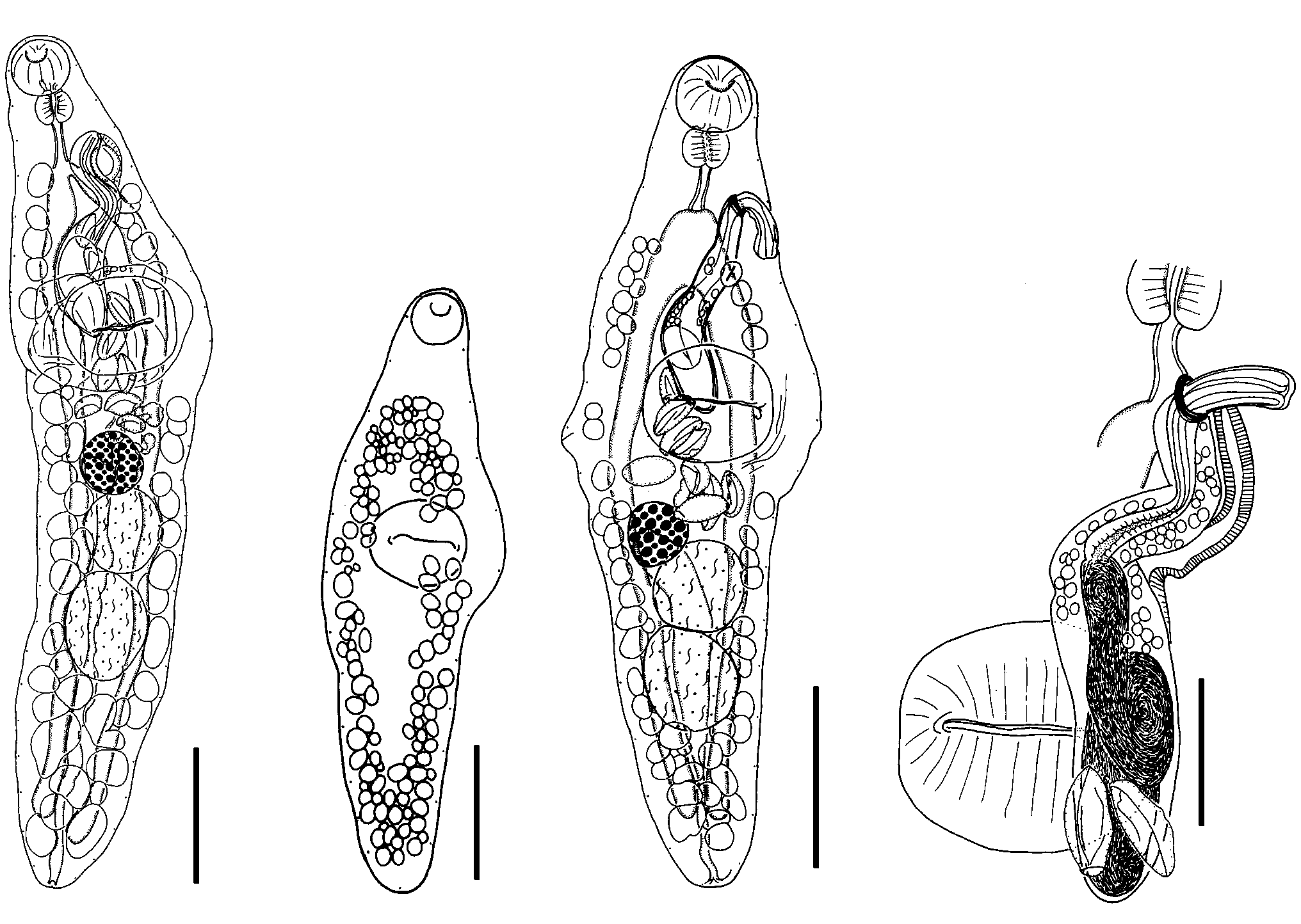
Description ( Figs 33-36 View Figs 33-36 )
Based on 8 unflattened, whole-mount specimens and 1 set of serial sagittal sections and measurements of 5 gravid unflattened whole-mount worms.
Body elongate, spindle shaped, maximum width in region of ventral sucker, 1,140 -1,578 (1,315) × 302-363 (331); width to length ratio 1:3.4-4.3 (4.0). Oral sucker globular, opening ventrally subterminal, 88-104 (95) × 98-110 (113). Ventral sucker transversely oval, in anterior third of body, 158-179 (167) × 181-213 (101); sucker width ratio 1:1.8-1.9 (1.9). Forebody 358-456 (396) long, 27-35 (30)% of body-length. Prepharynx short, dorsal to oral sucker. Pharynx subglobular, 48-59 (55) × 60-70 (65); pharynx to oral sucker width ratio 1:1.5-1.6 (1.6). Oesophagus distinct, short. Intestinal bifurcation in forebody, 107-166 (134) anterior to ventral sucker. Caeca terminate blindly close to posterior extremity. Excretory pore ventrally subterminal.
Testes 2, oval, entire, contiguous to slightly separated, tandem, in posterior half of body, anterior 115-161 (136) × 121-145 (131), posterior 147-193 (166) × 121-159 (143). Post-testicular area 213-387 (300) long, 19-27 (23)% of body-length. Cirrussac elongate, clavate, thick walled, extends from point just posterior to posterior margin of pharynx, overlaps ventral sucker dorsally to its aperture (n=2), sometimes to level of (n=2) or posterior to posterior margin of ventral sucker (n=2), 303-412 (371) × 61-92 (74). Internal seminal vesicle tubular, sinuous, broadest at posterior end, fills broad posterior portion of cirrus-sac, surrounded by gland cells anteriorly. Pars prostatica distinct, thick walled, surrounded by gland cells. Ejaculatory duct long, thick walled. Genital atrium small. Genital pore antero-sinistral to intestinal bifurcation,
midway between lateral margin and oesophagus, usually with cirrus protruding, (n=4), 189-209 (199) from anterior end, 13-18 (15)% of body-length.
Ovary entire, spherical, contiguously anterior to or antero-dextral to anterior testis, 73-117 (89) × 78-115 (95). Mehlis’ gland indistinct, usually anterior to ovary, occasionally sinistral (n=1) to ovary. Canalicular seminal receptacle saccular, usually dorsal (n=4), sometimes antero-dextral (n=1), or sinistral to ovary, overlapping left side of ovary and anterior portion of anterior testis (n=1). Laurer’s canal present, opens dorso-sinistrally to ovary. Uterus coils intercaecally between anterior testis and ventral sucker, sometimes overlaps caeca ventrally, and ovary and testis dorsally, then passes to genital pore without coiling. Metraterm distinct, thick walled, overlaps left caecum. Eggs few, large, operculate, oval, 61-78 (70) × 28-50 (37). Vitelline follicles extend from 182-202 (194) from anterior extremity, 19-27 (23)% of body-length, to 11-38 (26) from posterior extremity; lateral fields may be continuous (n=3) or interrupted in ventral sucker area (n=3), ventral fields separate in forebody, and posteriorly to posterior margin of posterior testis, confluent or almost in post-testicular area; dorsal field confluent in forebody and post-testicular area always with continuous medial and sometimes bilateral or unilateral interruption in ventral sucker, uterine and gonad areas; follicles lie lateral, ventral and dorsal to caeca; anterior limit sometimes level with posterior end of oesophagus (n=3) or more anteriorly to point roughly level with mid-way between anterior and posterior ends of oesophagus (n=3).
Excretory vesicle I-shaped, with narrow posterior end surrounded by few gland cells, passes anteriorly to point dorsal to posterior third of ovary.
Etymology: This species is named for the mother of the first author.
Comments: Macvicaria adomeae n. sp. can be accommodated in Group D as outlined above and can be distinguished from other species as follows:
M. antarctica has a smaller pharynx, a shorter forebody at 26% of the body-length, a smaller post-testicular area and distinctly smaller eggs 42-51 × 20-28.
M. georgiana has vitelline fields reaching to the pharynx, a saccular internal seminal vesicle, a shorter forebody (according to the illustrations in Zdzitowiecki, 1997) and small knobs on the anopercular pole of the eggs.
M. issaitschikowi has a shorter forebody at 27% of the body-length, a slightly shorter post-testicular area of 19% of the body-length, a smaller pharynx, a larger ventral sucker, with a sucker width ratio of 1:2.61 versus 1:1.8-1.9 (1.9), smaller eggs 57-63 × 38-40, and its caeca terminate at about the level of the posterior margin of the posterior testis ( Yamaguti, 1938) whereas they terminate well beyond the posterior margin of the posterior testis in M. adomeae .
M. muraenolepidis has a saccular internal seminal vesicle and smaller eggs (36-50 x 21-32) with small anopercular knobs.
M. heronensis can be distinguished by its slightly longer forebody at 35% of the body-length, a shorter post-testicular area at 19% of the body-length, longer and narrower eggs at 68-84 × 29-32 (76 × 31), the posterior extent of the uterus, the gonads which are situated more posteriorly and a genital pore closer to the anterior end.
| QM |
Queensland Museum |
No known copyright restrictions apply. See Agosti, D., Egloff, W., 2009. Taxonomic information exchange and copyright: the Plazi approach. BMC Research Notes 2009, 2:53 for further explanation.