
Polysyncraton pedunculatum Kott, 2001
|
publication ID |
https://doi.org/ 10.1080/00222930310001647334 |
|
DOI |
https://doi.org/10.5281/zenodo.4653940 |
|
persistent identifier |
https://treatment.plazi.org/id/A1678788-FF96-FF04-8108-45BFFD45A3F2 |
|
treatment provided by |
Carolina |
|
scientific name |
Polysyncraton pedunculatum Kott, 2001 |
| status |
|
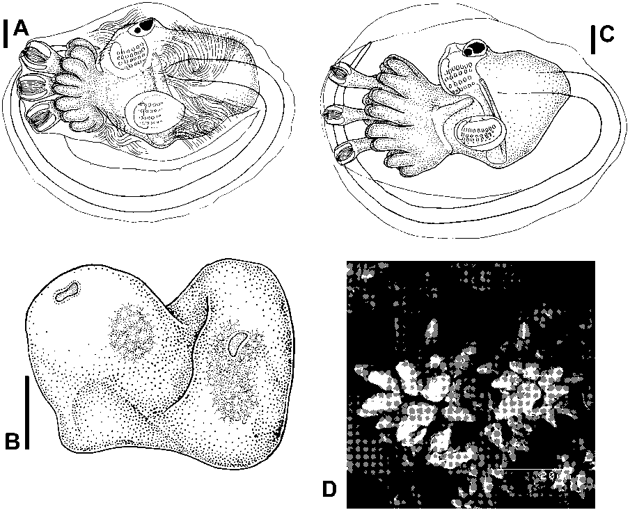
Polysyncraton pedunculatum Kott, 2001
( figure 7A View FIG )
Polysyncraton pedunculatum Kott, 2001: 121 and synonymy.
Distribution. New records: South Australia (Top Gallant I, QM GH962; Ward I., QM GH928; Franklin I., QM GH2290). Previously recorded (see Kott, 2001): South Australia.
Description. Newly recorded colonies are firm, slightly flattened, inverted cones, narrowing to short, thick, fleshy stalks, or spherical or mushroom-shaped heads with the stalk from the centre of the under surface. Although Kott (2001) refers to an extensive posterior common cloacal cavity separating the zooid layer from the central test, this cavity occurs only in a specimen from the Yorke Peninsular (QM G302867). Zooids are along each side of thoracic primary common cloacal canals that form a network with the surface depressed slightly over these canals. There are no posterior abdominal cavities. Colonies are reported to have been dark blue or purple in life.
Zooids are long, as previously described, with a short narrow atrial lip, long narrow thorax, retractor muscle from halfway down a long oesophageal neck, and four loose coils of the vas deferens around six or seven loose testis follicles.
Larvae are in the central test of specimens collected in March (QM GH962). The large (0.8 mm long), robust trunk has the tail wound three-quarters of the way around it. Conspicuous striations are on the surface of the test. A spherical yolk mass is in the anterior part of the trunk behind six broad ectodermal ampullae on each side of the three antero-median adhesive organs. A waist separates the ampullae from the mass of the larval trunk in more advanced larvae but is not conspicuous. A thoracic blastozooid is at the base of the oozooid gut loop on the left. An external horizontal ampulla was not detected.
Remarks. The colonies resemble those of Polysyncraton rica , although that species has sessile colonies (without a stalk), spicules in a layer beneath the surface, a fine retractor muscle from the top of the oesophageal neck, only three coils of the vas deferens, sometimes a particularly large atrial tongue, a posterior abdominal cavity always separating the central test from the zooid layer and zooids in clumps rather than along each side of common cloacal canals. The larva of the present species previously was not known. Although it is the same length as that of P. rica and has a single blastozooid, it differs in its six (rather than eight) long, narrow lateral ampullae on each side of the antero-median adhesive organs, a less pronounced waist, striations on the larval test and it lacks a conspicuous horizontal ampulla on the left side of the trunk.
The appearance of living colonies of this species is not known, photographs of colonies of P. rica having been wrongly assigned to it ( Kott, 1997: pl. 85; Kott, 2001: pl. 6D; see below, P. rica ).
No known copyright restrictions apply. See Agosti, D., Egloff, W., 2009. Taxonomic information exchange and copyright: the Plazi approach. BMC Research Notes 2009, 2:53 for further explanation.
|
Kingdom |
|
|
Phylum |
|
|
Class |
|
|
Order |
|
|
Family |
|
|
Genus |
Polysyncraton pedunculatum Kott, 2001
| Kott, Patricia 2004 |
Polysyncraton pedunculatum Kott, 2001: 121
| KOTT, P. 2001: 121 |