
Acrorhinichthys poyatoi, Taverne & Capasso, 2015
|
publication ID |
https://doi.org/ 10.5852/ejt.2015.116 |
|
publication LSID |
lsid:zoobank.org:pub:DC7FCF48-1E85-4205-89CA-E1D49577F971 |
|
DOI |
https://doi.org/10.5281/zenodo.3795208 |
|
persistent identifier |
https://treatment.plazi.org/id/49D71C02-0276-483F-BC07-76B2FCD5897C |
|
taxon LSID |
lsid:zoobank.org:act:49D71C02-0276-483F-BC07-76B2FCD5897C |
|
treatment provided by |
Carolina |
|
scientific name |
Acrorhinichthys poyatoi |
| status |
gen. et sp. nov. |
Acrorhinichthys poyatoi gen. et sp. nov.
urn:lsid:zoobank.org:act:49D71C02-0276-483F-BC07-76B2FCD5897C
Figs 1–19 View Fig View Fig View Fig View Fig View Fig View Fig View Fig View Fig View Fig View Fig View Fig View Fig View Fig View Fig View Fig View Fig View Fig
Diagnosis
Primitive pycnodontiform fish with a deep body, a rounded ventral margin and an angular dorsal profile. Head triangular in shape, with a rectilinear frontal profile and a long preorbital region. Pointed snout, with reduced jaws. Mouth gape obliquely oriented. Premaxilla bearing 3 incisiform teeth. Dentary bearing 5 incisiform teeth. Vomer covered by numerous small, rounded teeth arranged in regular rows. Prefrontal present. Few bony plates (= tesserae) present in the gular region. Parietal without brush-like process. No temporal fenestra. Dermohyomandibula fused to hyomandibula. Preopercle much larger than the exposed part of hyomandibula. A small ectopterygoid. Tubular infraorbitals. Cleithrum with a broad ventral branch. Pectoral fin with 16 or 17 rays. Ventral fin with 4 rays. Dorsal and anal fins striplike. Dorsal fin origin located just behind dorsal apex. Dorsal fin with 51 to 55 pterygiophores. Anal fin with 36 to 41 pterygiophores. Neural and haemal arches not completely surrounding the notochord. 24 to 27 vertebral segments in front of epichordal series. 14 haemal spines in front of hypochordal series. Neural and haemal spines with anterior, wing-like expansion. One postzygapophysis on the neural and haemal arches in the caudal region. 9 to 10 pairs of broad ribs. Caudal peduncle very short. 4 or 5 epichordals. 10 or 11 hypochordals, of which 3 posterior ones slightly broadened. 2 urodermals. Caudal fin double emarginated, with 22 or 23 principal rays. Dorsal ridge containing 8 spiny scutes. The eighth dorsal scute larger and forming a small prominence at the dorsal apex. Ventral keel with 13 scutes, 2 of them postcloacal. Very large first ventral scute. A row of complete scales associated with the dorsal scutes. Scales only in the abdominal region, with scale bars in the dorsal part and complete scales in the ventral part. 2 imbricated scales above the cloaca, 1 small, triangle-shaped ventral one and 1 larger, dorsal one with a broad and concave ventral margin.
Etymology
The species name of the new Lebanese fossil fish is dedicated to the Spanish palaeontologist Francesco Poyato-Ariza, who has greatly improved our knowledge of pycnodontiform fishes.
Holotype
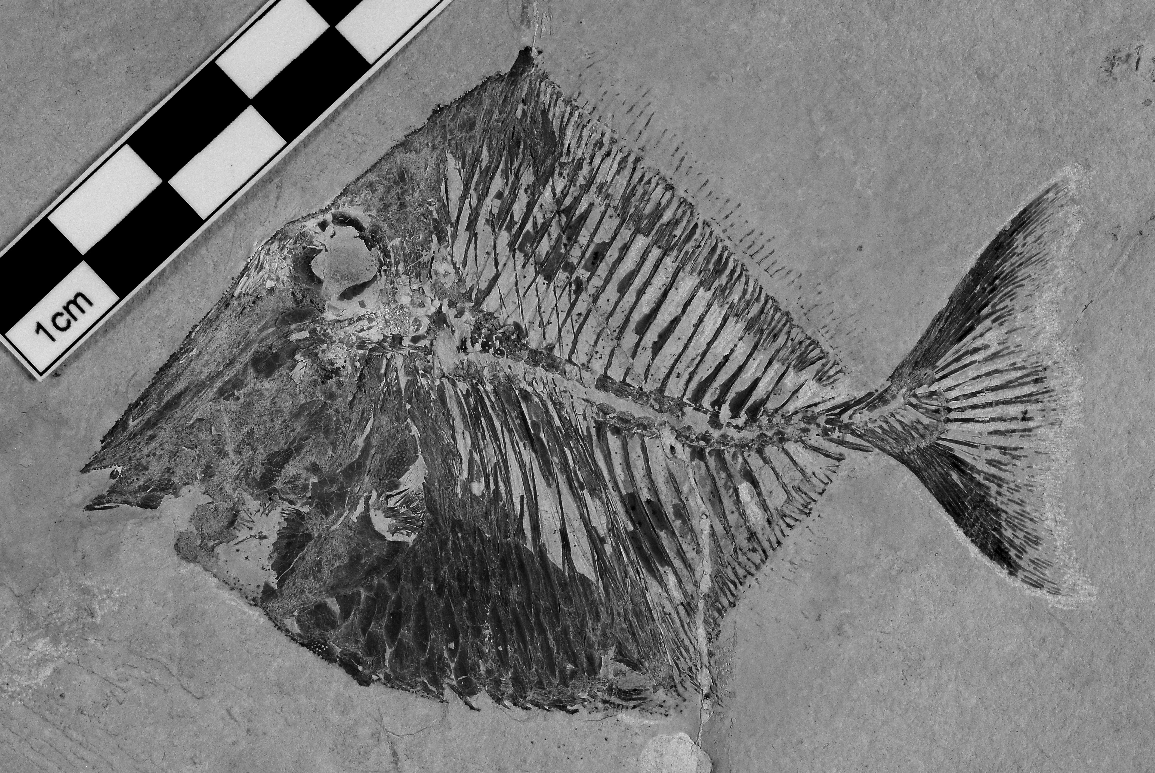
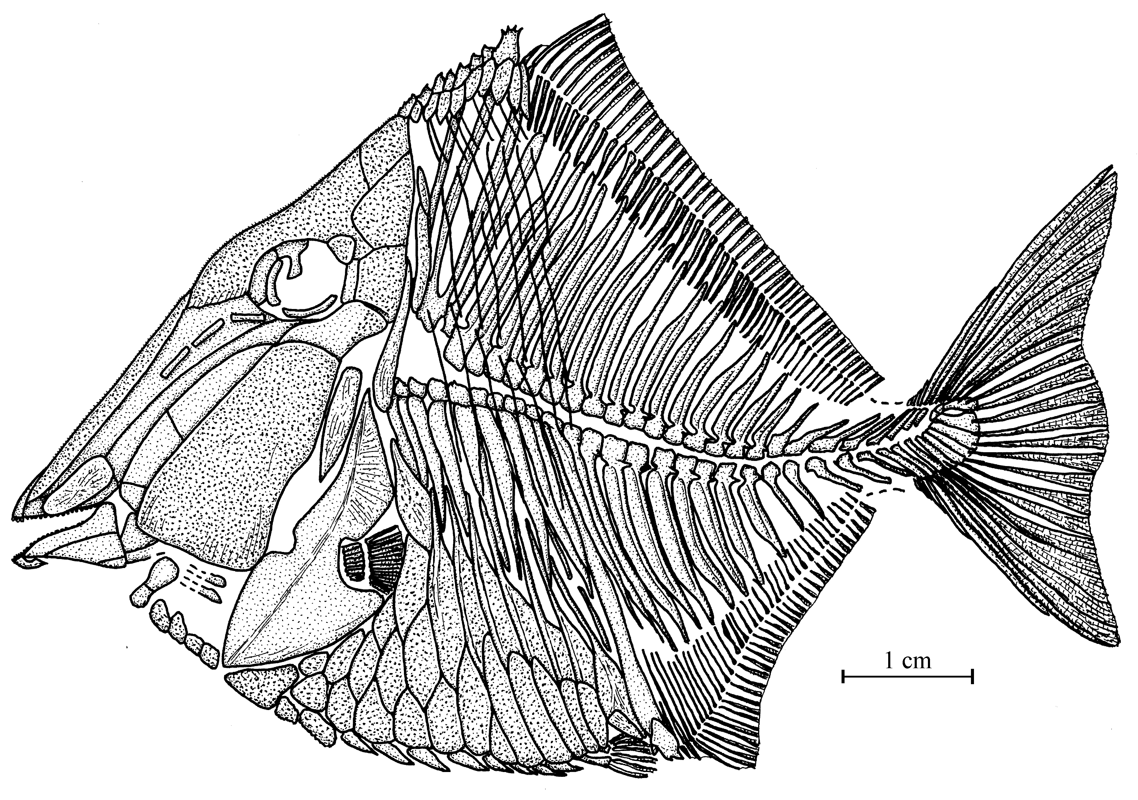
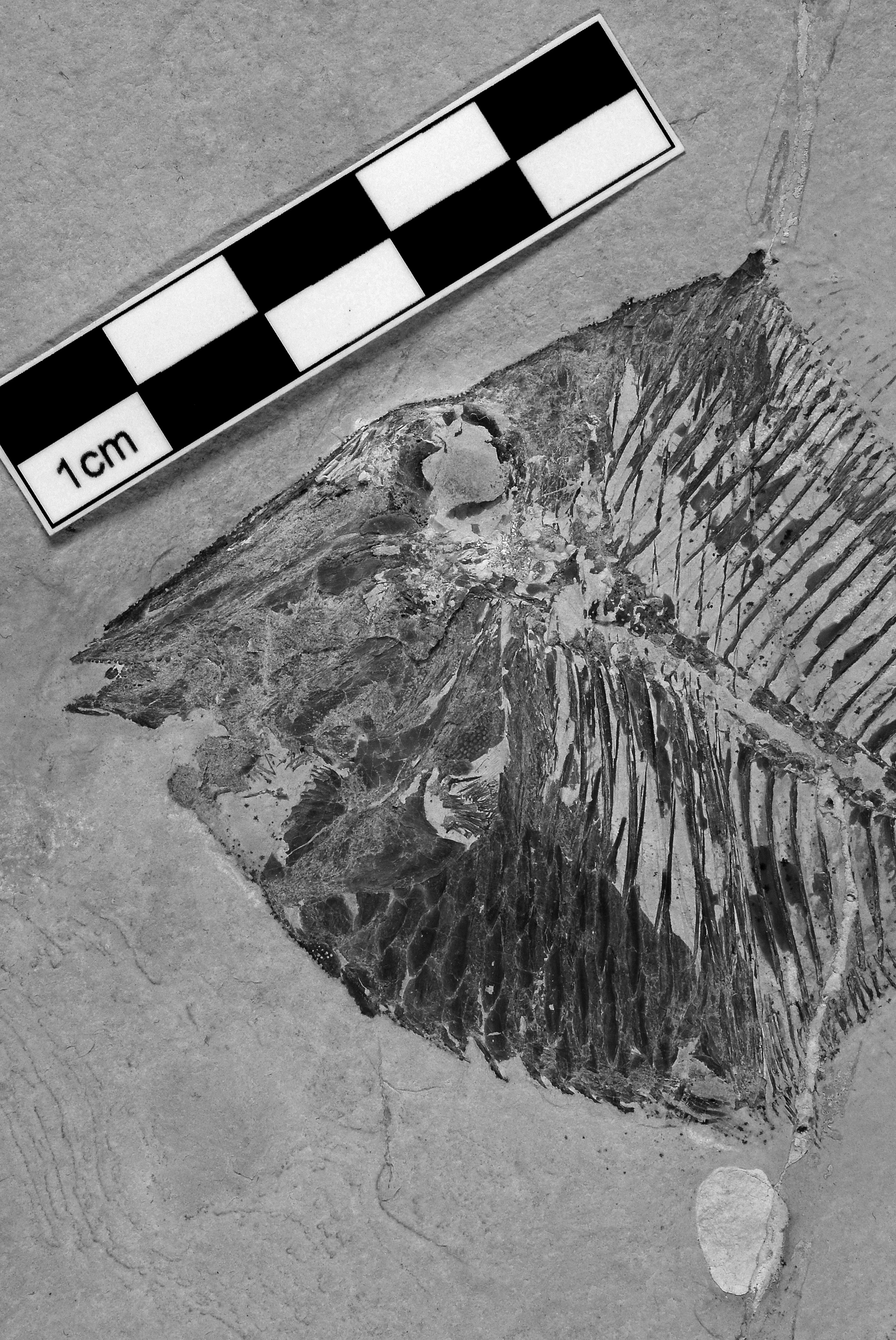
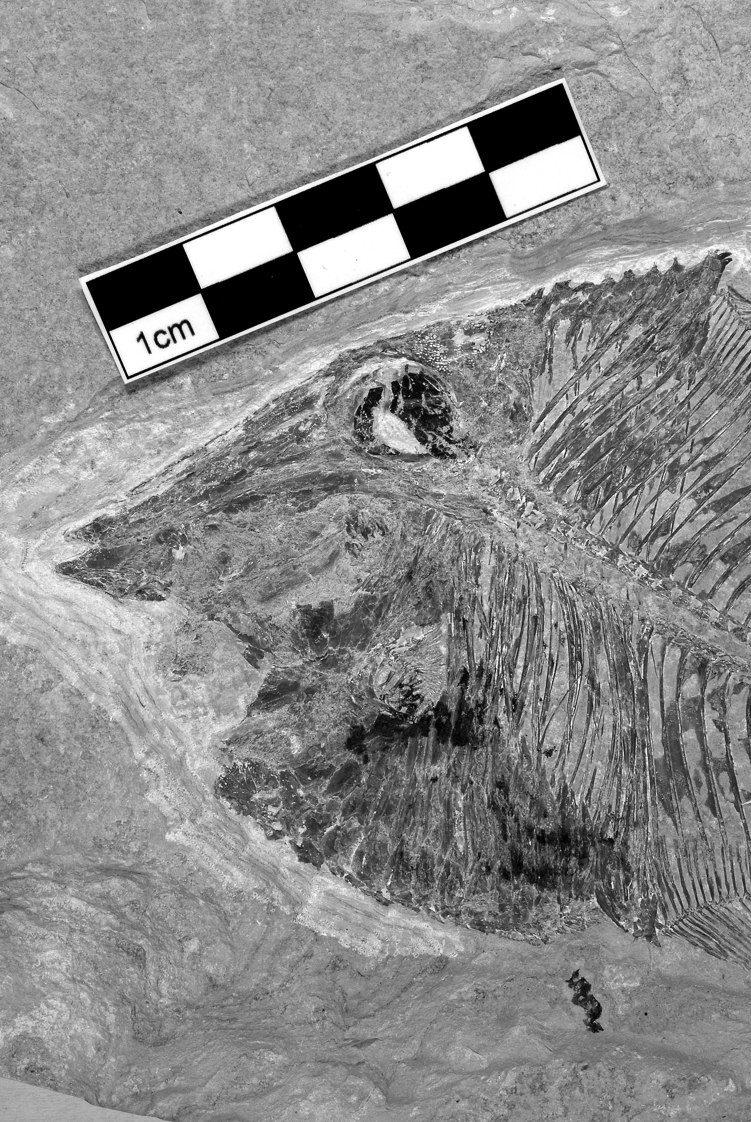
Sample CLC S-630a, b, part and counterpart of a complete specimen from Ein Namoura, Lebanon ( Figs 1–2 View Fig View Fig ). Total length: 87 mm. Standard length: 72 mm.
Paratypes
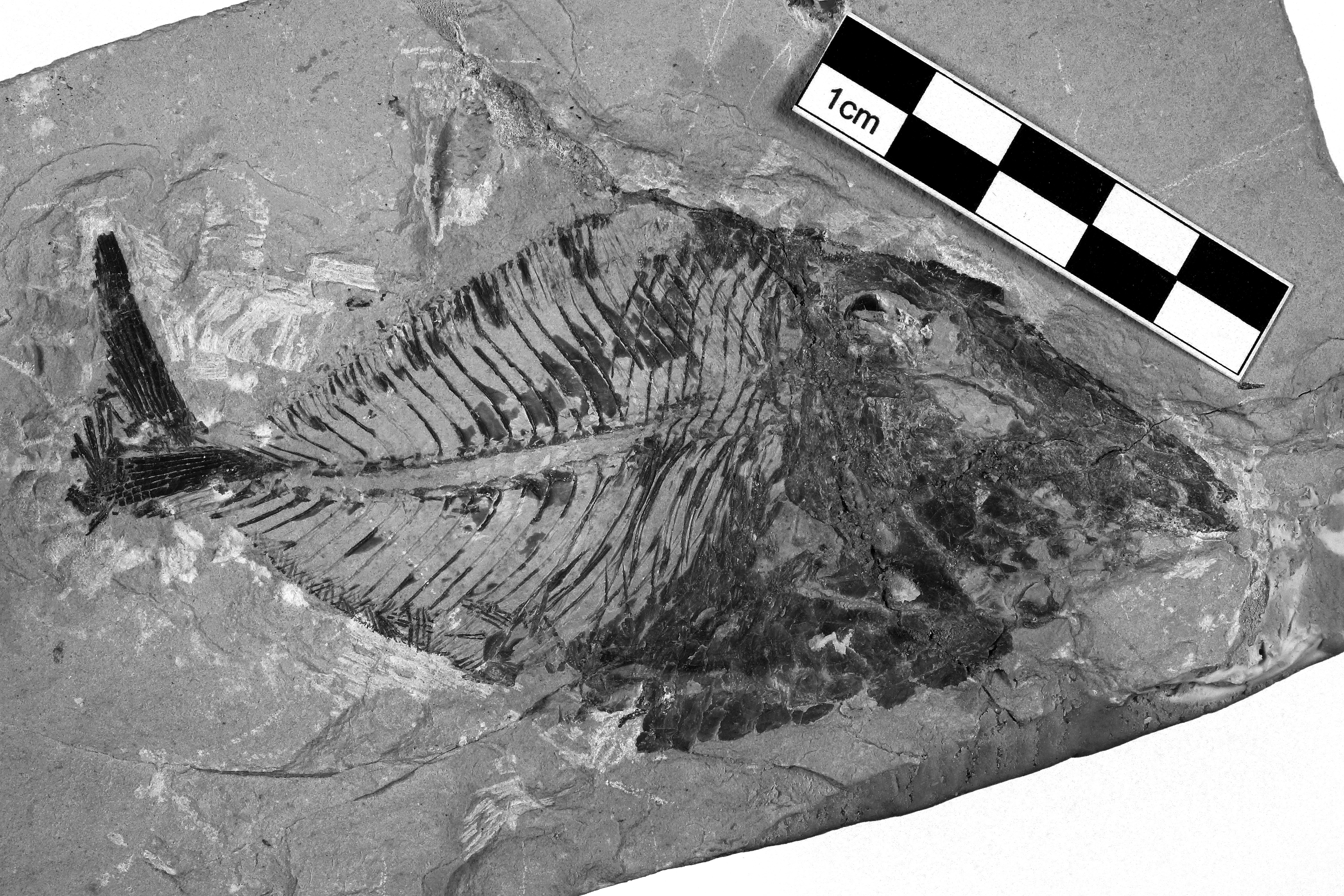
Sample CLC S-461, a complete specimen from Ein Namoura, Lebanon (Fig. 3). Total length: 114 mm. Standard length: 98 mm.
Sample CLC S-1098, a nearly complete specimen from Haqel, Lebanon ( Fig. 4 View Fig ). The caudal fin is crushed. Total length: 115 mm. Standard length: 102 mm.
Formation and locality
Marine Upper Cenomanian deposits of Haqel and Ein Namoura, Lebanon.
General morphology and morphometric data ( Figs 1–5 View Fig View Fig View Fig View Fig )
Acrorhinichthys poyatoi gen. et sp. nov. is a small pycnodontiform. The total length of the specimens does not exceed 12 cm. The fish is high-bodied but not discoid in shape. The dorsal profile is angular, with a well marked apex located midway between the snout and the caudal peduncle. This apex, built with the enlarged posterior dorsal scute, forms a small dorsal prominence. The ventral margin of the body is more or less rounded.
The morphometric data of the holotype (CLC S-630a, b) and of one of the paratypes (CLC S-461) are given in % of their standard length, 72 mm and 98 mm respectively:
CLC S-630 CLC S-461 Length of the head (from the snout to the occipital region) …………………42.4 % …….. 38.4 % Depth of the head (including the pectoral girdle) ……………………………58.5 % …….. 50.5 % Maximum depth of the body (at the dorsal apex level) ………………………80.5 % …….. 70.3 % Prepelvic length ………………………………………………………………61.0 % …….. 55.2 % Predorsal length ………………………………………………………………55.6 % …….. 56.0 % Basal length of the dorsal fin …………………………………………………53.7 % …….. 47.4 % Preanal length …………………………………………………………………2.7 % …….. 66.8 % Basal length of the anal fin …………………………………………………33.2 % …….. 31.9 % Depth of the caudal peduncle …………………………………………………7.3 % ……… 7.3 %
Osteology
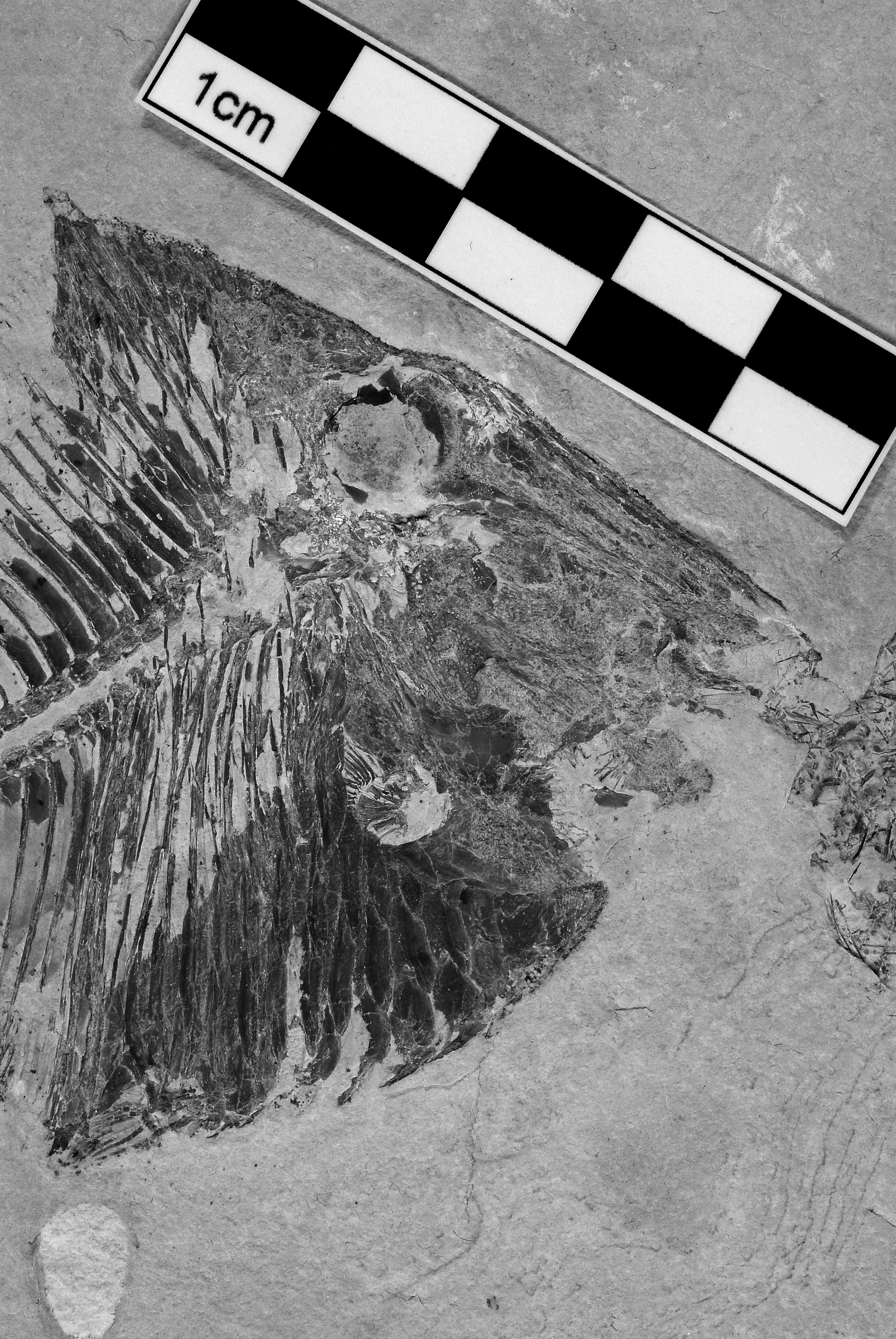
1. The skull ( Figs 6 View Fig –11)
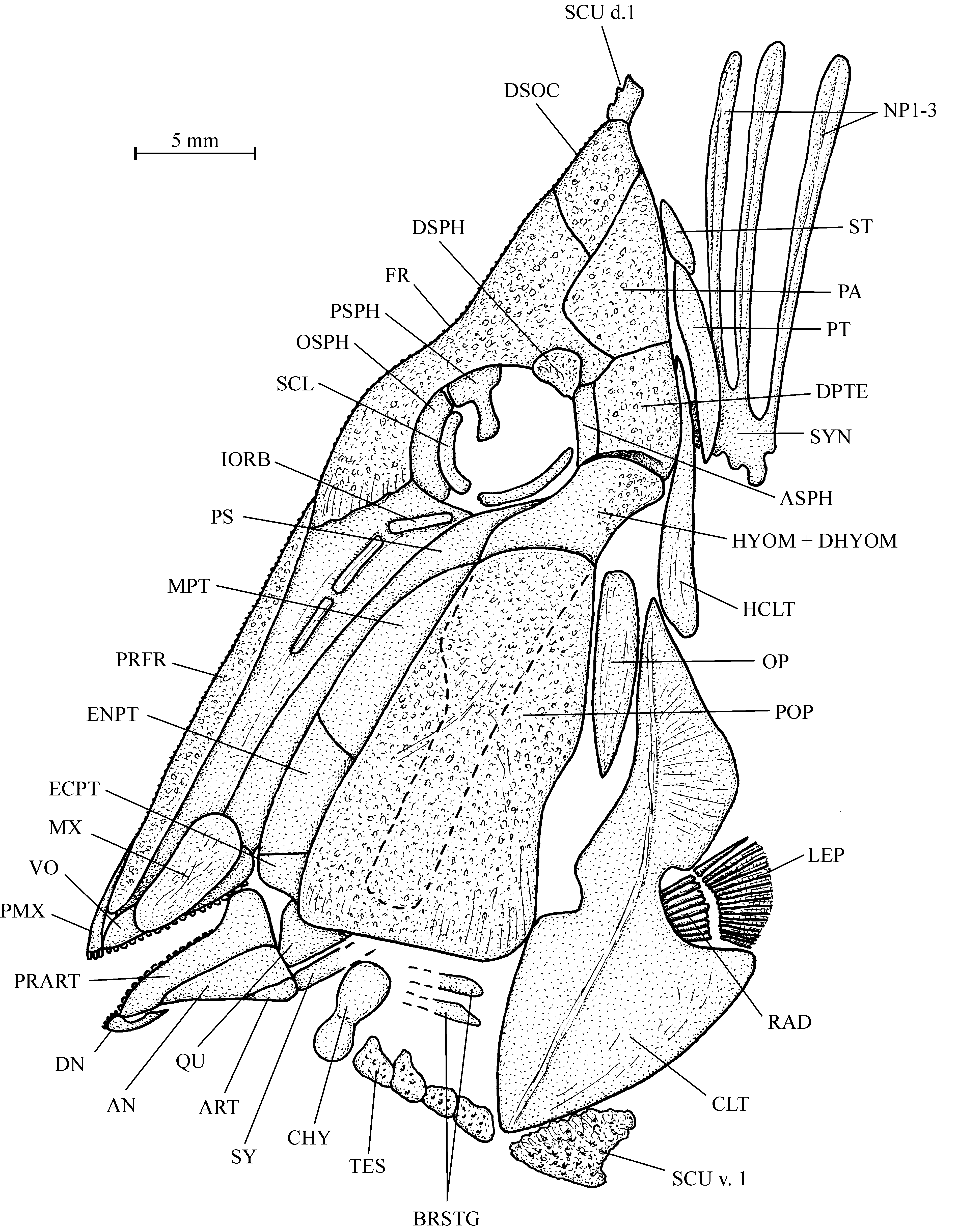
The overall shape of the head is triangular in lateral view, with a very long preorbital region, a large orbit, reduced jaws and a more or less pointed snout. The frontal profile between the occipital area and the snout is almost rectilinear. The dermal bones of the skull are ornamented with small tubercles and very thin ridges.
The mesethmoid is long and narrow. Its dorsal margin is covered by a pair of elongated and narrow prefrontals. The vomer is long and toothed, but it is not possible to determinate the number of tooth rows. Only one row is visible in the holotype. It contains 16 small, rounded teeth, the last ones being a little longer than the others.
The skull roof is formed by one dermosupraoccipital and paired frontals, parietals and dermopterotics. There is no temporal fenestra. The frontal is short and rather narrow, except in its posterior part. It covers the orbital area and exceeds this region only a little anteriorly and posteriorly. The dermosupraoccipital is small and the occipital region is conical in shape. The parietal is triangle-shaped, with a broad ventral margin and a pointed upper corner. The bone is devoid of a brush-like process. The dermopterotic is deeper than broad. A small supratemporal (= extrascapular) is located behind the parietal on the holotype CLC S-630a.
The posterior bones of the endocranium are not visible, except the autosphenotic that is partly covered by the dermosphenotic and the exoccipital that seems to be fused with a synarcual.
The very long edentulous parasphenoid is inflected downwards below the orbit. The anterior part of the bone is not enlarged. The orbitosphenoid and pleurosphenoid are preserved in the orbit of the holotype and in paratype CLC S-461. The orbitosphenoid is pressed against the mesethmoid. The pterosphenoid is a small bone. The presence of a basisphenoid is uncertain.
The jaws are very small when compared to the skull size. The mouth gape is obliquely oriented. As preserved in the holotype, the premaxilla bears 3 incisiform teeth and the dentary, reduced to its ventral branch, 5 teeth. The dentary is much shorter than the premaxilla. The maxilla, visible in paratype CLC S-1098, is longer than deep. The prearticular is triangle-shaped, with a well developed coronoid process. The bone bears small, rounded teeth, similar to those of the vomer, and they seem to be irregularly arranged. The angular partially covers the prearticular. The articular forms the posterior ventral corner of the mandible.
Both the quadrate and the symplectic articulate with the lower jaw. A small ectopterygoid and parts of the metapterygoid and entopterygoid are visible between the parasphenoid and the anterior border of the preopercle. The entopterygoid and ectopterygoid are toothless as is usual in Pycnodontiformes .
The dermosphenotic is a small, plate-like bone. Fragments of tubular infraorbitals are preserved on the holotype and paratype CLC S-461. A sclerotic ring is present and is composed of two elements.
The preopercle is by far the largest bone of the skull and covers practically the complete cheek. The small, deep, narrow, dorsally rounded and ventrally pointed opercle is wedged between the preopercle and the dorsal branch of the cleithrum. The posterior parts of two branchiostegal rays are preserved in paratype CLC S-461.
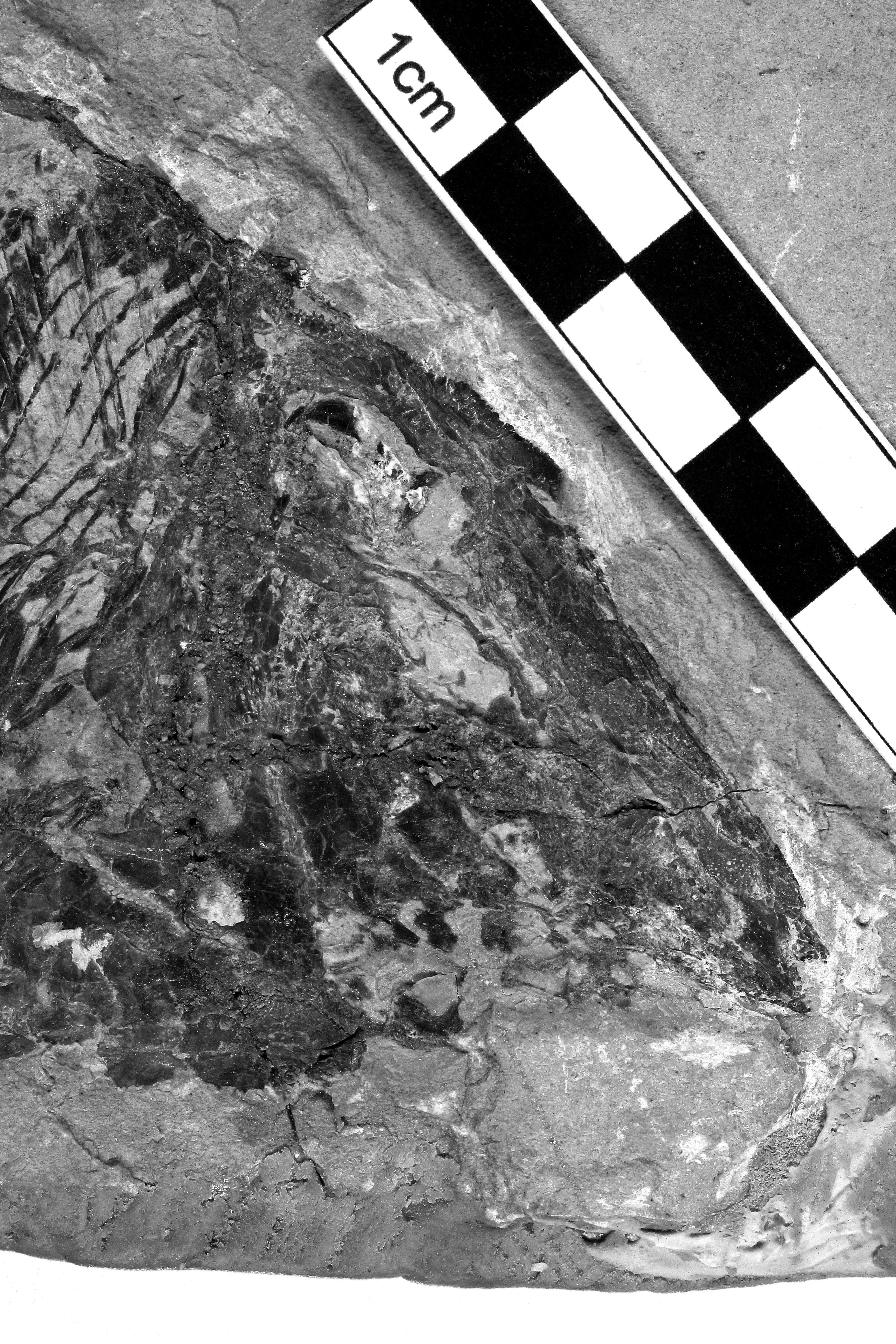
Fragments and impressions of a few ornamented bony tesserae are visible in the gular region of the holotype CLC S-630a.
The exposed part of the hyomandibula-dermohyomandibula is much smaller than the preopercle. The long and broad ventral branch of the hyomandibula is visible under the preopercle in paratype CLC S-461 and in the holotype. The hyomandibula is devoid of an opercular process. Traces of a rather large ceratohyal bar are visible in paratype CLC S-1098. The holotype shows a few long and thin branchiospines.
2. The girdles ( Figs 6–10 View Fig View Fig View Fig View Fig View Fig , 19 View Fig )
The cleithrum is very similar to the one of Proscinetes elegans (Agassiz, 1833) as figured by Nursall (1996: fig. 11b). The two branches of the bone are separated by a large posterior concavity in which the pectoral fin is inserted. The anteroventral branch is more or less triangular, rather short but very broad and ornamented with a median crest. The dorsal branch is also triangle-shaped, but somewhat narrower than the ventral one and with an acuminate upper extremity. The posttemporal and the deep and narrow hypercleithrum (= supracleithrum) are visible in paratype CLC S-1098. The endochondral part of the girdle is not visible. The short pectoral fin is supported by 8 or 9 pterygiophores and starts with 1 dorsal spine followed by 16 or 17 short segmented rays.
The reduced pelvic girdle is well preserved on the holotype. Each pelvic bone bears 4 segmented rays.
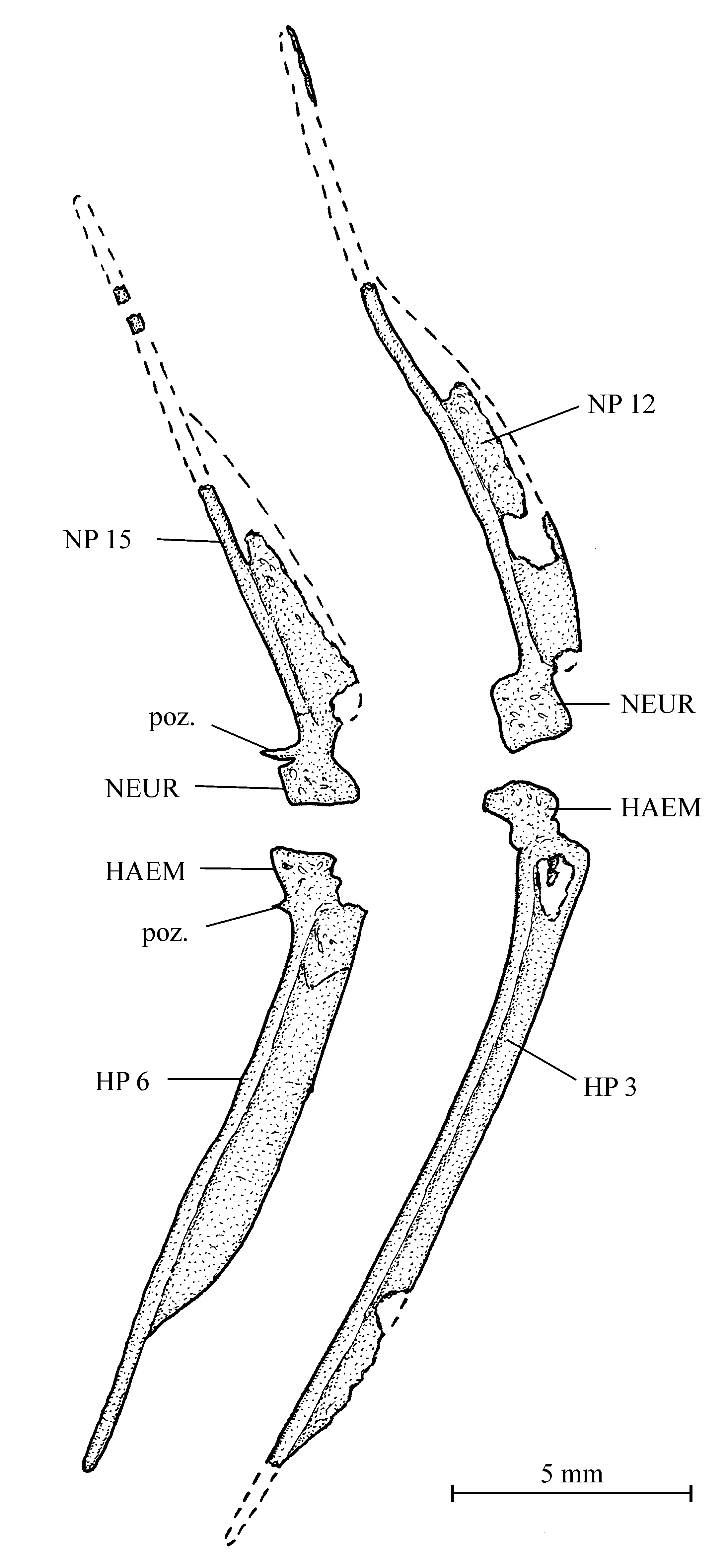
3. The axial skeleton ( Figs 1–10 View Fig View Fig View Fig View Fig View Fig View Fig View Fig View Fig View Fig )
Samples CLC S-630 (a and b), S-461 and S-1098, respectively, possess 24, 25 and 27 neural spines in front of the epichordal series. All three specimens have 14 haemal spines in front of the hypochordal elements. There are 9 or 10 pairs of long ribs that are broadened in their upper part and so become
contiguous. There are no autogenous neural spines. The first three neural spines are fused to a synarcual that is articulated to the rear of the skull and also seems to include the exoccipital. The neural and haemal spines bear an anterior sagittal flange, except the few ones preceding the epichordal and hypochordal pieces. The neural and haemal arches are well developed, but they do not completely surround the notochord. At the level of the first vertebrae, the neural arches are simply in contact. At the end of the abdominal region and in the caudal region, most neural and haemal arches present a more complex contact by means of one single, small postzygapophysis.
4. The dorsal and anal fins ( Figs 1–5 View Fig View Fig View Fig View Fig )
The shape of the dorsal and anal fins is strip-like (type A2 of Poyato-Ariza & Wenz 2002: fig. 34). The holotype and paratype CLC S-461 have 51 and 55 pterygiophores in the dorsal fin and 36 and 41 pterygiophores in the anal fin, respectively. The dorsal and anal fins of paratype CLC S-1098 are incomplete. The dorsal fin begins just after the dorsal apex. Each dorsal pterygiophore bears a ray. The first ray is reduced to a short spine. The two following rays are long, segmented and pointed. The remaining dorsal rays are segmented and branched. The origin of the anal fin is located at a more posterior level than the dorsal one. The first anal pterygiophore is short and bears a small, spiny ray. The second and the third pterygiophores support longer, segmented and pointed rays. The other anal rays are segmented and branched.
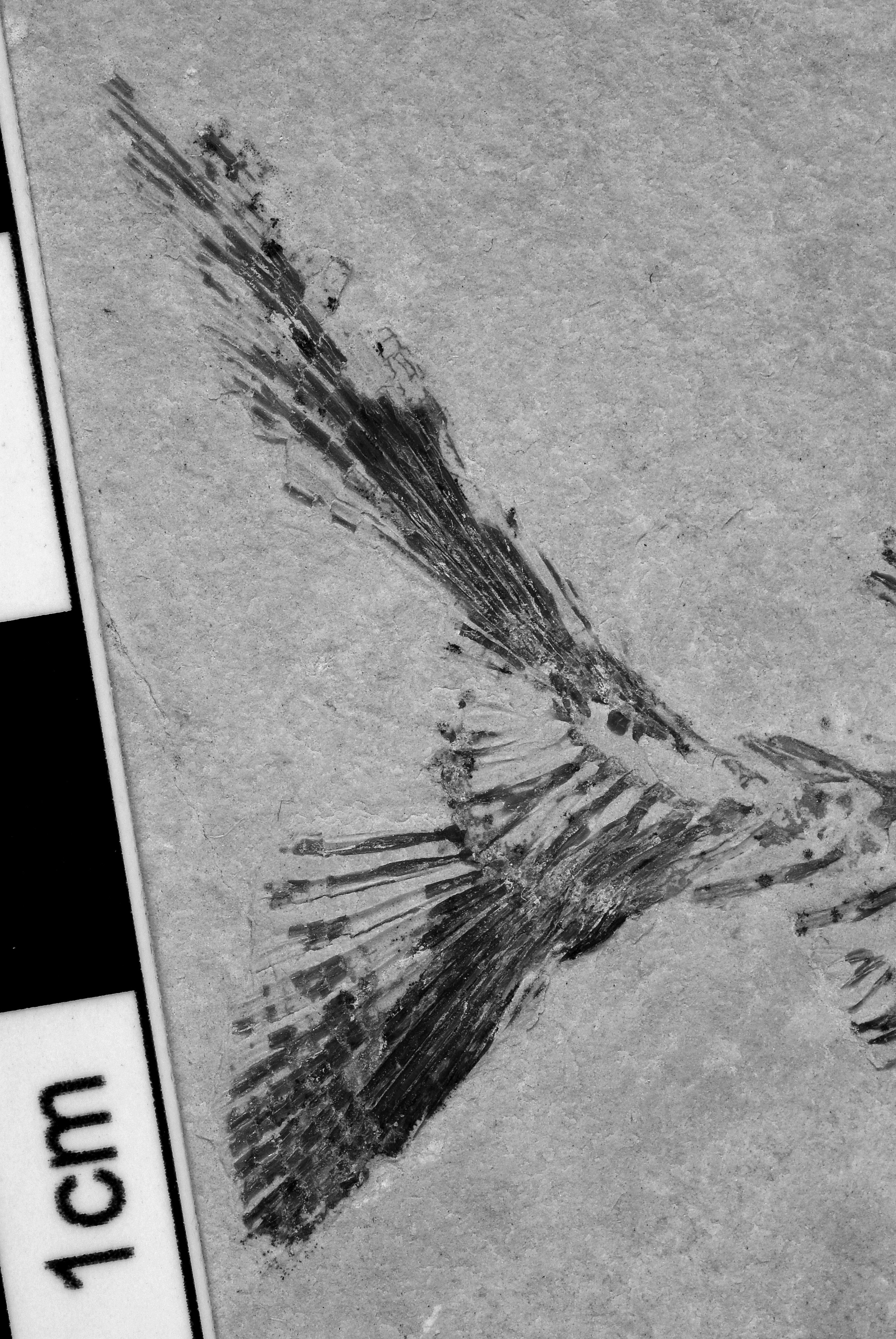
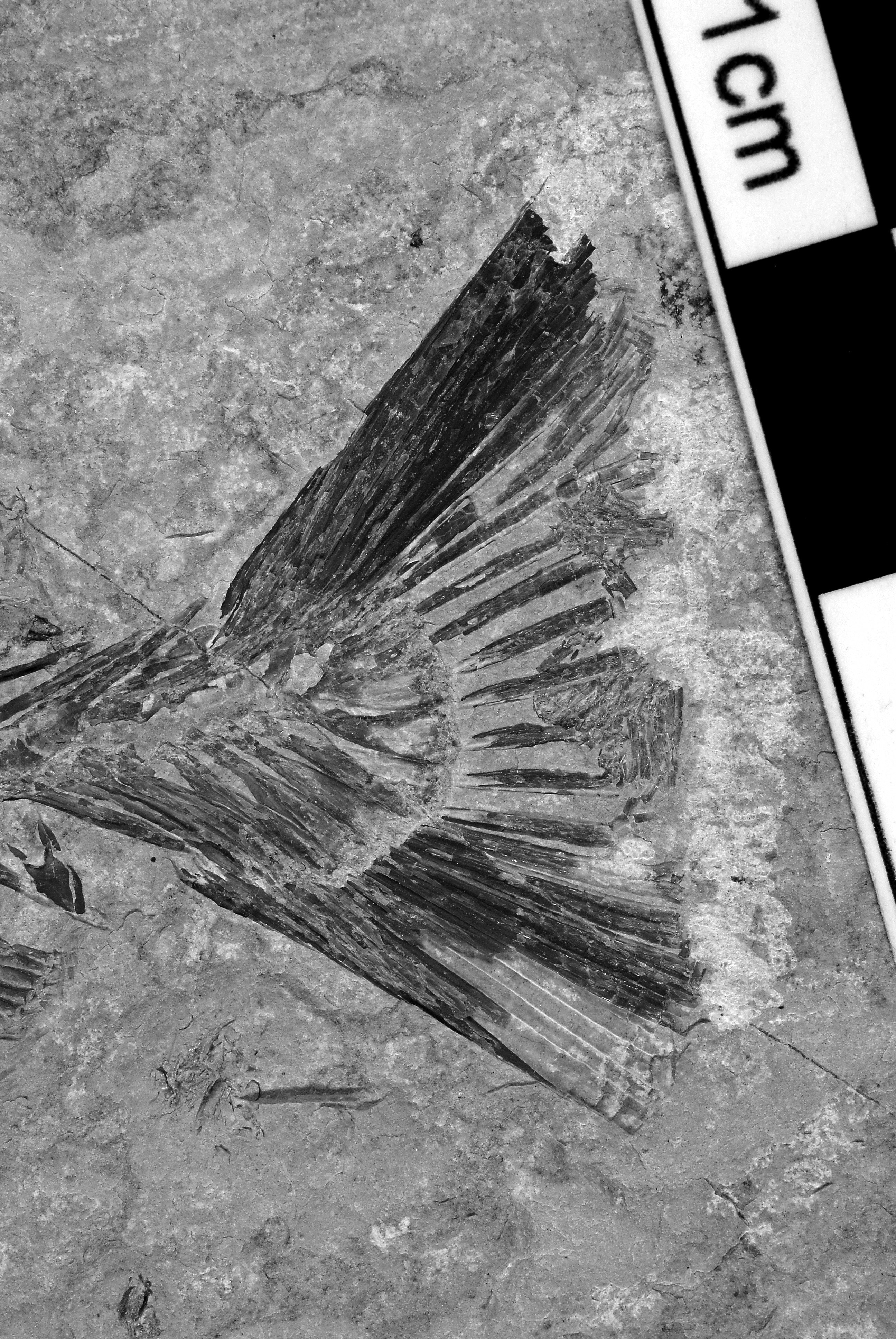
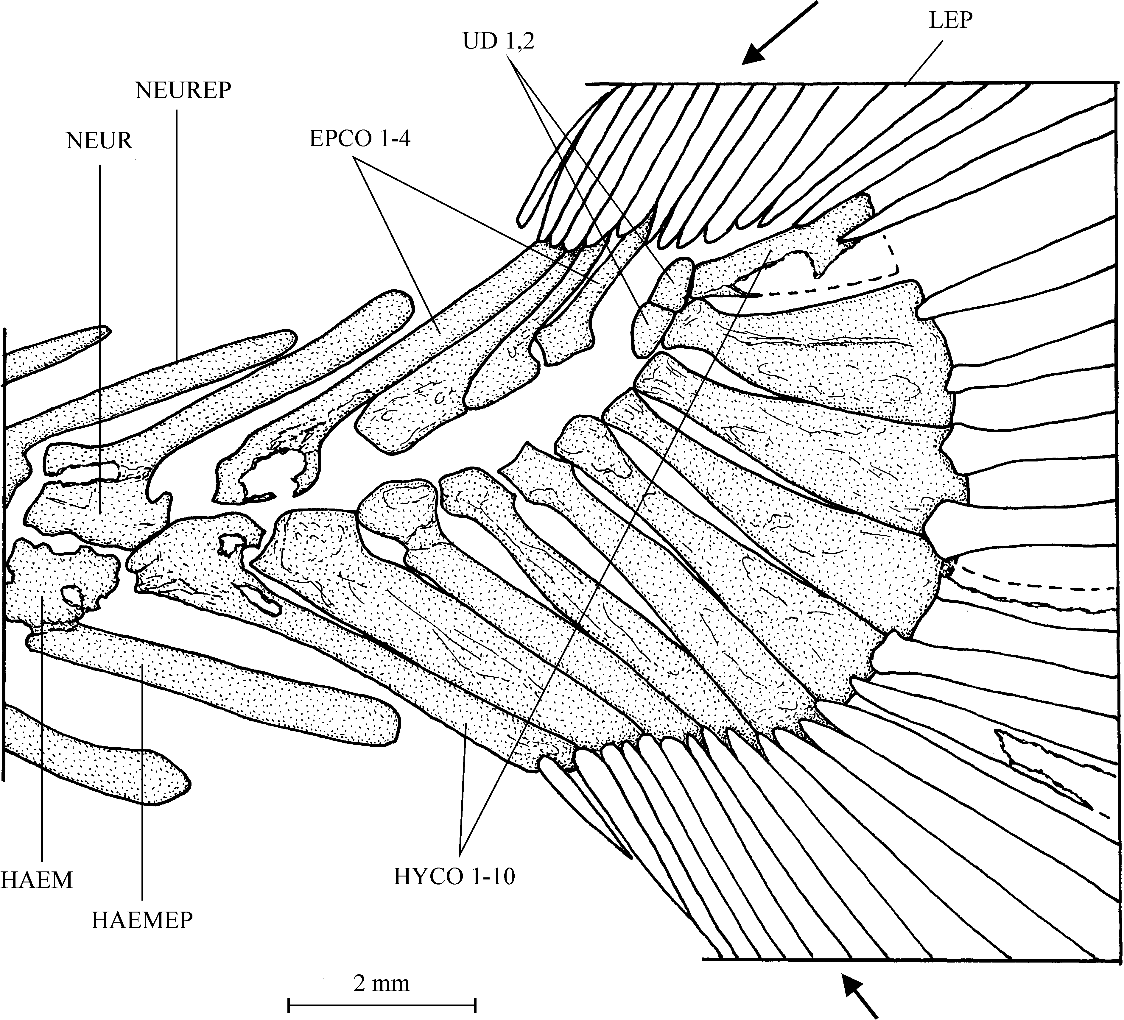
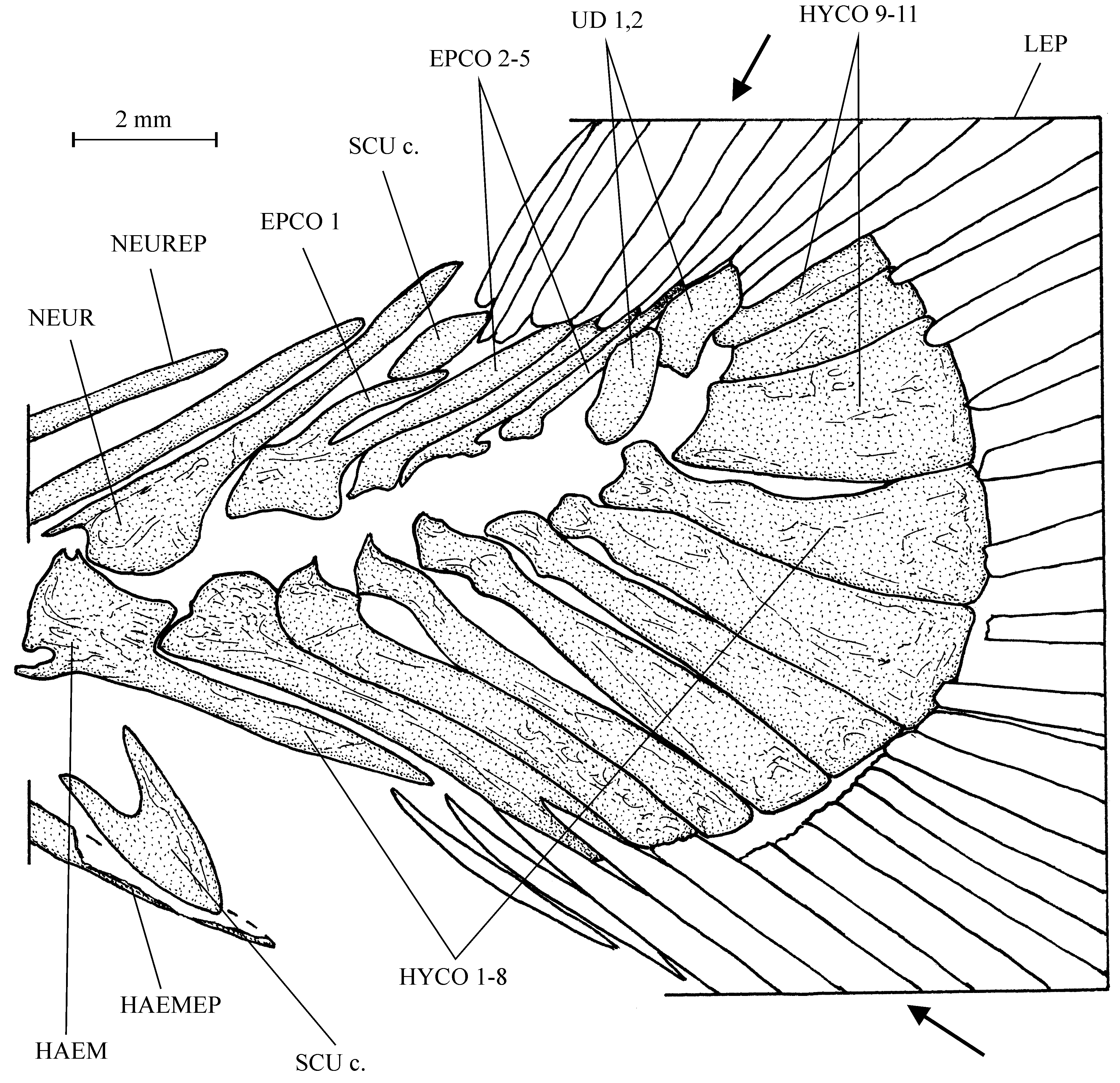
5. The caudal skeleton ( Figs 12–16 View Fig View Fig View Fig View Fig View Fig )
The caudal peduncle is very short because of the proximity of the dorsal and anal fins with the caudal fin. The caudal skeleton is composed of 4 or 5 epichordals, 10 or 11 hypochordals and 2 urodermals. Epichordals and hypochordals are rather long. The seventh, eighth and ninth hypochordals are slightly broadened, but there is no real hypertrophy.
The caudal fin is large and its contour is double emarginated ( Poyato-Ariza & Wenz 2002: fig. 36E). There are 22 or 23 principal caudal rays, 4 to 6 dorsal and 5 to 6 ventral procurrent rays. The most external dorsal and ventral principal rays are segmented and pointed. The other principal rays are segmented and branched.
In paratype CLC S-461, there are a small dorsal and a larger ventral scute preceding the caudal fin.
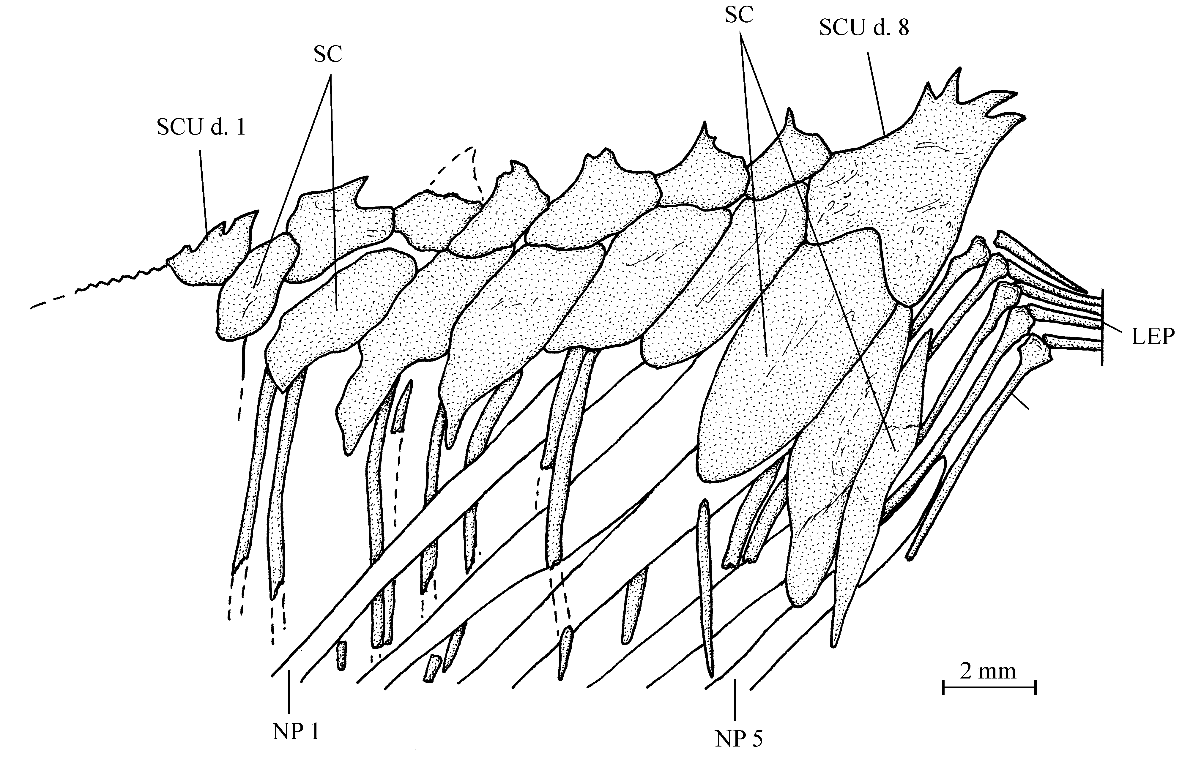
6. Squamation ( Figs 5 View Fig , 17–18 View Fig View Fig )
The dorsal ridge is perfectly preserved in paratype CLC S-461. It is composed of 8 spiny scutes. The first one bears three small spines and rests on the dermosupraoccipital. The last one bears four spines and is much larger than the preceding scutes. This large eighth scute forms a small prominence at the dorsal apex level.
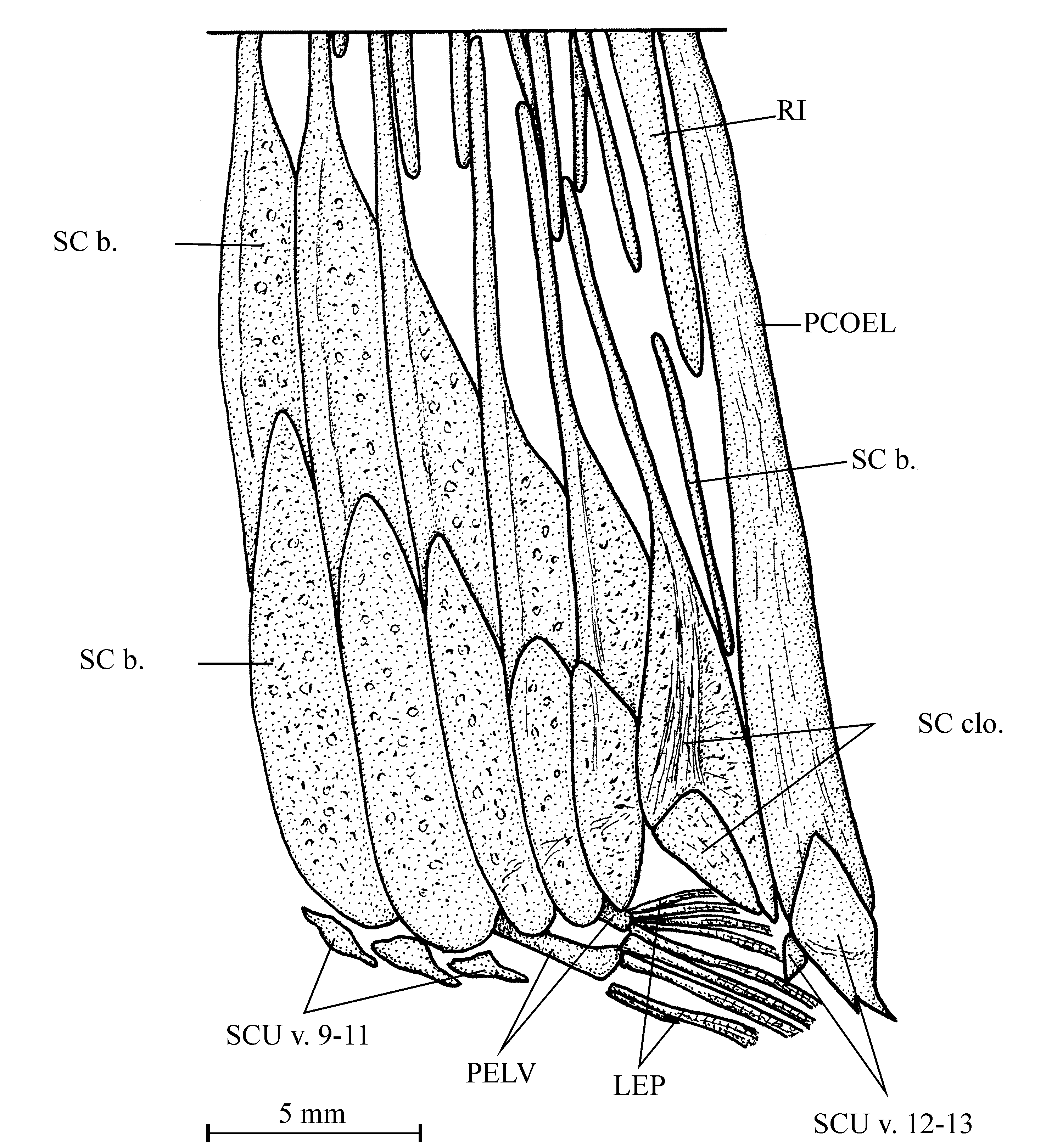
The holotype (CLC S-630a, b) exhibits the best preserved ventral keel. There are 13 scutes, of which 11 are located between the pectoral girdle and the cloaca, and 2 posterior to the cloaca. They are ornamented with small tubercles. The first one is triangularly shaped, located just below the cleithrum, and is by far the largest of the series. The second and third ones are smaller and rectangular in shape. The eight other
precloacal scutes are smaller still, with a pointed posterior extremity. The two postcloacal scutes are associated with the ventral extremity of the postcoelomic bone. The first one is very small. The second one is much larger and bears two small spines.
The flank scales are restricted to the abdominal region of the fish. A row of complete dorsal scales is associated with the dorsal ridge scutes. The two or three last scales of this series overlap the first dorsal pterygiophores. The scales are also complete in the ventral part of the abdominal region. The other flank scales are reduced to their bar-like component.
Two complete scales are located just above the cloaca. They are imbricated, one overlapping the other. The ventral one is small and triangular. The dorsal one is much deeper, with a lateral wing and a well developed bar-like component. Its ventral margin is broad and concave.


No known copyright restrictions apply. See Agosti, D., Egloff, W., 2009. Taxonomic information exchange and copyright: the Plazi approach. BMC Research Notes 2009, 2:53 for further explanation.
|
Kingdom |
|
|
Phylum |
|
|
Class |
|
|
Order |
|
|
Family |
|
|
Genus |